|
Exhibition Hall 11:00 - 12:00 |
|
|
|
Computer # |
|
3262.
 |
25 |
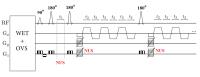
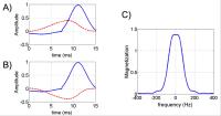
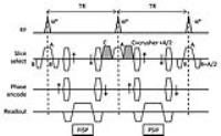
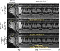
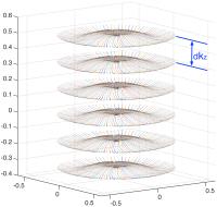
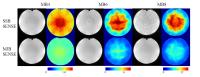
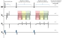
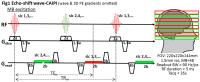
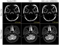
Improving Time Efficiency for T2-weighted Fat-Water Imaging by
Using Multiband Simultaneous Multi-Slice Accelerated TSE Dixon -
Video Not Available
Dingxin Wang1,2, Xiufeng Li2, Xiaoping
Wu2, and Kamil Ugurbil2
1Siemens Healthcare, Minneapolis, MN, United
States, 2Center
for Magnetic Resonance Research, University of Minnesota,
Minneapolis, MN, United States
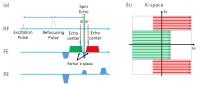
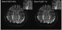
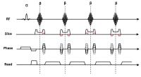
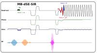
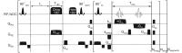
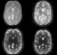
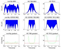
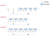
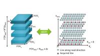
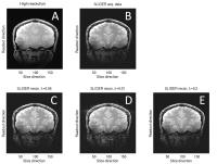
Dixon technique requires at least two images with different
echo times, which increases TSE echo spacing and TR, and
therefore prolongs total acquisition time. Slice
acceleration may help improve imaging efficiency of TSE
fat-water Dixon imaging. In this study, we develop a
multiband slice accelerated TSE Dixon sequence and
demonstrate the feasibility of SMS TSE Dixon acquisition for
efficient T2-weighted fat-water imaging.
|
|
3263.
 |
26 |
High Performance Volumetric 3T Breast Acquisition: A Foundation
for Multi-Parametric Imaging 
Jorge E Jimenez1, Kevin M Johnson1,
Leah C Henze Bancroft1, Diego Hernando2,
Roberta M Strigel1,2, Scott B Reeder2,3,
and Walter F Block1,2,3
1Department of Medical Physics, University of
Wisconsin-Madison, Madison, WI, United States, 2Department
of Radiology, University of Wisconsin School of Medicine and
Public Health, Madison, WI, United States, 3Department
of Biomedical Engineering, University of Wisconsin-Madison,
Madison, WI, United States
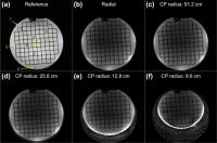
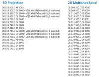
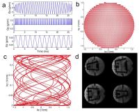
We present a 3D T1-Weighted radial trajectory suited to work
with IDEAL fat/water separation. Some relevant
characteristics are: rapid acquisition, reliable fat
suppression, and high resolution despite significant data
undersampling. The method is demonstrated in 3T bilateral
breast MR imaging where isotropic resolution of 0.8 mm is
achieved. In addition, we show the value of high count
channel array for breast imaging.
|
|
3264.
 |
27 |
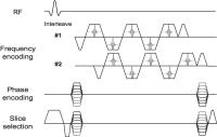
Bipolar time-interleaved multi-echo gradient echo imaging for
high-resolution water-fat imaging
Stefan Ruschke1, Holger Eggers2,
Hendrik Kooijman3, Houchun H. Hu4,
Ernst J. Rummeny1, Axel Haase5, Thomas
Baum1, and Dimitrios C. Karampinos1
1Department of Diagnostic and Interventional
Radiology, Technische Universität München, Munich, Germany, 2Philips
Research, Hamburg, Germany, 3Philips
Healthcare, Hamburg, Germany, 4Radiology,
Phoenix Children’s Hospital, Phoenix, AZ, United States, 5Zentralinstitut
fu¨r Medizintechnik, Technische Universität München,
Garching, Germany
As the spatial resolution of a multi-echo gradient-echo
imaging sequence increases, the echo time step increases
resulting in an increased echo time step and an echo time
selection that degrades the noise performance of water-fat
separation. Time-interleaved gradient echo imaging combined
with bipolar (flyback) gradients can achieve reasonable echo
time steps at high resolution without dramatically
increasing scan time. However, bipolar gradients are
associated with known phase errors problems, which can lead
to fat quantification errors. The present work develops a
methodology for acquiring bipolar time interleaved
multi-echo gradient echo data and for correcting the
relevant phase errors.
|
|
3265.
 |
28 |
Silicone, fat, and water separation using a single-pass 3D Dixon
acquisition
Ken-Pin Hwang1, Jingfei Ma1, Lloyd
Estkowski2, Ann Shimakawa2, Kang Wang2,
Daniel Litwiller2, Zachary Slavens2,
Ersin Bayram2, and Bruce Daniel3
1Department of Imaging Physics, The University of
Texas M.D. Anderson Cancer Center, Houston, TX, United
States, 2MR
Applications and Workflow, GE Healthcare, Waukesha, WI,
United States,3Department of Radiology, Stanford
University, Stanford, CA, United States
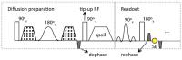
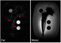
Separation of silicone, fat, and water is performed by using
a two-step Dixon processing algorithm and a bipolar
triple-echo readout in a 3D FSE sequence. The echoes are
spaced for conventional water-fat separation, but the first
and last echoes are also used to generate a phase map with
double the phase evolution for resolving fat from silicone,
which are relatively close in terms of chemical shift.
Individual images of each of the three species are
reconstructed in phantom and human data. The proposed method
demonstrates improved SNR efficiency and robustness to field
inhomogeneity compared to conventional saturation and
inversion recovery techniques.
|
|
3266.
 |
29 |
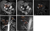
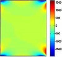
Incorporation of Prior Knowledge of Main Field Inhomogeneity in
Dixon Methods
Holger Eggers1, Liesbeth Geerts-Ossevoort2,
Gert Mulder2, and Clemens Bos3
1Philips Research, Hamburg, Germany, 2Philips
Healthcare, Best, Netherlands, 3Imaging
Division, University Medical Center Utrecht, Utrecht,
Netherlands
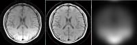
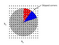
The robustness of Dixon methods often deteriorates close to
large main field inhomogeneity. To resolve this problem, the
exploitation of prior knowledge of magnet imperfections is
considered in this work. Magnet-induced main field
inhomogeneity is modeled to predict and correct spatial
variations of the phase in single-echo images before the
separation of water and fat signal. Improved fat suppression
is demonstrated with this approach in first-pass peripheral
angiography, in particular in the corners of the large FOVs.
|
|
3267.
 |
30 |
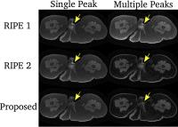
Comparison of Gradient Echo MRI Water-Fat separation and single
voxel 1H MRS for liver fat fraction measurements in a dietary
intervention study at 3T
Stephen Bawden1,2, Carolyn Chee3,
Caroline Hoad1, Guruprasad Aithal2,
Ian Macdonald3, Richard Bowtell1, and
Penny Gowland1
1Sir Peter Mansfield Imaging Centre, University
of Nottingham, Nottingham, United Kingdom, 2NIHR
Nottingham Digestive Diseases Research Unit, University of
Nottingham, Nottingham, United Kingdom, 3School
of Life Sciences, University of Nottingham, Nottingham,
United Kingdom
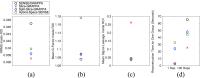
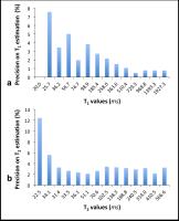
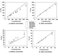
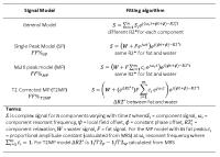
This study compared hepatic fat fraction measured using MRS
and gradient echo MRI at 3T. Fitting algorithms using a
single fat peak and multiple fat peaks were compared with
MRS data at TE=20ms and also T2-corrected MRS. Individual
differences between T2 values of water and fat calculated
from MRS were also used to estimate the difference between
R2*-water and R2*-fat and included in the multi peak fitting
per subject. The results showed a good correlation between
multi-peak and MRS data (R2 =
0.9), but applying T2-correction to MRS increased the
scatter (R2 =
0.67) and systematic error (gradient = 1.34). Using the new
R2* corrected fitting algorithm resulted in similar scatter
(R2 =
0.66) but improved systematic error (gradient = 1.09). The
results from this study indicate the dual-R2* fitting is
important at 3T and further developments should be made to
optimize these methods.
|
|
3268.
 |
31 |
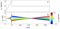
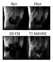
Path based phase estimation for fat suppression near metal
implants
Laura Jane King1, Rick Millane1, Hans
Weber2, Brian Hargreaves2, and Phil
Bones1
1Electrical and Computer Engineering, University
of Canterbury, Christchurch, New Zealand, 2Radiology,
Stanford University, Stanford, CA, United States
Being able to perform robust Dixon imaging near metal
implants would allow for improved contrast-enhanced fat
suppression. This requires accurate calculation of the phase
shift due to the B0 field variation. We present a new
technique where the phase is first estimated at the outer
edges of the image. The method works inwards along a set of
adjacent paths, finishing at the boundary of the implant.
The described method is used to successfully separate fat
and water in the vicinity of a titanium hip replacement,
with phantom results shown.
|
|
3269.
 |
32 |
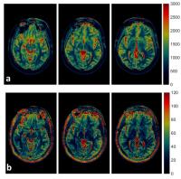
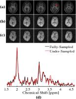
Improving Parameter Mapping in MRI Relaxometry and Multi-Echo
Dixon using an Automated Spectral Denoising
Felix Lugauer1,2, Dominik Nickel3,
Stephan A. R. Kannengiesser3, Samuel Barnes4,
Barbara Holshouser4, Jens Wetzl1,
Joachim Hornegger1, and Andreas Maier1,2
1Pattern Recognition Lab, Department of Computer
Science, Friedrich-Alexander Universität Erlangen-Nürnberg,
Erlangen, Germany, 2Research
Training Group 1773 “Heterogeneous Image Systems”, Erlangen,
Germany, 3MR
Applications Predevelopment, Siemens Healthcare GmbH,
Erlangen, Germany, 4Department
of Radiology, Loma Linda University Medical Center, Loma
Linda, CA, United States
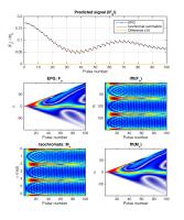
Magnitude-based parameter fitting is commonly used for
relaxometry and multi-echo Dixon but introduces a bias for
relaxation and fat fraction estimates, particularly for a
low signal-to-noise ratio and high relaxation. The
application of an automated, patchwise denoising to the
multi-echo image series prior to parameter fitting,
considerably increased the SNR; thus, reducing the bias and
standard deviation in the estimates of the fit. Our findings
were validated on both numerical and in-vivo experiments.
|
|
3270.
 |
33 |
Two-point fat water separation using safest path region growing
with self-feeding phasor estimation algorithm
Chuanli Cheng1,2, Chao Zou1, Hairong
Zheng1, and Xin Liu1
1Shenzhen Institutes of Advanced Technology,
Chinese Academy of Sciences, Shenzhen, China, People's
Republic of, 2University
of Chinese Academy of Sciences, Beijing, China, People's
Republic of
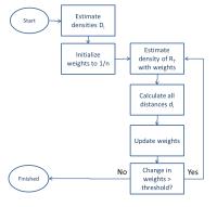
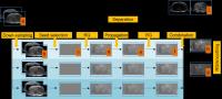
A novel two-point fat water separation method using safest
path region growing with self-feeding phasor estimation
algorithm is proposed. The phasor map is estimated by
multiresolution region growing scheme where the seed pixels
identification and region growing scheme is performed
independently between coarser resolutions, avoiding the
erroneous propagation between resolutions. The
“self-feeding” mechanism when merging the phasor maps
ensures the reliability of seed pixels selection at the
finest resolution. The algorithm was tested on c-spine and
abdomen data and shown to be robust in fast varying
field and disjoint areas.
|
|
3271.
 |
34 |
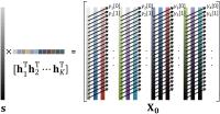
Increased measurement precision for fatty acid composition
mapping by parameter reduction
Johan Berglund1, Henric Rydén1, and
Mikael Skorpil1
1Karolinska University Hospital, Stockholm,
Sweden
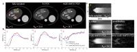
An imaging method for mapping Fatty Acid Composition using
three triglyceride spectrum model parameters (FAC3) was
modified into mapping Fatty Acid Composition with only one
spectrum model parameter (FAC1), namely the average number
of double bonds per triglyceride (ndb). Images of a
patient with a lipoma were reconstructed using both methods.
The FAC3 ndb map
showed large-scale variation from left to right, giving
significant variation between subcutaneous adipose tissue
locations. Measurements from the FAC1 ndb map
were consistent within the adipose tissue, offering a higher
level of confidence and more precise measurements.
|
|
3272.
 |
35 |
A Fast and Globally Optimal 3-D Graph Search Algorithm for Phase
Unwrapping in MRI with Applications in Quantitative
Susceptibility Mapping (QSM)
Chen Cui1, Abhay Shah1, Cameron M.
Cushing2, Vince Magnotta3, Xiaodong Wu1,2,
and Mathews Jacob1
1Electrical and Computer Engineering, University
of Iowa, Iowa City, IA, United States, 2Radiology
Oncology, University of Iowa Hospitals and Clinics, Iowa
City, IA, United States, 3Radiology,
University of Iowa Hospitals and Clinics, Iowa City, IA,
United States
Phase wrapping is a classic problem in many fields of study.
In MRI, Unwrapping the phase in the estimation of B0 field
inhomogeneity maps is challenging commonly due to the
presence of a large field inhomogeneity, anatomical
discontinuities or low SNR in certain regions of the map
(e.g. boundaries). We propose a general phase unwrapping
method that exploits the smoothness of the field map in
three spatial dimensions. The method is solved using a
linear-convex constrained graph search algorithm that
provides the globally guaranteed optimal solution without
over-smoothing effect. The proposed scheme aims to produce a
robust solution for field map estimation that will further
improve the quality of quantitative susceptibility mapping
(QSM).
|
|
3273.
 |
36 |
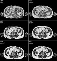
Analysis of Bias with SNR in Multi-echo Chemical Shift Encoded
Fat Quantification
James H Holmes1, Diego Hernando2, Kang
Wang1, Ann Shimakawa3, Nate Roberts2,
and Scott B Reeder2,4,5,6
1MR Applications and Workflow, GE Healthcare,
Madison, WI, United States, 2Radiology,
University of Wisconsin-Madison, Madison, WI, United States, 3MR
Applications and Workflow, GE Healthcare, Menlo Park, CA,
United States, 4Medical
Physics, University of Wisconsin-Madison, Madison, WI,
United States, 5Biomedical
Engineering, University of Wisconsin-Madison, Madison, WI,
United States,6Emergency Medicine, University of
Wisconsin-Madison, Madison, WI, United States
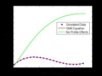
Multi-echo chemical shift encoded techniques provide
accurate fat quantification over a broad range of
fat-fractions and acquisition parameters. In order to be
accurate, these techniques must correct for all relevant
confounding factors. Emerging applications (eg: fat
quantification with high spatial resolution or in iron
overloaded tissues) can result in data with significantly
lower signal-to-noise ratio (SNR) compared to previously
established applications. In this work, we characterize the
accuracy of current noise bias correction techniques for fat
quantification in the low SNR regime and show simple
modifications may enable accurate fat quantification for a
wider range of SNR.
|
|
3274.
 |
37 |
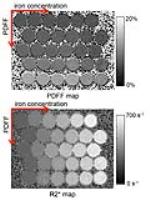
DEVELOPMENT AND MULTI-CENTER VALIDATION OF A NOVEL
WATER-FAT-IRON PHANTOM FOR JOINT FAT AND IRON QUANTIFICATION
Samir D. Sharma1, Diego Hernando1,
Takeshi Yokoo2, Mustafa R. Bashir3,4,
Jean Shaffer3,4, Qing Yuan2, Stefan
Ruschke5, Dimitrios C. Karampinos5,
Jean H. Brittain1, and Scott B. Reeder1,6
1Radiology, University of Wisconsin - Madison,
Madison, WI, United States, 2Radiology,
UT Southwestern Medical Center, Dallas, TX, United States, 3Radiology,
Duke University, Durham, NC, United States, 4Center
for Advanced Magnetic Resonance Development, Duke
University, Durham, NC, United States, 5Diagnostic
and Interventional Radiology, Technische Universität
München, Munich, Germany, 6Medical
Physics, University of Wisconsin-Madison, Madison, WI,
United States
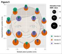
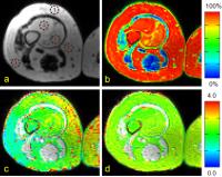
The need for rapid and non-invasive assessment of fat and
iron deposition has become increasingly important given the
high prevalence of obesity and obesity-related comorbidities
as well as the need for monitoring chelation treatment in
patients with iron overload. Recent advances in
gradient-echo imaging have enabled the simultaneous
quantification of fat and iron concentrations throughout the
body. To ensure fidelity of these quantitative techniques,
validation studies must be performed, ideally with initial
testing in phantoms. In this work, we report on the
development of a water-fat-iron MRI phantom that exhibits
single-R2* decay, with controllable proton-density fat
fraction (PDFF) and iron concentration. The purpose of this
work is: 1) to describe the development of the
water-fat-iron phantom, and 2) to assess the multi-center,
multi-vendor reproducibility of joint fat and iron
quantification using this phantom.
|
|
3275.
 |
38 |
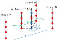
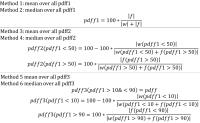
A Novel Phase Unwrapping Method Based on Pixel Clustering and
Local Surface Fitting with Application to Water-Fat Separation
Junying Cheng1,2, Yingjie Mei2,3,
Biaoshui Liu2, Xiaoyun Liu1, Ed. X. Wu4,5,
Wufan Chen1,2, and Yanqiu Feng2,4,5
1School of Automation Engineering, University of
Electronic Science and Technology of China, Chengdu, China,
People's Republic of, 2School
of Biomedical Engineering and Guangdong Provincial Key
Laboratory of Medical Image Processing, Southern Medical
University, Guangzhou, China, People's Republic of, 3Philips
Healthcare, Guangzhou, China, People's Republic of, 4Laboratory
of Biomedical Imaging and Signal Processing, The University
of Hong Kong, Hong Kong SAR, China, People's Republic of, 5Department
of Electrical and Electronic Engineering, The University of
Hong Kong, Hong Kong SAR, China, People's Republic of
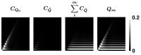
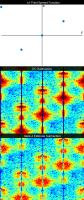
Current phase-unwrapping algorithms are challenged by rapid
phase variations, noise and disconnected regions. We propose
a novel phase-unwrapping method based on the observation the
phase local difference (pLD) and complex local difference
(cLD) maps. The proposed algorithm first clusters pixels
into disconnected regions by thresholding the cLD map and
then performs local polynominal surface fitting (LPSF) to
unwrap phase with the knowledge of wrapping locations
identified by thresholding the pLD map. Both simulation and
in vivo results demonstrate that the proposed method can
correctly unwrapped phase even in the presence of rapid
phase variation, low SNR, and disconnected regions, and has
great potential application to phase-related MRI in
practice.
|
|
3276.
 |
39 |
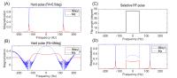
Modified Single-Echo Dixon Imaging for Improved SNR and CNR in
Contrast-Enhanced MRA
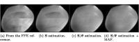
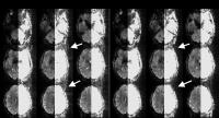
Holger Eggers1
1Philips Research, Hamburg, Germany
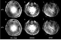
Chemical shift encoding-based water-fat imaging, or Dixon
imaging, is of recent interest in MRA. Single-echo Dixon
methods are particularly appealing for this application
because of their potential for decreasing scan times. This
work suggests modifications to existing single-echo Dixon
methods and demonstrates their benefits, primarily
improvements in SNR and CNR, in contrast-enhanced peripheral
MRA.
|
|
3277.
 |
40 |
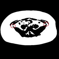
Separation of Abdominal Subcutaneous Adipose Tissue (SAT) and
Visceral Adipose Tissue (VAT) based on Wheel-Template in MRI
Steve Cheuk Ngai Hui1, Teng Zhang1,
Defeng Wang1, and Winnie Chiu Wing Chu1
1Dept. of Imaging and Interventional Radiology,
The Chinese University of Hong Kong, Hong Kong, Hong Kong
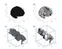
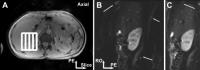
This abstract introduces the use of wheel-template to
perform segmentation on subcutaneous adipose tissue (SAT)
and visceral adipose tissue (VAT) at the abdominal region.
The proposed method detects narrow regions between SAT and
VAT, and uses line cut to separate two types of tissues
based on MRI data. It performs well on obese individuals and
the obtained results are correlated to those obtained from a
semi-automatic method. A quantitative measurement of SAT and
VAT is important as they are developed from different
pathways and suggested to be related to different chronic
diseases.
|
|
3278.
 |
41 |
Analytical Three-Point Dixon Method Using a Global Graph Cut
Jonathan Andersson1, Filip Malmberg1,
Håkan Ahlström1, and Joel Kullberg1
1Radiology, Uppsala University, Uppsala, Sweden
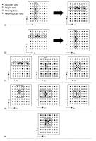
When reconstructing chemical shift encoded water-fat images
so called water-fat swaps due to incorrect phase maps can be
a significant problem, limiting clinical assessment and
quantitative measurements. A method is presented for solving
the problem in the case of 3 echoes, assuming only a fix
intra-echo spacing. Two possible solutions are analytically
calculated for each voxel. The correct global solution is
then found using a graph-cut method. The method succeeds
where a region-growing reference method fails at low SNR.
The presented method runs quickly and uses only one
parameter that can be set automatically, which should allow
for online implementation.
|
|
3279.
 |
42 |
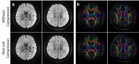
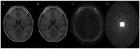
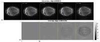
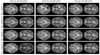
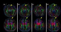
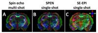
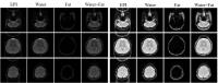
Fat Residue Removal by SENSE in EPI and DW-EPI
Victor B. Xie1,2, Mengye Lyu1,2,
Yilong Liu1,2, Yangqiu Feng1,2, Hua
Guo3, and Ed X. Wu1,2
1Laboratory of Biomedical Imaging and Signal
Processing, The University of Hong Kong, Hong Kong SAR,
China, People's Republic of, 2Department
of Electrical and Electronic Engineering, The University of
Hong Kong, Hong Kong SAR, China, People's Republic of, 3Center
for Biomedical Imaging Research, Department of Biomedical
Engineering, School of Medicine, Tsinghua University,
Beijing, China, People's Republic of
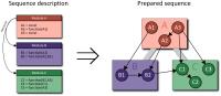
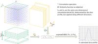
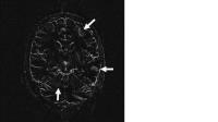
In this abstract, we proposed to use parallel imaging method
to remove the fat residue in EPI applications. In EPI, fat
is shifted along the phase encoding direction and can be
treated as another simultaneously exited slice
with controlled aliasing together with the water image. By
applying SENSE, water and fat residue can be effectively
separated. We have presented this simple method to separate
water and fat in EPI images and successfully applied it to
remove fat residue in the brain and abdomen fat-suppressed
EPI and DW-EPI images.
|
|
3280.
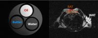 |
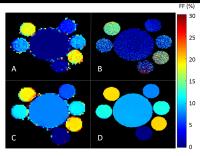
43 |
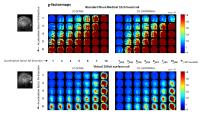
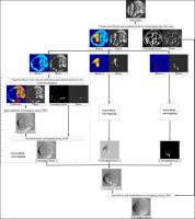
Characterization of Brown Adipose Tissue using
Multi-Varying-Peak MR Spectroscopy (MVP-MRS)
Gregory Simchick1, Jinjian Wu2,
Guangming Shi2, Hang Yin3, and Qun
Zhao1
1Bioimaging Research Center, University of
Georgia, Athens, GA, United States, 2Xidian
University, Xian, China, People's Republic of, 3Department
of Biochemistry and Molecular Biology, University of
Georgia, Athens, GA, United States
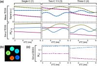
Differentiation of brown adipose tissue (BAT) from white
adipose tissue (WAT) using magnetic resonance imaging (MRI)
is clinically important to treat obesity, diabetes and heart
disease. In contrast to the traditional Dixon model of
single fat- and water-peak or fixed multi-fat-peak models,
we propose a Multi-Varying-Peaks MR Spectroscopic (MVP-MRS)
model, based on MR imaging data acquired with multiple
echoes, to better characterize chemical structures of the
fatty acid (saturated vs. unsaturated). Compared with
traditional classification of BAT and WAT using fat fraction
or proton relaxation time, the proposed MVP-MRS model can
achieve a correct classification rate of 95% between BAT and
WAT for in vivo mouse data implying great potentials in
future longitudinal imaging of BAT and WAT.
|
|
3281.
 |
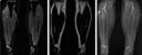
44 |
High-resolution imaging of muscular fat fraction - comparison of
chemical shift-encoded imaging and T2-based imaging
Lena Trinh1, Emelie Lind1, Pernilla
Peterson1, and Sven Månsson1
1Dept. of Translational Medicine, Medical
Radiation Physics, Skåne University Hospital, Lund
University, Malmö, Sweden
Chemical shift-encoded imaging is a quantitative method
commonly used to estimate fat fraction (FF) in various body
parts. However, for a reliable assessment, this technique
requires short inter echo spacing which can be challenging
if high spatial resolution is desirable. An alternative
quantitative method, based on the difference in
T2-relaxation time between fat and water, was examined and
compared to the chemical shift-encoded imaging method. This
T2-based technique successfully estimated FF in phantoms at
high resolution and large matrix size, when the chemical
shift-encoded method failed.
|
|
3282.
 |
45 |
Resolving Phase Ambiguity in Two-point Dixon Imaging Using a
Projected Power Method
Tao Zhang1,2, Yuxin Chen3, Marcus
Alley1, Brian Hargreaves1,2, John
Pauly2, and Shreyas Vasanawala1
1Radiology, Stanford University, Stanford, CA,
United States, 2Electrical
Engineering, Stanford University, Stanford, CA, United
States, 3Statistics,
Stanford University, Stanford, CA, United States
Dixon techniques offer robust water/fat separation in the
presence of static field inhomogeneity. Two-point Dixon
imaging with flexible echo times is desirable because of its
high scan efficiency and flexibility. A major challenge in
two-point Dixon imaging is how to estimate the phase error
resulting from field inhomogeneity. In this work, we
formulate a binary quadratic optimization and propose a fast
projected power method to resolve the phase ambiguity in
two-point Dixon imaging.
|
|
3283.
 |
46 |
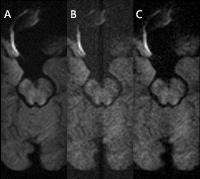
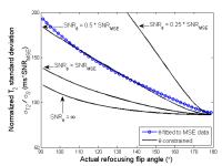
A comprehensive approach for effective motion artifact reduction
in Dixon
Gabriele Beck1, Alan Huang1, Adri
Duijndam1, and Lars van Loon1
1Philips Healthcare, Best, Netherlands
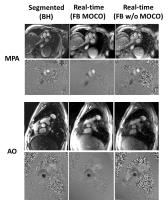
While Dixon provides superb fat suppression over large
imaging volumes, motion can be a challenge in specific
anatomies. This work evaluates a comprehensive approach to
reduce motion artifacts in Dixon TSE and FFE sequences,
combining a novel Dixon decorrelation approach, partial
averaging, modulus in-phase (IP) – out-phase (OP)
combinations and saturation of the physiological motion
artifact sources by the means of saturation pulses and
variable refocusing flip angle sweeps. We are able to show
that this approach allows us to effectively remove motion
artifacts improving the diagnostic quality of Dixon scans.
|
|