|
Exhibition Hall 13:30 - 14:30 |
|
|
|
Computer # |
|
3380.
 |
73 |
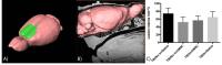
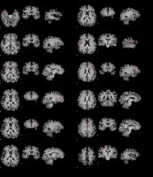
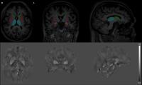
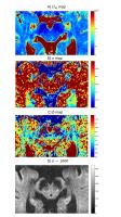
Does tau pathology play a role in abnormal iron deposition in
Alzheimer’s Disease? A quantitative susceptibility mapping study
in the rTg4510 mouse model of Tauopathy 
James O'Callaghan1, Holly Holmes1,
Nicholas Powell1, Jack Wells1, Ozama
Ismail1, Ian Harrison1, Bernard Siow1,
Michael O'Neill2, Emily Catherine Collins3,
Karin Shmueli4, and Mark Lythgoe1
1Centre for Advanced Biomedical Imaging,
University College London, London, United Kingdom, 2Eli
Lilly & Co. Ltd, Surrey, United Kingdom, 3Eli
Lilly and Company, Indianapolis, IN, United States,4Department
of Medical Physics and Biomedical Engineering, University
College London, London, United Kingdom
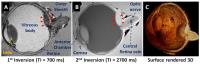
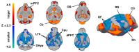
In this work, quantitative susceptibility mapping (QSM) and
T2* mapping were used to investigate iron accumulation both in-vivo and ex-vivo in
a mouse model of Alzheimer's Disease exhibiting tau
pathology for the first time. Magnetic susceptibility
increases relative to controls were identified in grey
matter and white matter brain regions and may indicate
sensitivity to tissue iron content. QSM in this mouse model
may therefore provide a non invasive method by which to
dissect the relationship between iron and tau pathology in
Alzheimer's Disease.
|
|
3381.
 |
74 |
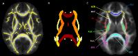
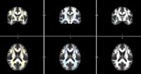
T2-weighted imaging of substantia nigra pars compacta shows
increased iron deposition in ventral lateral tier - Permission Withheld
Jason Langley1, Jan Sedlacik2, Daniel
E Huddleston3, Xiaoping Hu1, Jens
Fiehler2, and Kai Boelmans4
1Department of Biomedical Engineering, Emory
University & Georgia Tech, Atlanta, GA, United States, 2Department
of Neuroradiology, University Medical Center
Hamburg-Eppendorf (UKE), Hamburg, Germany, 3Department
of Neurology, Emory University, Atlanta, GA, United States, 4Department
of Neurology, University Medical Center Würzburg, Würzburg,
Germany
Recent results found that the SN seen in neuromenalnin
sensitive MRI and iron sensitive T2-weighted
contrasts is located in disparate spatial positions in
controls. Since iron is known to be deposited in the SN
after onset of Parkinson's disease(PD), we re-examine iron
sensitive measurements with respect to these new findings in
this abstract. Specifically, we find that the SN seen in T2-weighted
contrasts is enlarged in inferiorly and medially when
compared to controls. Most of this discrepancy happens in
the NM SN and we found the overlap to be an incredibly
sensitive marker for PD (p<10-15).
|
|
3382.
 |
75 |
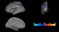
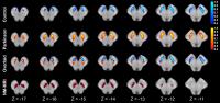
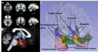
Association of Brain Iron Deposition in Parkinson’s Disease with
Comorbidities of Visual Hallucinations: An ROI-based
Quantitative Susceptibility Mapping Study
Darrell Ting Hung Li1, Edward Sai Kam Hui1,
Queenie Chan2, Nailin Yao3, Siew-eng
Chua4, Grainne M. McAlonan4,5, Shu
Leong Ho6, and Henry Ka Fung Mak1
1Department of Diagnostic Radiology, The
University of Hong Kong, Hong Kong, Hong Kong, 2Philips
Healthcare, Hong Kong, Hong Kong, 3Department
of Psychiatry, Yale University, New Haven, CT, United
States, 4Department
of Psychiatry, The University of Hong Kong, Hong Kong, Hong
Kong, 5Department
of Forensic and Neurodevelopmental Science, King’s College
London, London, United Kingdom, 6Department
of Medicine, The University of Hong Kong, Hong Kong, Hong
Kong
Abnormal iron accumulation in the brain may cause
oxidative-stress-induced neurodegeneration, which is one of
the hypothesis of nigral cell death in PD. It was also
believed that non-motor symptoms of PD patients are
associated with the increased of brain iron content. This
study examined the iron concentration in several subcortical
structures of PD patients with visual hallucinations by
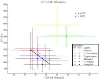
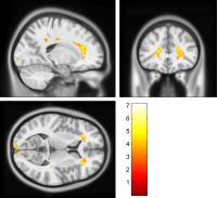
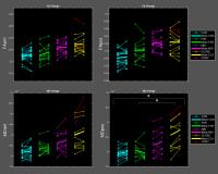
using the QSM technique. Higher magnetic susceptibility was
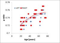
observed in the hippocampus of this patient group. The
result supported the hypothesis that hippocampal abnormality
could induce visuospatial memory impairment which may be the
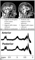
cause of visual hallucination in PD patients.
|
|
3383.
 |
76 |
Evaluation of gray matter degeneration in Parkinson’s disease by
using neurite-orientation dispersion and density imaging:
Analysis by gray matter-based spatial statistics
Koji Kamagata1, Kouhei Tsuruta2, Taku
Hatano3, Keigo Shimoji4, Masaaki Hori1,
Ayami Okuzumi3, Misaki Nakazawa2, Syo
Murata2, Ryo Ueda2, and Shigeki Aoki1
1Department of Radiology, Juntendo University,
Tokyo, Japan, 2Department
of Radiological Sciences, Tokyo Metropolitan University,
Tokyo, Japan, 3Department
of Neurology, Juntendo University, Tokyo, Japan, 4Department
of Diagnostic Radiology, Tokyo Metropolitan Geriatric
Hospital, Tokyo, Japan
In this study, neurite-orientation dispersion and density
imaging were used to estimate structural changes of neurites
in the gray matter of the brain, which is the earliest
pathological change in patients with PD. The results showed
a significant decrease in the intracellular volume fraction
in the right amygdala and right putamen in PD, suggesting a
decrease in neurite density that may reflect actual
pathological change. Given that NODDI could detect
pathological changes at the earliest stages in PD, it may be
useful for early diagnosis.
|
|
3384.
 |
77 |
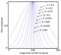
Evaluation of different high-pass filter on the susceptibility
in patients Parkinson’s disease and controls
Gerd Melkus1,2, Santanu Chakraborty1,2,
and Fahad A Essbaiheen 1,2,3
1Medical Imaging, The Ottawa Hospital, Ottawa,
ON, Canada, 2Radiology,
University of Ottawa, Ottawa, ON, Canada, 3King
Saud University, Riyadh, Saudi Arabia
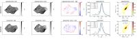
Quantitative susceptibility mapping (QSM) was found as a
useful method to evaluate neurodegenerative diseases such as
Parkinson’s disease. For QSM reconstruction background field
removed phase data is needed, but for retrospective studies
only high-pass filtered data might be available. In this
study we analyzed the influence of different high-pass
filtered phase images on the susceptibility assessment for
volunteers and Parkinson’s disease patients and compared the
results to QSM estimation using background field removed
phase data. With increasing high-pass filter strength
consistently lower susceptibility results, but up to a
certain filter strength differences in susceptibility can
still be distinguished.
|
|
3385.
 |
78 |
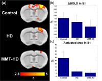
Imaging effects of memantine treatment in a mouse model of
Huntington disease using evoked and resting-state fMRI
Wei-Tang Chang1, Fiftarina Puspitasari1,
Ling-Yun Yeow1, Hui-Chien Tay1, Marta
Garcia Miralles2, Katrianne Bethia Koh2,
Liang-Juin Tan2, Mahmoud POULADI2,3,
and Kai-Hsiang Chuang1
1SBIC, A*STAR, Singapore, Singapore, 2TLGM,
A*STAR, Singapore, Singapore, 3Department
of Medicine, National University of Singapore, Singapore,
Singapore
Huntington disease (HD) is an incurable neurodegenerative
disease. Recently, memantine (MMT) was found to be effective
delaying the progression of disease phenotypes in a mouse
model of HD. Here we applied resting-state fMRI to evaluate
functional connectivity in HD and the MMT treatment effect
and its behavioral correlates. The results of forepaw
stimulation reduced evoked responses though significance was
hampered by large individual variation. Interestingly,
functional connectivity outside of DMN, but not within DMN,
was decreased by HD. With MMT treatment, the connectivity
increased in general. The FC relevant to the behavioral test
also showed behavioral correlates.
|
|
3386.
 |
79 |
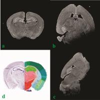
Developmental White Matter Alterations in Monkey Brains with
Huntington’s Disease
Yuguang Meng1, Anthony W.S. Chan2,3,
and Xiaodong Zhang1,3
1Yerkes Imaging Center, Yerkes National Primate
Research Center, Emory University, Atlanta, GA, United
States, 2Department
of Human Genetics, School of Medicine, Emory University,
Atlanta, GA, United States, 3Division
of Neuropharmacology and Neurologic Diseases, Yerkes
National Primate Research Center, Emory University, Atlanta,
GA, United States
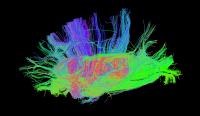
This study examined the developmental changes of white
matter integrity in rhesus monkey brains with the HD gene
mutation using diffusion tensor imaging (DTI). Widespread
developmental alterations are seen in striatum, and the
frontal, motor, sensory and visual brain areas. The findings
reveal the temporal-spatial evolution of abnormal white
matter maturation during the development of the brain with
the Huntington’s Disease, and suggest altered neural
substrates associated with motor and cognitive dysfunctions
in HD patients.
|
|
3387.
 |
80 |
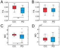
Diffusion tensor imaging of the substantia nigra in Parkinson’s
disease revisited
Jason Langley1, Daniel E Huddleston2,
Michael Merritt1, Xiangchuan Chen1,
Rebecca McMurray2, Michael Silver2,
Stewart A Factor2, and Xiaoping Hu1
1Department of Biomedical Engineering, Emory
University & Georgia Tech, Atlanta, GA, United States, 2Department
of Neurology, Emory University, Atlanta, GA, United States
Inconclusive results from prior diffusion tensor
imaging-based studies can be attributed to variability in
location of regions of interest used to define the
substantia nigra and its subcomponents. We apply recent
findings from neuromelanin sensitive MRI to standardize
regions of interest for the substantia nigra. Differences in
fractional anisotropy and mean diffusivity were found in the
neuromelanin sensitive substantia nigra but not in the
substantia nigra defined in the b0 image.
|
|
3388.
 |
81 |
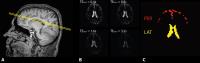
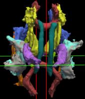
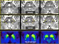
Diagnosis of Parkinsonism Using Nigrosome 1 Imaging at 3T:
Comparison of Interobserver Agreement between GRE Magnitude
Images (MEDIC) and Susceptibility Map-weighted Images (SMWI)
Eung Yeop Kim1, Yoon Ho Nam2, Young
Noh3, Young Hee Sung3, Byeong Ho Goh1,
Joon Hyung Ann1, and Jongho Lee4
1Radiology, Gachon University Gil Medical Center,
Incheon, Korea, Republic of, 2Radiology,
Seoul St. Mary Hospital, Seoul, Korea, Republic of, 3Neurology,
Gachon University Gil Medical Center, Incheon, Korea,
Republic of, 4Electrical
and Computer Engineering, Seoul National University, Seoul,
Korea, Republic of
Susceptibility map-weighted imaging (SMWI) improves both CNR
and SNR in comparison with conventional SWI. In this work,
we compared SMWI with MEDIC (a multi-echo GRE imaging that
combines magnitude images via sum of squares) for nigrosome
1 imaging at 3T in terms of interobserver agreement and
diagnostic performance in 74 subjects (44 with Parkinson’s
disease). Two experienced and two less-experienced reviewers
visually assessed both imaging sets separately. Compared
with MEDIC, SMWI showed higher kappa values and larger AUCs,
regardless of level of experience. As a result, nigrosome 1
imaging using SMWI significantly improved diagnostic
performance as well as interobserver agreement.
|
|
3389.
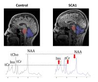 |
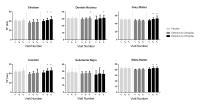
82 |
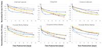
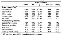
Sensitivity of volumetric MRI and MRS to onset and progression
of neurodegeneration
Dinesh K Deelchand1, James M Joers1,
Adarsh Ravishankar2, Tianmeng Lyu3,
Uzay Emir1,4, Diane Hutter1,
Christopher M Gomez5, Khalaf O Bushara6,
Christophe Lenglet1, Lynn E Eberly3,
and Gulin Oz1
1Center for Magnetic Resonance Research,
University of Minnesota, Minneapolis, MN, United States, 2School
of Physics and Astronomy, University of Minnesota,
Minneapolis, MN, United States,3Division of
Biostatistics, University of Minnesota, Minneapolis, MN,
United States, 4University
of Oxford, Oxford, United Kingdom, 5Department
of Neurology, University of Chicago, Chicago, IL, United
States, 6Department
of Neurology, University of Minnesota, Minneapolis, MN,
United States
The goal of this study was to combine MRS with volumetric
MRI to determine the sensitivity of these two techniques to
onset and progression of neurodegeneration in patients with
early-moderate spinocerebellar ataxia type 1. Subjects were
scanned at baseline and followed up at ~18 and ~36 months at
3T. Both MRI and MRS measures were found to be more
sensitive to disease progression than standardized clinical
scores. This study shows that volumetric MRI was most
sensitive to disease progression while MRS might be more
sensitive to detect the disease’s early stage.
|
|
3390.
 |
83 |
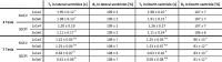
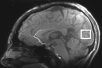
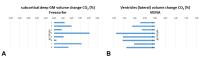
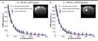
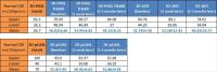
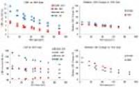
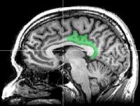
QSM and T2* Mapping in a Six Month Placebo-Controlled Clinical
Trial of the Iron Chelator Deferiprone in Parkinson’s Disease
Rexford Newbould1, Courtney Bishop1,
Antonio Martin-Bastida2, and David Dexter2
1Imanova Centre for Imaging Sciences, London,
United Kingdom, 2Imperial
College London, London, United Kingdom
A double-blind placebo-controlled clinical trial of an iron
chelation therapy was performed in 25 subjects with
Parkinson’s disease (PD) and 12 healthy matched controls.
Two MRI measures of brain iron at each of three visits at
baseline, 3 months, and 6 months were derived from a high
resolution 3D multiecho volume: T2* and QSM maps. T2* values
significantly lengthened in six of nine pre-defined ROIs by
the final visit in the highest dose group. QSM values ,
however, did not change with treatment. This differential
trajectory between relaxation and susceptibility may result
from ferritin’s complex relaxation behavior.
|
|
3391.
 |
84 |
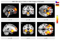
Characterizing neurodegeneration in progressive supranuclear
palsy using VBM and SVM classification
Karsten Mueller1, Robert Jech2,3,
Cecilia Bonnet2,3, Jaroslav Tintera4,
Harald E Möller1, Klaus Fassbender5,
Jan Kassubek6, Markus Otto6, Evžen
Ružicka2,3, and Matthias L Schroeter1,7
1Max Planck Institute for Human Cognitive and
Brain Sciences, Leipzig, Germany, 2Department
of Neurology and Center of Clinical Neuroscience, Charles
University in Prague, Prague, Czech Republic,31st
Faculty of Medicine and General University Hospital in
Prague, Prague, Czech Republic, 4Institute
for Clinical and Experimental Medicine, Prague, Czech
Republic, 5Clinic
and Polyclinic for Neurology, Saarland University Homburg,
Homburg, Germany, 6Clinic
and Polyclinic for Neurology, University of Ulm, Ulm,
Germany, 7Clinic
for Cognitive Neurology, University Hospital Leipzig,
Leipzig, Germany
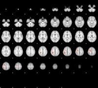
Structural brain differences were investigated between
patients with progressive supranuclear palsy (PSP) and
healthy controls with T1-weighted images (MP-RAGE) acquired
at four centers with different 3T scanners (Siemens). Using
voxel-based morphometry, we found a major decline in gray
matter density in brainstem, insula, striatum, and
frontomedian regions that is in line with the current
literature. Support-vector-machine classification provided a
high sensitivity of disease detection when using relevant
brain regions in feature selection.
|
|
3392.
 |
85 |
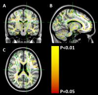
Robust global and widespread local white matter abnormalities in
a longitudinal study of juvenile neuronal ceroid lipofuscinosis
(CLN3)
Ulrika Roine1, Timo Roine2,3, Antti
Hakkarainen3, Anna Tokola3, Marja H.
Balk3, Minna Mannerkoski4, Tuula
Lönnqvist5, and Taina Autti3
1Department of Neuroscience and Biomedical
Engineering, Aalto University, Espoo, Finland, 2iMinds-Vision
Lab, Department of Physics, University of Antwerp, Wilrijk
(Antwerp), Belgium, 3HUS
Medical Imaging Center, Radiology, University of Helsinki
and Helsinki University Hospital, Helsinki, Finland, 4Child
Psychiatry, University of Helsinki and Helsinki University
Hospital, Helsinki, Finland,5Department of Child
Neurology, Children's Hospital, University of Helsinki and
Helsinki University Hospital, Helsinki, Finland
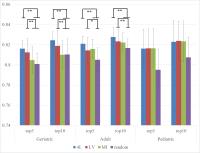
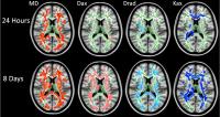
Juvenile neuronal ceroid lipofuscinosis (CLN3), is a
progressive neurodegenerative lysosomal storage disease of
the childhood, which manifests with loss of vision,
seizures and loss of cognitive and motor functions, and
leads to premature death. We investigated global and local
white matter microstructure with diffusion MRI in 14
children with CLN3 imaged at two time points. Robust global
analysis was performed using whole-brain tractography and
white matter tract skeleton. Local microstructural
abnormalities were investigated using tract-based spatial
statistics. Significantly decreased fractional anisotropy
and increased diffusivity values were found in subjects with
CLN3 both at the global and local scale.
|
|
3393.
 |
86 |
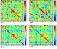
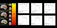
Structural brain connectome and cognitive impairment in
Parkinson’s disease - Permission Withheld
Sebastiano Galantucci1, Federica Agosta1,
Elka Stefanova2, Silvia Basaia1,
Martijn van den Heuvel3, Tanja Stojkovic2,
Elisa Canu1, Iva Stankovic2, Vladana
Spica2, Vladimir S. Kostic2, and
Massimo Filippi1,4
1Neuroimaging Research Unit, San Raffaele
Scientific Institute, Vita-Salute San Raffaele University,
Milan, Italy, 2Clinic
of Neurology, Faculty of Medicine, University of Belgrade,
Belgrade, Yugoslavia,3Department of Psychiatry,
Rudolf Magnus Institute of Neuroscience, University Medical
Center Utrecht, Utrecht, Netherlands, 4Department
of Neurology, San Raffaele Scientific Institute, Vita-Salute
San Raffaele University, Milan, Italy
To date, MRI biomarkers have been demonstrated extremely
useful for detecting and monitoring the neurodegenerative
processes. However, brain network analysis seems the most
powerful approach to quantitatively describe the topological
organization of the brain connectome even at early stages of
neurodegenerative diseases. This study provided promising
biomarkers to detect features of neurodegeneration in
PD-MCI, being able to distinguish it from PD without MCI.
This study shows that the presence of subtle cognitive
deficits not causing a dementia, produces a huge alteration
of brain networks suggesting the importance of the study of
connectomics in the investigation of neurodegenerative
diseases.
|
|
3394.
 |
87 |
fMRI reveals plasticity compensating for early dopaminergic loss
at corticostriatal synapse
Chiao-Chi Chen1, Yi-Hua Hsu1, Nai-Wei
Yao1, and Chen Chang1
1Institute of Biomedical Sciences, Academia
Sinica, Taipei, Taiwan
The dopaminergic system possesses striking plasticity
compensating for motor aberrations from neuronal loss.
Little is known regarding the compensation mechanism during
dopaminergic loss, preventing the aberration from being
arrested and treated early. Here we present in vivo imaging
evidence from functional magnetic resonance imaging showing
that, after dopaminergic depletion, the dorsolateral
striatum (DOLS) exhibited an early and transient
vasodilation cluster in response to specific forepaw
stimulation. Activation of DOLS NMDA receptors causes this
vasodilation, protects dopaminergic fibers from denervation,
and counteracts motor deficits. The findings have clinical
implications for early detection and intervention in brain
disorders such as Parkinson’s disease.
|
|
3395.
 |
88 |
Dopaminergic therapy modulates cortical perfusion in Parkinson’s
disease with and without dementia according to ASL perfusion MRI
Chien-Yuan Eddy Lin1, Wei-Che Lin2,
Pei-Chin Chen2, Yung-Cheng Huang3,
Nai-Wen Tsai4, Hsiu-Ling Chen2,
Hung-Chen Wang5, Tsu-Kung Lin4,
Kun-Hsien Chou6, Meng-Hsiang Chen2,
Yi-Wen Chen2, and Cheng-Hsien Lu4
1GE Healthcare, Taipei, Taiwan, 2Department
of Diagnostic Radiology, Kaohsiung Chang Gung Memorial
Hospital, Kaohsiung, Taiwan, 3Department
of Nuclear Medicine, Kaohsiung Chang Gung Memorial Hospital,
Kaohsiung, Taiwan, 4Department
of Neurology, Kaohsiung Chang Gung Memorial Hospital,
Kaohsiung, Taiwan, 5Department
of Nuerosurgery, Kaohsiung Chang Gung Memorial Hospital,
Kaohsiung, Taiwan, 6Brain
Research Center, National Yang-Ming University, Taipei,
Taiwan
We examined the cerebral perfusion differences among 17
Parkinson’s disease (PD) patients, 17 PD with dementia (PDD)
patients, and 17 healthy controls and used noncontrast
arterial spin labelling MRI to assess the effects of
dopaminergic therapies on perfusion in the patients. We
demonstrated progressive widespread cortical hypoperfusion
in PD and PDD and robust effects for the dopaminergic
therapies. These patterns of hypoperfusion could be related
to cognitive dysfunctions and disease severity. Furthermore,
desensitization to dopaminergic therapies in terms of
cortical perfusion was found as the disease progressed,
supporting the concept that long-term therapies are
associated with the therapeutic window narrowing.
|
|
3396.
 |
89 |
Aberrant interhemispheric structural and functional connectivity
in amyotrophic lateral sclerosis: converging evidences from DTI
and resting-state fMRI - Permission Withheld
Jiuquan Zhang1,2, Bing Ji2,3, Zhihao
Li2, Jun Hu4, Jian Wang1,
Mingze Xu5, and Xiaoping Hu2
1Department of Radiology, Southwest Hospital,
Chongqing, China, People's Republic of, 2Department
of Biomedical Engineering, Emory University & Georgia
Institute of Technology, Atlanta, GA, United States, 3University
of Shanghai for Science & Technology, Shanghai, China,
People's Republic of, 4Department
of Neurology, Southwest Hospital, Chongqing, China, People's
Republic of, 5Department
of Biomedical Engineering, Perking University, Beijing,
China, People's Republic of
The corpus callosum (CC) involvement is a consistent feature
of Amyotrophic lateral sclerosis (ALS), thus suggesting a
pathopysiology of reduced interhemispheric neural
connectivity. In the current study, we directly examined the
interhemispheric functional and structural connectivities in
ALS. In terms of functional connectivity, extensive
alterations in voxel mirrored homotopic connectivity were
found in ALS. With structural connectivity, while there were
widespread reductions in DTI metrics, only the fiber
probability index through CC subregion III in the ALS
patients was significantly decreased compared with the
controls. These findings provide further evidence for
structural and functional interhemispheric connectivity
impairment in ALS.
|
|
3397.
 |
90 |
Investigation of inter-hemispheric functional connectivity in
Parkinson's disease with asymmetric onset using Voxel-Mirrored
Homotopic Connectivity
Yong Zhang1, Naying He2, Hua-Wei Lin2,
Ajit Shankaranarayanan3, Zhenyu Zhou1,
and Fu-Hua Yan2
1MR Research China, GE Healthcare, Beijing,
China, People's Republic of, 2Radiology,
Ruijin Hospital, Shanghai Jiaotong University School of
Medicine, Shanghai, China, People's Republic of, 3GE
Healthcare, Menlo Park, CA, United States
This preliminary study used voxel-mirrored homotopic
connectivity (VMHC), a novel resting-state fMRI parameter to
investigate inter-hemispheric functional connectivity in
Parkinson’s Disease (PD) with asymmetric onset. Fifteen left
side onset (LPD) patients, sixteen right side onset (RPD)
patients and nineteen healthy controls were recruited for
comparison. Both of LPD and RPD patients showed decreased
VMHC in post-central gyrus responsible for motor functions.
The decreased VMHC in the cuneus and middle occipital gyrus
in LPD patients might affect visual processing function. For
RPD patients, VMHC changes in the middle and superior
frontal gyrus could be relevant to advanced cognitive
impairment.
|
|
3398.
 |
91 |
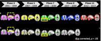
Can longitudinal diffusion-weighted imaging of the basal ganglia
be used as a surrogate marker in preclinical Huntington’s
disease?
Chris Patrick Pflanz1, Marina Charquero-Ballester2,
Adnan Majid3, Anderson Winkler1,
Emmanuel Vallee1, Mark Jenkinson1,
Adam Aron3, and Gwenaelle Douaud1
1FMRIB Centre, University of Oxford, Oxford,
United Kingdom, 2Oxford,
United Kingdom, 3San
Diego, CA, United States
Huntington’s disease is a monogenetic,
neurodegenerative movement disorder that is amenable to
predictive genetic testing. Here, we investigated whether
longitudinal diffusion-weighted imaging of the basal ganglia
could be used to detect early microstructural changes in
participants with presymptomatic Huntington’s disease
(preHD). We found the first results showing significant
longitudinal changes in the microstructure of the basal
ganglia within a
preclinical HD population. We further showed that, while FA
and MD might be less sensitive to longitudinal changes than
volumetric measures, they provide mechanistic insights into
the underlying physiopathological process that are
complementary to the monotonic, non-specific changes in the
volume of the basal ganglia.
|
|
3399.
 |
92 |
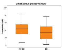
Brain Iron Deficiency in Restless Legs Syndrome Measured by
Quantitative Susceptibility and its Relation to Clinical
Features
Xu Li1,2, Hongjun Liu1,2, Richard P
Allen3, Christopher J Earley3, Richard
A.E. Edden1,2, Peter B Barker1,2,
Tiana Cruz3, and Peter C.M. van Zijl1,2
1F.M. Kirby Research Center for Functional Brain
Imaging, Kennedy Krieger Institute, Baltimore, MD, United
States, 2Radiology,
Johns Hopkins University School of Medicine, Baltimore, MD,
United States,3Neurology, Johns Hopkins
University School of Medicine, Baltimore, MD, United States
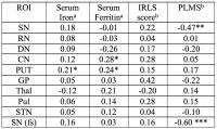
Possible brain iron deficiency was assessed using
quantitative susceptibility mapping at 7T in restless legs
syndrome (RLS) and analyzed with clinical measurements
including IRLS severity score, serum iron, serum ferritin
and periodic limb movement during sleep (PLMS). Using
magnetic susceptibility as a brain iron index and compared
to control group, significantly decreased iron was found in
RLS patients in dentate nuclei and thalamus, and in
substantia nigra in a subset of RLS patients with severe
clinical symptoms with PLMS larger than 100 times per hour.
Significant correlation between PLMS and brain iron was only
found in substantia nigra in RLS.
|
|
3400.
 |
93 |
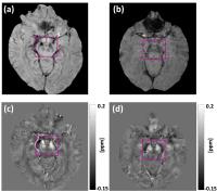
Quantitative Susceptibility Mapping of the “Swallow tail” in
Parkinson disease
Santanu Chakraborty1,2, Gerd Melkus1,2,
Fahad Essbaiheen1,2,3, David A Grimes4,
and Tiago Mestre4
1Medical Imaging, The Ottawa Hospital, Ottawa,
ON, Canada, 2Radiology,
University of Ottawa, Ottawa, ON, Canada, 3King
Saud University, Riyadh, Saudi Arabia, 4Neurology,
The Ottawa Hospital, Ottawa, ON, Canada
Parkinson disease (PD) continues to be diagnosed based on
clinical findings. Recently, in SWI images, the loss of
‘swallow tail’ appearance in dorsolateral substantia nigra
in PD patients yielded high diagnostic accuracy. In our
study we measured susceptibility values using QSM in the
‘swallow tail’ area in seven Parkinson’s disease patients
and compared to five control subjects. The susceptibility in
the swallow tail area was higher in the PD group (0.072 vs.
0.058). This likely suggests increased iron deposition
causing a masking effect that contributes along with
dopaminergic neurons loss to the disappearance of the
‘swallow tail’ in PD patients.
|
|
3401.
 |
94 |
MRI signatures in the brain of patients with PD and iRBD
Silvia Mangia1, Philip Burton1, Alena
Svatkova2,3, Igor Nestrasil4,
Alejandra Sierra Lopez5, Karin Shmueli6,
Lynn Eberly7, Michael Howell4, Paul
Tuite4, and Shalom Michaeli1
1CMRR, Department of Radiology, University of
Minnesota, Minneapolis, MN, United States, 2Department
of Pediatrics, University of Minnesota, Minneapolis, MN,
United States, 3Central
European Institute of Technology (CEITEC), Masaryk
University, Brno, Czech Republic, 4Department
of Neurology, University of Minnesota, Minneapolis, MN,
United States, 5A.I.Virtanen
Institute for Molecular Sciences, University of Eastern
Finland, Kuopio, Finland, 6Department
of Medical Physics and Biomedical Engineering, University
College London, London, United Kingdom, 7Division
of Biostatistics, University of Minnesota, Minneapolis, MN,
United States
The idiopathic rapid eye movement sleep behavior disorder
(iRBD) is a condition that often evolves into Parkinson’s
disease (PD), therefore by monitoring iRBD one can track the
neurodegeneration of individuals that may progress to PD.
Here we used a battery of MRI contrasts to characterize
brain tissue properties such as microstructural integrity,
iron loads, and functional connectivity in 10 iRBD, 10 PD
and 10 age-matched healthy subjects. Rotating frame
relaxation methods adiabatic T1,2ρ and
RAFFn, along with DTI and rsfMRI detected heterogeneous
abnormalities in several subcortical structures of PD and
iRBD subjects.
|
|
3402.
 |
95 |
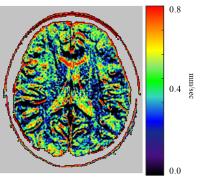
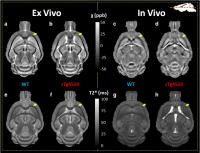
A High b-Value Diffusion Study of Brainstem Abnormality in
Patients with Parkinson's Disease Using a CTRW Model
Zheng Zhong1,2, Muge Karaman1, Douglas
Merkitch3, Jennifer Goldman3, and
Xiaohong Joe Zhou1,4
1Center for MR Research, Chicago, IL, United
States, 2Bioengineering,
University of Illinois at Chicago, Chicago, IL, United
States, 3Neurological
Sciences, Rush University Medical Center, Chicago, IL,
United States, 4Radiology,
Neurosurgery and Bioengineering, University of Illinois
Hospital & Health Sciences system, Chicago, IL, United
States
It has been known that the substantia nigra of brain stem
shows structural abnormalities with the progression of
Parkinson’s disease. While high b-value diffusion imaging
has the potential to reveal such structural changes,
single-shot EPI suffers from unwanted geometric distortion
which may result in poor analysis of the diffusion data. In
this study, we use a recently developed reduced field of
view imaging technique and analyze the abnormalities
occurring in the substantia nigra of the Parkinson’s disease
patients by using the continuous-time random-walk (CTRW)
model.
|
|
3403.
 |
96 |
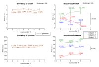
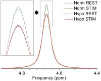
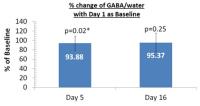
The Effect of Primidone on Gamma-Aminobutyric Acid Concentration
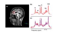
in the Dentate Nucleus in Patients with Essential Tremor
Ulrike Dydak1,2, Ruoyun Ma1,2, Nora
Hernandez3, Johnathan P Dyke4, and
Elan Louis3,5,6
1School of Health Sciences, Purdue University,
West Lafayette, IN, United States, 2Department
of Radiology and Imaging Sciences, Indiana University School
of Medicine, Indianapolis, IN, United States,3Department
of Neurology, Yale School of Medicine, New Heaven, CT,
United States, 4Department
of Radiology, Weill Cornell Medical College, New York, NY,
United States, 5Center
for Neuroepidemiology and Clinical Neurological Research,
Yale School of Medicine, New Heaven, CT, United States, 6Department
of Chronic Disease Epidemiology, Yale School of Public
Health, New Heaven, CT, United States
Whether current use of the medication primidone affects
dentate γ-aminobutyric acid (GABA) concentrations is
unknown. Yet, this may be an important confounder in studies
of the pathophysiology of essential tremor (ET). Using the
MEGA-PRESS J-editing sequence, we found no difference in
dentate GABA levels between patients taking primidone and
patients not taking primidone. Furthermore, there was no
association between daily primidone dose and dentate GABA
concentration. These data suggest that it is not necessary
to exclude ET patients on primidone from MRS studies of
dentate GABA concentration.
|
|