ISMRM 24th Annual Meeting & Exhibition • 07-13 May 2016 • Singapore

Electronic Poster Session: Diffusion 2

3428 -3451 Diffusion: Other
3452 -3475 Diffusion: Body
3476 -3499 Diffusion Applications

Exhibition Hall 

14:30 - 15:30

    Computer #

 
3428.   
25 Does reduced FOV Diffusion Weighted Imaging inherently yield lower ADC ?
Suchandrima Banerjee1, David Aramburu-Nunez2, Ramesh Paudyal2, Thomas Chenevert3, Michael Boss4, and Amita Shukla-Dave2,5
1Global MR Applications & Workflow, GE Healthcare, Menlo Park, CA, United States, 2Department of Medical Physics, Memorial Sloan-Kettering Cancer Center, New York, NY, United States, 3Department of Radiology, University of Michigan Health System, Ann Arbor, MI, United States, 4Applied Physics Division, National Institute of Standards and Technology, Boulder, CO, United States, 5Department of Radiology, Memorial Sloan-Kettering Cancer Center, New York, NY, United States
The benefits of reduced field-of-view (rFOV) imaging with the single-shot echoplanar diffusion sequence such as lower distortion and better discrimination of tumor from benign tissue have been demonstrated in several anatomies. In most of these published works, lower ADC was reported using rFOV compared to the standard full FOV (fFOV) method, irrespective of the technique by which rFOV was achieved. In this work we conducted controlled experiments in 3 phantoms to avoid some of the confounding factors present in vivo and investigated if there is a systemic underestimation of ADC in rFOV DWI compared to fFOV DWI. 


3429.   
26 Diffusion Tensor Imaging of the Cervical and Thoracic Spinal Cord in Pediatric Subjects using an inner FOV 2D RF pulse sequence.
Sona Saksena1, Devon M Middleton2, Laura Krisa3, Pallav Shah2, Scott H Faro2, Rebecca Sinko3, John Gaughan4, Jürgen Finsterbusch5, M J Mulcahey3, and Feroze B Mohamed1
1Department of Radiology, Thomas Jefferson University, Philadelphia, PA, United States, 2Department of Radiology, Temple University, Philadelphia, PA, United States, 3Department of Occupational Therapy, Thomas Jefferson University, Philadelphia, PA, United States, 4Biostatistics Consulting Center, Temple University School of Medicine, Philadelphia, PA, United States, 5Department of Systems Neuroscience, University Medical Center Hamburg-Eppendorf, Hamburg, Germany
This is the first study in pediatric subjects investigating the DTI values and it’s reproducibility along the entire cervical and thoracic spinal cord (SC). DTI data was acquired from 22 typically developing (TD) children and 15 patients with spinal cord injury (SCI) using an inner field-of-view DTI sequence. Regions of interest were manually drawn on whole cord at every axial slice along the cervical and thoracic SC. Fractional anisotropy and radial diffusivity values were significantly different between TD and SCI suggesting that these appear to be the most sensitive parameter in assessing the state of SC in chronic phase of SCI. This study demonstrates that DTI has a potential to be used as an imaging biomarker for evaluating the extent of injury, which may be useful to prognosticate as well as monitor patients with SCI


3430.   
27 Time-dependent diffusion for assessment of treatment response in GL261 murine glioma model
Olivier Reynaud1, Jin Zhang1, Kerryanne Veronica Winters1, Dmitry Novikov1, and Sungheon Gene Kim1
1Center for Biomedical Imaging, Department of Radiology, NYU School of Medicine, New York, NY, United States
In this study, we investigate the feasibility of using the recently proposed diffusion MRI method, POMACE (Pulsed and Oscillating gradient MRI Assessment of Cell size and Extracellular space) for characterization of tissue changes induced by chemotherapy. Surface-to-volume ratio (S/V), cell size (Rcell) and extracellular space volume fraction (ECS) are estimated prior to and after 5FU and bevacizumab treatment in GL261 murine glioma model. Preliminary results indicate an early ECS decrease two days after 5FU/bevacizumab administration. By combining OGSE and PGSE, POMACE provides non-invasive and quantitative metrics regarding tumor microstructure that can be applied to evaluate treatment response.


3431.   
28 Diffusion Tensor Imaging and Neuropsychological Study: Pilot Findings in HIV Adults
Rajakumar Nagarajan1, Manoj K Sarma1, Edward Xu1, Paul M Macey2, Mario Guerrero 3, Vanessa Correa 3, Eric S Daar3, and M.Albert Thomas1
1Radiological Sciences, University of California Los Angeles, Los Angeles, CA, United States, 2UCLA School of Nursing, University of California at Los Angeles, Los Angeles, CA, United States, 3Department of Medicine, Harbor-UCLA Medical Center, Torrance, CA, United States
Our current study examined whether neuroimaging abnormalities in HIV infected adults are associated with neuropsychological changes as measured by diffusion tensor imaging (DTI) and HIV Dementia scale (HDS). Several brain regions demonstrated significantly decreased values of fractional anisotropy and mean diffusivity. Also there was a significant decline of HDS scores observed in the construction abilities of HIV infected adults compared to healthy controls. This study indicates that DTI is a sensitive tool for correlating neuroanatomic pathologic features with specific cognitive deficits in patients with HIV infection.


3432.   
29 Temporal evolution of diffusion indices in white matter and grey matter of macaque brains with ischemic stroke - Permission Withheld
Chun-Xia Li1, Yumei Yan1, Frank Tong2, Doty Kempf1, Stuart Zola1,3,4, Leonard Howell1,3, and Xiaodong Zhang 1
1Yerkes National Primate Research Center, Emory University, Atlanta, GA, United States, 2Department of Radiology, School of Medicine, Emory University, Atlanta, GA, United States, 3Department of Psychiatry and Behavioral Sciences, School of Medicine, Emory University, Atlanta, GA, United States, 4Atlanta Veterans Affairs Medical Center, Decatur, GA, United States
In the present study, adult macaque monkeys were utilized to characterize the temporal changes of DTI-derived diffusion indices in grey matter (GM) and white matter (WM) after ischemic stroke. Our reults showed that mean diffusivity (MD), radial diffusivity (RD) and axial Diffusivity (AD)  decreased significantly in GM and WM  immediately post stroke while fractional anisotropy (FA) remained unchanged until 48-hour post stroke.  The results demonstrate the sensitivity and robustness of MD, AD and RD to access the ischemic injury of GM and WM during hyperacute stroke, and also reveal the temporal evolutiom of the WM and GM microstructure following stroke onset.


3433.   
30 Clinical Evaluation of Simultaneous Multi-Slice Diffusion-weighted Magnetic Resonance Imaging of the Prostate - Permission Withheld
Jakob Weiss1, Jana Taron1, Ahmed E. Othman1, Petros Martirosian1, Sascha Kaufmann1, Ulrich Kramer1, Konstantin Nikolaou1, and Mike Notohamiprodjo1
1Diagnostic and Interventional Radiology, University of Tuebingen, Tuebingen, Germany
To evaluate the clinical performance of simultaneous multi-slice (sms) diffusion-weighted imaging (DWI) of the prostate. A prototype sms-DWI sequence was utilized and compared to conventional DWI sequences. Sms acquisition is based on the recently introduced principle of simultaneously exciting and acquiring multiple slices by a single radiofrequency (RF) pulse, thus accelerating image acquisition directly by the number of simultaneously excited slices (MB-factor). Image analyses revealed similar image quality and lesion detection as compared to conventional DWI sequences at a substantially decreased acquisition time. Sms-DWI seems a valuable alternative to conventional DWI sequences for prostate imaging.


3434.   
31 Comparison of six different diffusion weighted MRI models in pancreatic cancer patients
Oliver J. Gurney-Champion1,2, Remy Klaassen3, Martijn Froeling1,4, Jaap Stoker1, Johanna W. Wilmink3,5, Arjan Bel2, Hanneke W.M. van Laarhoven3, and Aart J. Nederveen1
1Radiology, Academic Medical Center, Amsterdam, Netherlands, 2Radiation Oncology, Academic Medical Center, Amsterdam, Netherlands, 3Medical Oncology, Academic Medical Center, Amsterdam, Netherlands,4Radiology, University Medical Center Utrecht, Utrecht, Netherlands, 5Internal Medicine, Academic Medical Center, Amsterdam, Netherlands
We compare the performance of six models for diffusion weighted MRI (DWI) data signal intensity as a function of b-values. We obtained three DWI-datasets spread over two sessions from 9 pancreatic cancer patients. To characterize the fit parameters’ sensitivity to abnormalities, we compared the difference in fit parameter values between tumorous and healthy tissue. To characterize repeatability, we obtained the coefficient of variation. Every model had exactly one parameter that was significantly sensitive to tumorous tissue (p<0.05). From these parameters, the most sensitive parameter was f2 from the tri-exponential model, whereas the best repeatable measure was the ADC from the mono-exponential model.


3435.   
32 Intravoxel incoherent motion analysis of renal allograft diffusion with clinical and histopathological correlation in pediatric kidney transplant patients
Clare B Poynton1, Marsha Lee2, Yi Li1, Zoltan Laszik3, John D Mackenzie1, Pauline W Worters4, and Jesse Courtier1
1Department of Radiology and Biomedical Imaging, University of California, San Francisco, San Francisco, CA, United States, 2Department of Pediatrics, University of California, San Francisco, San Francisco, CA, United States, 3Department of Pathology, University of California, San Francisco, San Francisco, CA, United States, 4GE Healthcare, Menlo Park, CA, United States
This study investigated the use of intravoxel incoherent motion (IVIM) analysis for characterizing diffusion in renal allografts of pediatric transplant recipients.  Patients were separated into two groups according to whether or not a renal allograft biopsy resulted in a change in clinical management. Patients requiring a change in management (i.e., increase in immunosuppression) showed statistically significant differences in tissue diffusivity in the region of the biopsy relative to those that did not require any change. These results suggest IVIM analysis may be a useful non-invasive tool for guiding clinical management of pediatric kidney transplant patients.


3436.   
33 Tract-profiling and bundle statistics: a test-retest validation study
Martin Cousineau1, Eleftherios Garyfallidis1, Marc-Alexandre Côté1, Pierre-Marc Jodoin1, and Maxime Descoteaux1
1Computer Science, Université de Sherbrooke, Sherbrooke, QC, Canada
Tractometry is a hot topic in population analysis that combines diffusion MRI metrics along white matter bundles. Using a dataset of 11 healthy subjects with 3 different acquisitions collected within the same week, and a state of the art processing pipeline that removes outliers and splits bundles in separate parts by computing their centroid, we perform a test-retest study which validates that tractometry results are reproducible in terms of shape, volume, average metric value, and tract profiles.


3437.   
34 Methodological considerations on graph theoretical analysis of structural brain networks
Timo Roine1, Ben Jeurissen1, Daniele Perrone2, Jan Aelterman2, Wilfried Philips2, Jan Sijbers1, and Alexander Leemans3
1iMinds-Vision Lab, Department of Physics, University of Antwerp, Wilrijk (Antwerp), Belgium, 2Ghent University-iMinds/Image Processing and Interpretation, Ghent, Belgium, 3Image Sciences Institute, University Medical Center Utrecht, Utrecht, Netherlands
We studied the reproducibility of whole-brain structural brain connectivity networks reconstructed with constrained spherical deconvolution based probabilistic fiber tractography. Our main finding is that a low spherical harmonics order decreases the reproducibility of graph measures in connectomics. This is most likely caused by the wider peaks in the fiber orientation distributions, which increase the variation in orientations sampled by the tractography algorithm. Based on our observations, we recommend using spherical harmonics decomposition with an order of at least eight whenever the data allows so. In addition, threshold value was important for binary networks, and some network properties were highly intercorrelated.


3438.   
35 Diffusion-Weighted Hyperpolarized 13C-Urea in a Murine Model of Liver Fibrosis
Irene Marco-Rius1, Jeremy A Gordon1, Peder EZ Larson1, Romelyn delos Santos1, Robert A Bok1, Aras Mattis2,3, Jacquelyn Maher3,4, Daniel B Vigneron1,3, and Michael A Ohliger1,3
1Department of Radiology and Biomedical Imaging, University of California San Francisco, San Francisco, CA, United States, 2Department of Pathology, University of California San Francisco, San Francisco, CA, United States, 3Liver Center, University of California, San Francisco, San Francisco, CA, United States, 4Department of Medicine, Division of Gastroenterology, University of California San Francisco, San Francisco, CA, United States
Diffusion weighted MRI has been widely used to measure the movement of water molecules and study tissue microstructure in order to characterize both diffuse and focal liver disease. In liver fibrosis, for instance, increased collagen formation is associated with restricted diffusion of water. However, the majority of water within the liver is either in the vascular or intracellular space, making the diffusion of water a potentially poor marker for fibrosis, which is an extracellular process. Here, we investigated applying diffusion-weighted MRI with an exogenously injected extracellular agent, hyperpolarized 13C-urea, as a potentially more sensitive probe of the extracellular space in the liver in a mouse model of liver fibrosis induced with CCl4


3439.   
36 Diffusion parameters derived from multi b-value DWI-data as surrogate marker for kidney function
Oliver J. Gurney-Champion1,2, René van der Bel3, Martijn Froeling1,4, C.T. Paul Krediet3, and Aart J. Nederveen1
1Radiology, Academic Medical Center, Amsterdam, Netherlands, 2Radiation Oncology, Academic Medical Center, Amsterdam, Netherlands, 3Internal Medicine, Academic Medical Center, Amsterdam, Netherlands,4Radiology, University Medical Center Utrecht, Utrecht, Netherlands
In this study, we show the potential of the tri-exponential intravoxel incoherent motion (IVIM) model parameters as surrogates for kidney function. We show (n=8) that f1, f2 and D (fit parameters) changed as function of angiotensin-II dose (0, 0.3, 0.9 and 3 ng/kg/min). The changes in f1 and f2 correlated (correlation coefficient r=0.42 and -0.62; p=0.01 and p<0.001) to changes in effective renal plasma flow (ERPF) and glomerular filtration rate (GFR) deduced from a 125I-thalamate and 131I-hippuran clearing test. Finally, we showed (n=6) high inter-session and intra-session repeatability with coefficients of variation below 15% for all fit parameters.


3440.   
37 Inter-modality correlation of prostatic microenvironmental tissue stiffness and water diffusivity using quantitative functional imaging techniques. - Video Not Available
Hugh Harvey1, Jeremie Fromageau2, Veronica Morgan1, Liz Bancroft3, Ros Eeles3, Jeff Bamber2, and Nandita deSouza1
1Radiotherapy & Imaging, The Institute of Cancer Research and Royal Marsden NHS Foundation Trust, London, United Kingdom, 2The Institute of Cancer Research, London, United Kingdom, 3Oncogenetics, The Institute of Cancer Research and Royal Marsden NHS Foundation Trust, London, United Kingdom
Location-matched ROI analysis of tissue stiffness (SWE) and water diffusivity (ADC) in 9 normal prostates demonstrated that both techniques can adequately differentiate between peripheral and transitional zonal microenvironments, and that there is a weak negative correlation between tissue stiffness and water diffusivity in the peripheral zone. This suggests that factors such as microvascularity, cell size, extracellular matrix and macromolecules may have a differential effect on tissue stiffness and diffusivity. Transitional zone stiffness is too heterogeneous to demonstrate significant inter-modality correlation.


3441.   
38 Dependence of DTI Measures on SNR in a Multicenter Clinical Trial
Xiaopeng Zhou1, Ken Sakaie1, Josef Debbins2, Robert Fox1, and Mark Lowe1
1Cleveland Clinic, Cleveland, OH, United States, 2Barrow Neurological Institute, Phoenix, AZ, United States
The dependence of DTI metrics on SNR was investigated in a longitudinal multicenter clinical trial. We presented a combined simulation and experimental analysis of the effect of SNR on DTI measures using 27 scanners over 15 months.  Although the dependence of DTI measures on SNR is different for each type of scanner, we demonstrate the inverse relation between mean diffusivity and SNR and FA decreasing slightly with SNR. A sufficient SNR is essential for a reliable estimation of DTI parameters generally and the dependence of DTI measures on SNR should be accounted for or corrected when comparing results among scanners.


3442.   
39 The effect of brain microbleeds on the structural brain network after stroke
Xiaopei Xu1, Henry KF Mak1, Raja Rizal Azman2, Kui-Kai Lau3, and Edward S Hui1
1Department of Diagnostic Radiology, The University of Hong Kong, Hong Kong, Hong Kong, 2Department of Bio-Medical Imaging, University of Malaya, Kuala, Malaysia, 3Department of Medicine, The University of Hong Kong, Hong Kong, Hong Kong
To explore the influences of brain microbleeds (BMBs) on structural brain network in stroke patients, we used DTI-based tractography and brain network analysis to investigate the brain structural network configuration in stroke patients with and without BMBs. Our results demonstrated disrupted balance between integrated and segregated process in global network, and enhanced specialized functions in stroke patients with BMBs. These findings suggested that the presence of BMBs is related to a disturbed brain network organization with imbalanced integrated and segregated information process ability, and brain network analysis is a sensitive tool to assess the impaired cognition caused by cerebral small vessel disease.


3443.   
40 DTI Association with Working Memory and Speed in Cognitive Network Pathways
Junyu Guo1, John O. Glass1, JungWon Hyun1, Yimei Li1, Conklin Heather1, Lisa Jacola1, Ching-Hon Pui1, Sima Jeha1, and Wilbrun E. Reddick1
1St Jude Children's Research Hospital, Memphis, TN, United States
We investigate the relationship of structural diffusion tensor imaging (DTI) metrics with working memory and decision speed performance in children treated for acute lymphoblastic leukemia (ALL). We built a core neurocognitive network including a central executive network, a salience network, and subcortical cortex based on previous fMRI findings. We generated structural connectivity pathways based on high-resolution DTI data from the human connectome project, and applied those in ALL patients to quantify DTI measures in each pathway.  We found that DTI measures in most  pathways were significantly associated with working memory and decision speed performance suggesting an essential structural neurocognitive network.


3444.   
41 Clinical Application of Simultaneous Multi-Slice DWI: A Multi-Body-Part Comparison with Standard EPI DWI - Permission Withheld
Man Sun1, Hongyan Ni2, Xu Yan3, and Tianyi Qian4
1First Central Clinical College, Tianjin Medical University, Tianjin, China, People's Republic of, 2Tianjin First Central Hospital, Tianjin, China, People's Republic of, 3MR Collaborations NE Asia, Siemens Healthcare, Shanghai, China, People's Republic of, 4MR Collaborations NE Asia, Siemens Healthcare, Beijing, China, People's Republic of
Applying simultaneous-multi-slice EPI (SMS EPI) sequences in diffusion-weighted imaging (DWI) can reduce scanning time greatly, but its clinical-routine image quality in different body parts has not been broadly tested. This study compared multi-site DWI images acquired with a prototype SMS EPI sequence with those acquired with a traditional EPI sequence, computed SNR and CNR and conducted paired t-test. The results show there are no significant differences of SNR and CNR between the two kinds of sequence (p>0.05). When the SMS EPI sequence was applied to the clinical DWI examination, it not only reduced scanning time, but also kept the SNR, CNR in the same level as standard EPI. The SMS sequence was able to satisfy the requirements of clinical disease diagnosis.


3445.   
42 Altered social communication network in boys with autism spectrum disorder
Yu-Chun Lo1, Yu-Jen Chen1, Yung-Chin Hsu1, Susan Shur-Fen Gau2, and Wen-Yih Isaac Tseng1
1Institute of Medical Device and Imaging, National Taiwan University College of Medicine, Taipei, Taiwan, 2Department of Psychiatry, National Taiwan University College of Medicine, Taipei, Taiwan
Impaired social communication skills have been consistently reported in autism spectrum disorder (ASD). We used diffusion spectrum imaging to measure white matter integrity of the social communication network, and investigated its relationships with social communication and social interaction in 62 ASD and 55 typically developing (TD) boys. ASD showed partially reduced white matter integrity of the social communication network as compared to TD. Positive correlations were found between white matter integrity and the social interaction in ASD. In conclusion, altered microstructural property of the social communication network might be a structural correlate of social communication deficits in ASD.


3446.   
43 The negative residuals of diffusion kurtosis model: a study on rat brain imaging
Wei-Cheng Lee1, Sheng-Min Huang 1, Cheng-He Li1, Kung-Chu Ho2, and Fu-Nien Wang1
1Department of Biomedical Engineering and Environmental Sciences, National Tsing Hua University, Hsinchu, Taiwan, 2Nuclear Medicine, Chang Gung Memorial Hospital, Taoyuan, Taiwan
The well-behaved residual of a successful fitting model should present as an independent random variable in its histogram, otherwise it will contain structure that is not accounted for in this model. In the study, we found that there are two groups of fitting residuals. And the spatial mapping of these smaller negative pixels reveals that the overestimation of kurtosis model is related to the contribution of vessel signal. Therefore, we proposed a semi-kurtosis model to calculate the restricted diffusion adequately and obtain correct DKI information in these regions with vascular interference.


3447.   
44 Spherical Deconvolution of High-Resolution 7T Whole-Head Diffusion Magnetic Resonance Images shows reduced radial anisotropic diffusion in human primary somatosensory cortex
Ralf Lützkendorf1, Robin M. Heidemann2, Thorsten Feiweier2, Michael Luchtmann3, Sebastian Baecke1, Joern Kaufmann4, Joerg Stadler5, Eike Budinger5, and Johannes Bernarding1
1Biometry and Medical Informatics, University of Magdeburg, Magdeburg, Germany, 2Siemens Healthcare GmbH, Erlangen, Germany, 3Department of Neurosurgery, University of Magdeburg, Magdeburg, Germany, 4Department of Neurology, University of Magdeburg, Magdeburg, Germany, 5Leibniz Institute for Neurobiology, Magdeburg, Germany
Diffusion anisotropy in cortical gray matter (GM) and adjacent white matter (WM) provides microanatomic information about the course of the neuronal structures within GM and when connecting to other brain regions. However, interwoven neuronal fiber orientations and complex folded structures render the analysis difficult. Ultra-high-field diffusion MR imaging (dMRI) overcomes these limitations as the improved SNR allowed acquiring 1.4 mm isotropic voxel with increased diffusion-weighting. Applying constrained spherical deconvolution (1) enabled resolving radial and tangential anisotropic diffusion in cortical gray matter confirming recent reports of reduced radial anisotropy in primary somatosensory cortex as compared to other cortical areas (2).


3448.   
45 Investigation of diffusion signal behavior at different diffusion times in a human hepatocellular carcinoma xenograft model - Permission Withheld
Mami Iima1,2, Tomomi Nobashi1, Hirohiko Imai3, Sho Koyasu1, Akira Yamamoto1, Yuji Nakamoto1, Masako Kataoka1, Tetsuya Matsuda3, and Kaori Togashi1
1Department of Diagnostic Imaging and Nuclear Medicine, Graduate Schoolof Medicine, Kyoto University, Kyoto, Japan, 2The Hakubi Center for Advancer Research, Kyoto University, Kyoto, Japan, 3Department of Systems Science, Graduate Schoolof of Informatics, Kyoto University, Kyoto, Japan
The relationship between diffusion time and diffusion parameters in a human hepatocellular carcinoma xenograft mouse model was investigated at 7T. There was an increase in K values and decrease in ADCo values in 27.6ms compared to 9.6 ms. Accordiginly a remarkable difference in a composite index (sADC) was also found. The investigation of the water diffusion behavior at different diffusion times may provide valuable information on the contribution of the different compartments or tissue components to the overall diffusion MRI.


3449.   
46 Altered microstructural integrity of the white matter tracts in strabismus: A systematic tract-specific analysis over the whole brain
Hsien-Te Su1, Tzu-Hsun Tsai2, Yao-Chia Shih3, Yu-Shiang Tzeng4, Chien-Chung Chen4, and Wen-Yih Isaac Tseng1,5
1Institute of Medical Device and Imaging, National Taiwan University College of Medicine, Taipei City, Taiwan, 2Department of ophthalmology, National Taiwan University Hospital, Taipei City, Taiwan, 3Institute of Biomedical Engineering,National Taiwan University, Taipei City, Taiwan, 4Department of Psychology, National Taiwan University College of Science, Taipei City, Taiwan, 5Molecular Imaging Center, National Taiwan University, Taipei City, Taiwan
To investigate whether the white matter tracts are altered in patients with strabismus, we used diffusion spectrum imaging to measure microstructural property of 78 major white matter tracts, and compared the property between 12 patients with strabismus adults and 24 matched controls. As compared to the controls, patients showed differences with large effect sizes in the left vertical occipital fasciculus, callosal fibers to bilateral precuneus, callosal fibers to bilateral inferior parietal lobules, and the left superior longitudinal fasciculus I. The altered white matter tracts support the hypothesis of underlying microstructural changes of the visual pathways in strabismus.


3450.   
47 Flip Angle Optimization in Diffusion-Weighted Imaging using Simultaneous Multi-Slice Acceleration
Wei Liu1 and Kun Zhou1
1Siemens Shenzhen Magnetic Resonance Ltd, Shenzhen, China, People's Republic of
In this study, an automatic optimization scheme was proposed in diffusion-weighted imaging using simultaneous multi-slice (SMS) acceleration to determine an appropriate flip angle based on a short TR, which is capable to obtain similar contrast of a pair of tissue type compared to the one with long TR scan. With this optimization scheme, minimal TR in SMS case is practically achievable and there is no obvious contrast deviation between images from long TR (FA = 900) and short TR (FA < 900).


3451.   
48 Investigation of Intravoxel Fiber Configuration Complexity in the Human Heart
Changyu SUN1, Lihui WANG2, Feng YANG3, and Yuemin ZHU1
1CREATIS; CNRS UMR 5220; Inserm U 1044; INSA of Lyon; University of Lyon;, Lyon, France, 2School of computer science and technology, Guizhou University, Guiyang, China, People's Republic of, 3School of Computer and Information Technology, Beijing JiaoTong University, Beijing, China, People's Republic of
Fiber crossing in the myocardium of the heart is controversial despite some reports on the heart of animals. The purpose of this study is to investigate the fiber configuration complexity in a voxel, such as fiber crossing, in the human heart. We applied two different reconstruction techniques commonly used in high angular resolution diffusion imaging (HARDI) to conventional diffusion tensor imaging (DTI) data of the human heart acquired on clinical MRI system. The results consistently demonstrated that the human myocardium exhibits some complex fiber configurations such as fiber crossing in a voxel.
Exhibition Hall 

14:30 - 15:30

    Computer #

 
3462.   
59 Optimization of Direct-coronal Diffusion-weighted Whole Body Imaging with Background Suppression (DWIBS) Using Single-shot Turbo Spin-echo (TSE) at 3T MRI.
Makoto Suzuki1, Tatsuya Kuramoto1, Yuki Hachiman1, Yu Nishina2, Satoru Morita2, Kayoko Abe2, and Masami Yoneyama3
1Department of radiological servece, Tokyo Women's Medical University Hospital, Tokyo, Japan, 2Department of Diagnostic Imaging and Nuclear Medicine, Tokyo Women's Medical University Hospital, Tokyo, Japan, 3IS Business Group, Philips electronics japan, Tokyo, Japan
Direct-coronal diffusion-weighted whole body imaging with background body signal suppression using single-shot turbo spin-echo (TSE-DWIBS) with b-values of 400, 600, 800, and 1000 mm2/s, and DWIBS using echo-planner imaging (EPI-DWIBS) with b-value of 1000mm2/s  were obtained using 3T MR scanner in 5 healthy volunteers. We investigated optimal b-value of TSE-DWIBS and compared image distortions, contrast, signal-to-noise ratio (SNR), and artifacts between TSE-DWIBS and EPI-DWIBS. Image contrast and SNR of TSE-DWIBS with b-value of 800 mm2/s were equivalent to those of EPI-DWIBS with b-value of 1000 mm2/s. TSE-DWIBS had less image distortions and artifacts than EPI-DWIBS. Direct-coronal TSE-DWIBS using 3T MRI can be applied for clinical cases.


3463.   
60 Repeatability on diffusion MRI measurements on the different numbers of b values and excitations of the breast and its comparison with lactating breasts - Permission Withheld
Mami Iima1,2, Masako Kataoka1, Shotaro Kanao1, Natsuko Onishi1, Makiko Kawai1, Akane Ohashi1, Rena Sakaguchi1, and Kaori Togashi1
1Department of Diagnostic Imaging and Nuclear Medicine, Graduate Schoolof Medicine, Kyoto University, Kyoto, Japan, 2The Hakubi Center for Advancer Research, Kyoto University, Kyoto, Japan
The effects of different numbers of b values and excitations on the non-Gaussian DWI parameter estimates in the breast of normal volunteers was assessed. A good agreement of Do, K and synthetic ADC200-1500 was observed among different number of b values as well as excitations. Lower Do and sADC200-1500 were found in women with lactation period. Higher K and fIVIM were observed in lactating volunteers with some overlap. A limited protocol using only 5 b values could be relevant in clinical setting, resulting in a remarkable reduction in acquisition time.


3464.   
61 Zoomed EPI-DWI of the Kidney Using Two-Dimensional Spatially-Selective Radiofrequency Excitation Pulses
Yong-Lan He1, Philipp Riffel2, Daniel Hausmann2, Stefan Schönberg2, and Ulrike Attenberger2
1Radiology, Peking Union Medical College Hospital, Beijing, China, People's Republic of, 2Department of Clinical Radiology and Nuclear Medicine, University Medical Center Mannheim, Heidelberg University, Mannheim, Germany
This is the first MR study to evaluate the use of zoomed EPI technology for diffusion-weighted imaging in the kidney. Zoomed diffusion-weighted EPI leads to substantial image quality improvements with reduction of susceptibility artifacts in renal DWI.  The combination of the two sequences for renal imaging might be recommended: a c-EPI scan to cover and screen the whole upper abdomen and a z-EPI scan focused on the targeted anatomical structure and suspected lesions. 


3465.   
62 Non-invasive quantification of prostate cancer using AMICO framework for VERDICT MRI
Elisenda Bonet-Carne1, Alessandro Daducci2,3, Eleftheria Panagiotaki1, Edward Johnston4, Nicola Stevens4, David Atkinson4, Shonit Punwani4, and Daniel C Alexander1
1Centre for Medical Image Computing, University College London, London, United Kingdom, 2Signal Processing Lab (LTS5), École Polytechnique Fédérale de Lausanne, Lausanne, Switzerland, 3University Hospital Center (CHUV) and University of Lausanne (UNIL), Lausanne, Switzerland, 4Centre for Medical Imaging, University College London, London, United Kingdom
The aim of this study is to extend the AMICO framework to the VERDICT model-based diffusion-weighted MRI (DW-MRI) technique and to evaluate its performance to prostate cancer imaging. DW-MRI was acquired for 4 subjects and the VERDICT model was fitted to the data using both fitting procedures. In both cases similar differences in parameter values between tumour and normal tissue were found. The AMICO formulation reduces the computation time for VERDICT and produces parameter maps that are more homogeneous than those obtained with the original fitting. The AMICO formulation reflects the microstructural differences in a clinically practical time.


3466.   
63 Assessing micro- and macro-circulation of graft kidneys by using IVIM-DWI and Non-contrast MRA
Yung-Chieh Chang1, Mu-Ch Chung2, Kuo-Hsiung Shu2, Hao-Chung Ho3, Yen-Chieh Ouyang4, Jyh-Wen Chai1, and ClaytonChi-Chang Chen1
1Department of Radiology, Taichung Veterans General Hospital, Taiwan, Taiwan, Taiwan, 2Division of Nephrology, Taichung Veterans General Hospital, Taiwan, Taiwan, Taiwan, 3Division of Urolog, Taichung Veterans General Hospital, Taiwan, Taiwan, Taiwan, 4Department of Electrical Engineering, National Chung Hsing University, Taiwan, Taiwan
The goal of the present study was to perform IVIM-DWI and non-contrast MRA of graft kidneys to evaluate the micro- and micro-structural status in 17 patients within 48 hours after transplantation. Five of them had a second scan in six months later.  The experimental results suggested that D*, D and f values in IVIM-DWI might considerably be useful in early detection of functional change graft kidneys and non-contrast MRA and PC-MRI could effectively assess patency and flow rates of graft vessels. Combination of both IVIM-DWI and non-contrast MRA would be potentially promising in clinical application for monitoring graft kidneys.


3467.   
64 The repeatability of IVIM with fuzzy clustering algorithm in liver imaging
Kaining Shi1, Ying Liu2, Yu Shi2, and Qiyong Guo2
1Imaging Systems Clinical Science, Philips Healthcare, Beijing, China, People's Republic of, 2Radiology department, Shengjing Hospital of China Medical University, Shenyang, China, People's Republic of
Fussy clustering technique (FCM) has been combined with IVIM to increase the stability and reduce the post-processing time of the nonlinear curve fitting in IVIM. Another problem of the widely used bi-exponential IVIM model is its poor repeatability. This work is to assess the repeatability of IVIM with FCM between two scans in healthy liver by calculating the coefficient of variation and the 95% Bland–Altman limits of agreements. Results proved that FCM could improve the repeatability of IVIM, especially for the parameter D*, which was the most unstable among total 3 parameters.


3468.   
65 Preliminary Investigation: Gaussian and Non-Gaussian Diffusion Method in Diagnostic Differentiation of Prostate Cancer from Prostatic Hyperplasia
Chen lihua1, Liu ailian1, Song qingwei1, wang heqing1, sun meiyu1, Li ye1, Chen ailian1, and Zheng dandan2
1The Affiliated Hospital of Dalian Medical University, Dalian, China, Dalian, China, People's Republic of, 2GE Healthcare, MR Research China, Beijing, Beijing, China, People's Republic of
Prostate cancer is the second most common cancer for men, and it has high leading cause of cancer death among men. In this study, DKI and DWI MR measurements were performed to investigate the correlation of the MK, Ka, Kr, MD, Da, Dr, FA and ADC values in ROIs of the prostate and benign prostatic hyperplasia. DKI working at present scanning hardware are capable to detect the pathophysiological changes unattainable to conventional MRI techniques. 


3469.   
66 Diffusion weighted imaging of prostate cancer: mathematical modeling of signal obtained using low b values
Harri Merisaari1, Parisa Movahedi1, Ileana Montoya1, Jussi Toivonen1, Marko Pesola1, Pekka Taimen2, Peter Boström2, Tapio Pahikkala1, Hannu Juhani Aronen1, and Ivan Jambor1
1University of Turku, Turku, Finland, 2Turku University Hospital, Turku, Finland
Eighty-one patients with historically confirmed PCa underwent two repeated 3T DWI examinations performed using 14 b-values in the range of 0-500 s/mm2 and diffusion time of 19.004 ms. Various fitting methods for IVIM and mathematical models were evaluated in the terms of fitting quality (Akaike information criteria), repeatability, and Gleason score prediction. Monoexponential model demonstrated the highest repeatability and clinical values in the regions-of-interest based analysis of PCa DWI, b-values in the range of 0-500 s/mm2.


3470.   
67 Characterizing breast lesions by Using Mono-exponential, Bi-exponential, and Stretched Exponential Diffusion-weighted MR Imaging and Diffusion Kurtosis MR Imaging - Video Not Available
Kun Sun1, Xu Yan2, and Fuhua Yan1
1Department of Radiology, Ruijin Hospital,Shanghai Jiao Tong University School of Medicine, Shanghai, China, People's Republic of, 2Application Dept, Siemens Healthcare, Shanghai, China, People's Republic of
DKI model may provide additional information and improve the characterizing of breast lesions compared with conventional diffusion parameters. The kurtosis and water molecular diffusion heterogeneity index derived from DKI and stretched Exponential DWI may be helpful for the preoperative differentiation of proliferative activity of breast cancer.


3471.   
68 Comparative analysis of diffusion models for glioma malignancy grade differentiation
Ivan I. Maximov1, Aram Tonoyan2, Daniel Edelhoff1, Igor Pronin2, and Dieter Suter1
1TU Dortmund University, Dortmund, Germany, 2Burdenko Neurosurgery Institute, Moscow, Russian Federation
Diffusion weighted imaging is very powerful technique allows one to diagnose and differentiate glioma malignancy in vivo. Recently, the non-Gaussian diffusion models exhibited their efficacy in a detection of microsctructure changes for low and high grade gliomas. In this study we perform a comparative analysis of four diffusion models (DTI, DKI, two- and three-compartment models) in ability to provide a quantitative measure of the gliomas malignancy in vivo.


3472.   
69 Optimization of Single-shot Turbo Spin-echo Diffusion-weighted Imaging with Parallel Imaging in Healthy Pancreas
Yu Nishina1, Satoru Morita1, Tatsuya Kuramoto2, Makoto Suzuki2, Hitoshi Tadenuma2, Yasuhiro Goto2, Masami Yoneyama3, and Shuji Sakai1
1Diagnostic Imaging and Nuclear Medicine, Tokyo Women's Medical University Hospital, Tokyo, Japan, 2Radiological Service, Tokyo Women's Medical University Hospital, Tokyo, Japan, 3Philips Electronics Japan, Tokyo, Japan
We compared the image quality of single-shot turbo spin-echo diffusion-weighted imaging (TSE-DWI) with parallel imaging and conventional echo planar DWI (EPI-DWI) in the pancreas of healthy volunteers. TSE-DWI with a sensitivity encoding factor (SF) of 2, 4, 6, and EPI-DWI with SF2 (b-values = 0, 500, 1000 s/mm2) were obtained using a 3T MRI scanner. Two radiologists evaluated overall image quality, susceptibility artifacts, and aliasing artifacts. TSE-DWI with moderate SF had better image quality than conventional EPI-DWI for a medium b-value because of less susceptibility artifacts.


3473.   
70 7T diffusion MRI of the forearm nerves
Wieke Haakma1,2,3, Jeroen Hendrikse1, Anneriet M. Heemskerk4, Peter R. Luijten1, Michael Pedersen3, Alexander Leemans4, and Martijn Froeling1
1Radiology, University Medical Center Utrecht, Utrecht, Netherlands, 2Forensic Medicine, Aarhus University, Aarhus, Denmark, 3Comparative Medicine Lab, Department of Clinical Medicine, Aarhus University, Aarhus, Denmark, 4Image Sciences Institute, University Medical Center Utrecht, Utrecht, Netherlands
In this work we present our results obtained at the 7T MRI scanner investigating the nerves in the forearm. We tested two DTI protocols and were able to visualize the median, ulnar and radial nerves with fiber tractography with resolution up to 0.75×0.75×2.0 mm3. We have demonstrated the potential of 7T to identify these nerves and quantify their diffusion characteristics in a reliable way. We expect that the use of high resolution DTI can be beneficial in the investigation of peripheral nervous tissue in the forearm and might aid in identifying changes due to pathology. 


3474.   
71 Comparison of TSE and EPI for renal DTI
Fabian Hilbert1, Tobias Wech1, Henning Neubauer1, Simon Veldhoen1, Thorsten Alexander Bley1, and Herbert Köstler1
1Department of Diagnostic and Interventional Radiology, University of Würzburg, Würzburg, Germany
Echo-planar imaging (EPI) is usually applied to acquire diffusion-weighted images. While EPI is fast in acquiring an entire image, it brings along distortion artifacts. Turbo spin echo (TSE) acquisitions require more time than EPI, but avoid distortions. Purpose of this study was to compare TSE and EPI for diffusion tensor imaging of the human kidney. We found similar mean diffusivity with EPI and TSE. Fractional anisotropy provides good corticomedullary contrast with both methods TSE and EPI. TSE is a practicable alternative to EPI for diffusion tensor imaging of the kidney.


3475.   
72 Imaging bio-markers of Intervertebral disc degeneration by T2  Relaxometry and DTI A prospective Study with Multi parametric Imaging correlation - Video Not Available
rammohan sai venkata vadapalli1, Raghav dutt sai venkata Mulukutla2, and abhinav sriiram Vadapalli3
1Imaging, Vijaya diagnostics and research, Hyderabad 500020, India, 2Orthopaedic and Spine surgery, Udai orthopaedic Institute, Hyderabad 500020, India, 3Imaging, GH, Hyderabad 500020, India
Surgeons want to know Predictive disc health proximal to the level of fusion or intervention. Conventional MR imaging using T2 weighted Imaging is insensitive to early morphological changes with no predictive quantitative biomarker profile of early degeneration. The Thompson scale or the Pfirrmann scale are discontinuous scales fail to demonstrate the Degeneration or regeneration of disc Objectively. Multiparametric Imaging using T2 relaxometry maps,DTI indices namely FA and qualitative FA maps offer both quantitative and qualitative biomarkers for predictive disc health


3452.   
49 Prediction of the development of distant metastases from nasopharyngeal carcinoma using Diffusion-Weighted Imaging
Qi Yong Ai1, Ann D. King1, Benjamin King Hong Law1, Lok Yiu Sheila Wong1, Kunwar S. Bhatia1, David Ka Wai Yeung2, and Brigette B.Y. Ma2
1Department of Imaging and Interventional Radiology, The Chinese University of Hong Kong, Shatin, Hong Kong, 2Department of Clinical Oncology, The Chinese University of Hong Kong, Shatin, Hong Kong
This study evaluated pre-treatment diffusion weighted imaging of primary nasopharyngeal carcinoma for the prediction of patients at risk of distant metastases, based on long-term follow-up for at least 5 years. Analysis was performed of ADC parameters using histogram analysis (ADCmean, ADCskewness and ADCkurtosis), together with stage and volume of the primary tumour and nodes. Multivariate analysis showed pre-treatment ADCskewness, T stage and nodal volume were significant ( p =0.026, 0.022 and 0.005, respectively), distant metastases being more likely in patients with tumours with ADC values skewed toward the higher ADC range, and higher T stage and nodal volume.


3453.   
50 Differentiation of prostate cancer in central gland with Histogram analysis of apparent diffusion coefficient
Xiaohang Liu1, Liangping Zhou1, Weijun Peng1, and Yong Zhang2
1Radiology, Fudan university shanghai cancer center, Shanghai, China, People's Republic of, 2MR Research China, GE healthcare, Beijing, China, People's Republic of
The diffusion weighted imaging (DWI) is commonly used in the diagnosis of Prostate cancer, but the cancer in Central gland (CG) is considered harder to detected on conventional DWI,but histogram analysis of apparent diffusion coefficient could potentially improve the differentiation of prostate cancer in CG. 


3454.   
51 Statistical evaluation of bi-exponential IVIM effect in healthy human liver and its dependence on the cut-off b-value for segmented biexponemtial fit - Permission Withheld
Oi Lei Wong1, Jing Yuan1, Gladys Goh Lo2, Thomas W. T. Leung3, Wai Ki Chung2, and Benny W. H. Ho2
1Medical Physics and Research Department, Hong Kong Sanatorium & Hospital, Hong Kong, Hong Kong, 2Department of diagnostic & interventional radiology, Hong Kong Sanatorium & Hospital, Hong Kong, Hong Kong, 3Comprehensive Oncology Center, Hong Kong Sanatorium & Hospital, Hong Kong, Hong Kong
The use of IVIM in assessing the microcapillary perfusion and true diffusion has attracted elevating attention. The segmented bi-exponential fitting has been widely adopted to calculate IVIM metrics, in which a cut-off b-value is pre-defined for perfusion and diffusion region separation. This study calculated the corrected Akaike Information Criterion (AICc) for bi-exponential IVIM model in the healthy human liver with varying cut-off b-values and then compared to the AICc for the mono-exponential model. The statistical preference of the bi-exponential model to mono-exponential model was demonstrated based on the lowest AICc. This bi-exponential model preference was independent of the choice of cut-off b-value and ROI location.


3455.   
52 Histogram analysis of apparent diffusion coefficient in characterizing solid pancreatic masses
Yoshihiko Fukukura1, Toshikazu Shindo1, Yuichi Kumagae1, Koji Takumi1, Hiroto Hakamada1, Masanori Nakajo1, and Takashi Yoshiura1
1Radiology, Kagoshima University Graduate School of Medical and Dental Sciences, Kagoshima, Japan
This study focused on the potential of ADC histogram analysis on DW imaging to characterize solid pancreatic masses. Among the ADC histogram parameters, the entropy of ADC with every b-value combination showed the highest area under the receiver operating characteristic curve for distinguishing neuroendocrine tumors from pancreatic adenocarcinomas and mass-forming autoimmune pancreatitis. The entropy of ADC might add helpful information in differentiating neuroendocrine tumors from pancreatic adenocarcinomas and mass-forming autoimmune pancreatitis, especially in patients with contraindication to contrast agents or with solid pancreatic masses showing atypical findings at dynamic CT or MRI.


3456.   
53 Histogram analysis of apparent diffusion coefficient for non-small cell lung cancer: correlation with pathological grade. - Permission Withheld
Mariko Doai1, Naoko Tsuchiya1, Hisao Tonami1, and Katsuo Usuda2
1Radiology, Kanazawa Medical University, Kahoku, Japan, 2Thoracic surgery, Kanazawa Medical Universiy, Kahoku, Japan
The purpose of this study is to investigate the application of histogram analysis of ADC in determining the pathological grade of non-small cell lung cancer (NSCLC). The study included 54 patients and the histogram parameters were correlated with pathological grades. For the parameters that were significantly different between high- and low-grade tumors, ROC curve analysis was performed. The 95th percentile of ADC was the most beneficial parameter for distinguishing high-from low-grade tumors. ADC histogram analysis on the basis of the entire tumor volume is useful for predicting the pathological grade of NSCLC.


3457.   
54 The superiority of high b value iZOOM with a Two Dimensional Radiofrequency Pulse (2D RF) Echo Planner Imaging (EPI) Sequence in Prostate Compared with regular ss-EPI DWI at 3.0 T
Kangjie Xu1, Xiaodong Zhang1, Chengyan Wang2, Hongxia Sun1, Yu Zhang3, Zhigang Wu4, Juan Wei5, and Xiaoying Wang1
1The Department of Radiology, Peking University First Hospital, Beijing, China, People's Republic of, 2Academy for Advanced Interdisciplinary Studies, Peking University, Beijing, China, People's Republic of, 3Philips Healthcare, Beijing, China, People's Republic of, 4Philips Healthcare (Suzhou) Co., Ltd (Suzhou), Suzhou, China, People's Republic of, 5Philips research China, Beijing, China, People's Republic of
This study aims to assess the clinical feasibility of high b value iZOOM on prostate and to validate whether it is superior to regular DWI at 3.0 T with 58 patients recruited in the study. The scan protocol included iZOOM and DWI with b value of 2000s/mm2. Subjective and objective evaluation was carried out from three aspects: the basic image quality, the display of lesions, and the overall image quality. Our study found that with the same high b value, iZOOM had better comprehensive image quality, less artifact and distortion yet similar contrast compared with regular DWI.


3458.   
55 Feasibility of vertebral bone Diffusion Weighted MR Imaging (DWI) for early diagnosis of hepatic osteodystrophy in Primary Sclerosing Cholangitis
Sarah Keller1, Fabian Kording1, Hendrick Kooijman2, Christoph Schramm3, Roland Fischer4, Adam Gerhard1, Ansgar Lohse3, Harald Ittrich1, and Jin Yamamura1
1Diagnostic and Interventional Radiology, University Medical Center Hamburg Eppendorf (UKE), Hamburg, Germany, 2MRI, Philips GmbH, Hamburg, Germany, 3Department of Internal Medicine, University Medical Center Hamburg Eppendorf (UKE), Hamburg, Germany, 4Biochemistry, University Medical Center Hamburg Eppendorf (UKE), Hamburg, Germany
Hepatic osteodystrophy is a frequent complication in patients with chronic cholestatic and non-choleatatic liver disease, affecting up to 20-30% of patients [1, 2]. Besides conventional dual x-ray absorbtiometry (DXA) scans, diffusion-weighted MRI (DWI) has been performed for the evaluation of osteoporosis and osteopenia in risk patients. Follow up MRI examinations of PSC patients, to exclude malignancy and identify bile duct stenosis, are frequently performed in clinical routine. The aim of this study was to test the feasibility of add-on DWI sequences during routine examination for the detection of early changes of the bone marrow density (BMD) in comparison to DXA T-score values of the vertebral bone and healthy controls.


3459.   
56 Readout-Segmented Echo-Planar Imaging Improves the Image Quality of Diffusion-Weighted MR Imaging in Rectal Cancer - Video Not Available
Chunchao Xia1, Panli Zuo2, David Porter3, Zhenlin Li4, and Bin Song4
1West China Hospital, Chengdu, China, People's Republic of, 2Beijing, China, People's Republic of, 3Erlangen, Germany, 4Chengdu, China, People's Republic of
Readout-segment EPI (rs-EPI) is a new approach to reduce susceptibility artifacts and blurring in DWI. In this study, we qualitatively and quantitatively compare the images quality of ss-EPI and rs-EPI for rectal cancer DWI


3460.   
 
57 Clinical application of quantitative DWI in the pathological grading of lipid-containing small HCC
Weimin An1, Jing Lei1, Changchun Liu1, and Jinghui Dong1
1302 military hospital of China, Bei Jing, China, People's Republic of
This study is designed to investigate whether ADC values of lipid-containing small HCC with different pathological grading are the same as well as the correlation of ADC values with the pathological differentiate degree. There was significant difference between low-grade group and moderate-grade group, as well as between low-grade group and high-grade group (P <0.05), and there was obviously  significant difference between moderate-grade and high-grade group (p <0.05). Meanwhile, there was moderately positive correlation between ADC value and pathological grading of lipid-containing small HCC (P=0.002, r=0.419) and the coefficient of association was 0.95.  This study demonstrated ADC value can distinguish different pathological grading of lipid-containing small HCC, and there is moderately positive correlation between them. 


3461.   
58 Implementing a fast MR screening protocol for pancreas in patients with mutations of BRCA1 and BRCA2: preliminary data on Diffusion-Weighted Images. - Permission Withheld
Andrea Agostini1, Mitchell Raeside1, Richard Do1, Amita Shukla-Dave2, David Aramburu Nunez2, Ramesh Paudyal2, Olga Smelianskaia1, Maggie Fung3, Monika Khan1, David Kelsen4, Gabriella Carollo5, and Lorenzo Mannelli1
1Department of Radiology, Memorial Sloan-Kettering Cancer Center, New York, NY, United States, 2Department of Medical Physics, Memorial Sloan-Kettering Cancer Center, New York, NY, United States, 3Global MR Applications and Workflow, GE Healthcare, New York, NY, United States, 4Department of Medicine, Memorial Sloan-Kettering Cancer Center, New York, NY, United States, 5Saint Vincent Ferrer High School, New York, NY, United States
The aim of the study is the qualitative and quantitative evaluation of Diffusion-weighted images (DWI) with reduced field-of-view (rFOV), that are incorporated into a fast rapid MR pancreas screening protocol (MRpsp). We are developing a fast rapid MRpsp to be performed in carriers of BRCA mutations after a breast MRI, using the built-in body coil of a 3T magnet without repositioning a patient who is in the prone position. 15 patients were scanned. For b-values <100 s/mm2, b=20 s/mm2 DWI had the best contrast-to-noise and signal-to-noise ratios. Among b-values >100 s/mm2, b=250 s/mm2 DWI showed the highest diagnostic information.
Exhibition Hall 

14:30 - 15:30

    Computer #

 
3476.   
73 
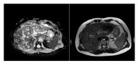
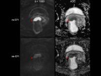
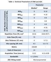
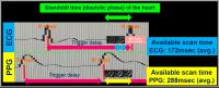
Simple and robust cardiac diffusion weighted imaging using single-shot Turbo Spin-Echo with peripheral pulse gating
Yasuhiro Goto1, Kenji Fukushima2, Masami Yoneyama3, Atsushi Takemura3, Hitoshi Tadenuma1, Mamoru Takeyama1, and Shuji Sakai2
1Department of Radioligical Service, Tokyo Women's Medical University Hospital, TOKYO, Japan, 2Department of Diagnostic imaging & Nuclear Medicine, Tokyo Women`s Medical University Hospital, Tokyo, Japan,3Philips Electronics Japan, Tokyo, Japan
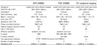
We evaluated the feasibility of optimal gating method and imaging parameters for Single-Shot Turbo Spin Echo (ssTSE) diffusion weighted image (DWI). As a result, The Peripheral pulse gating (PPG) synchronization method was significantly higher in visual scoring than that of the ECG synchronization method (p=0.02) and ssTSE-DWI using SENSE-factor 4.0 brought about the best image quality. Refocusing Flip Angle (RFA) tended to be higher in images with higher visual score. Visualizing cardiac DWI was feasible under above conditions. In conclusion, it is expected that ssTSE-DWI using PPG has a possibility to detect abnormal signal from myocardium by non-contrast MRI.


3477.   
74 
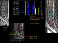
The Study of Vertebral Marrow Microstructure in Healthy Young Adults with Intravoxel Incoherent Motion Diffusion-weighted Imaging
Jinliang Niu1, Wenqi Wu1, Tong Gong1, Wenjin Li1, Dandan Zheng2, Zheng Zhong3, Hongwei Wang1, and Xiaohong Joe Zhou3
1The Second Hospital of Shanxi Medical University, Taiyuan, China, People's Republic of, 2GE Healthcare MR Research China, Beijing, China, People's Republic of, 3Center for Magnetic Resonance Research, University of Illinois Hospital &Health Sciences System, Chicago, IL, United States
MRI has become preferred over other imaging modalities in evaluating marrow compositions. Although the routine MRI can evaluate the cellularity of marrow according to signal intensity, it’s not quantitative analysis. IVIM provides both diffusion and perfusion quantification using a single imaging study at the same time, without intravenous contrast injection. IVIM has been applied in various diseases, but the application of IVIM in marrow composition is less. As the preliminary study, we will adopt the parameters of IVIM to assess the vertebral bone marrow microstructure, then to investigate gender-related cellular and capillary network of vertebral marrow in healthy young adults. 


3478.   
75 
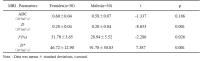
Corticospinal Tract Distribution in Motor Cortices of Adult Macaque Brains Revealed by High Angular Resolution Diffusion Imaging Tractography
Yuguang Meng1 and Xiaodong Zhang1,2
1Yerkes Imaging Center, Yerkes National Primate Research Center, Emory University, Atlanta, GA, United States, 2Division of Neuropharmacology and Neurologic Diseases, Yerkes National Primate Research Center, Emory University, Atlanta, GA, United States
      Non-human primates mimick most aspects of humans and are widely used in preclinical or medical studies. Understanding the structural connectivity in non-human primate brains can provide essential reference for translational research. The characterization of the corticospinal tracts plays a crucial role in motor function and has been well studied in human brain. However, it remains not fully understood in non-human primates. In this work, high angular resolution diffusion imaging (HARDI) tractography was utilized to evaluate the corticospinal tracts distribution in sub-regions of motor cortices of adult macaque monkeys, and high similarity to prior ex-vivo results was observed.


3479.   
76 
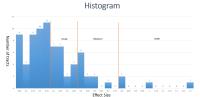
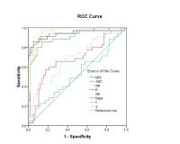
White matter lesions highly influence group comparison of diffusion tensor imaging metrics
Daniel Svärd1,2, Markus Nilsson3, Björn Lampinen4, Jimmy Lätt2, Pia Sundgren1,2, Erik Stomrud5, Lennart Minthon5, Katarina Nägga5, Oskar Hansson5,6, and Danielle van Westen1,2
1Diagnostic Radiology, Clinical Sciences, Lund University, Lund, Sweden, 2Center for Medical Imaging and Physiology, Skåne University Hospital, Lund, Sweden, 3Lund University Bioimaging Center, Lund University, Lund, Sweden, 4Department of Medical Radiation Physics, Lund University, Lund, Sweden, 5Clinical Memory Research Unit, Clinical Sciences, Lund University, Malmö, Sweden, 6Neurology, Clinical Sciences, Lund University, Lund, Sweden
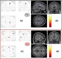
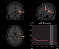
White matter lesions (WML) are common in cognitively healthy elderly and their presence in a brain region is associated with elevated mean diffusivity (MD) and reduced fractional anisotropy (FA). We compared patients with amnestic mild cognitive impairment (aMCI) to control groups with different prevalence of WML. Our results showed that including subjects with WML in the control group highly influence the outcome of statistical analysis of diffusion tensor imaging (DTI) metrics. We conclude that WML should be taken into consideration when designing and interpreting DTI studies.


3480.   
77 Quantitative Diffusion MRI of Hematopoietic Acute Radiation Syndrome using a Minipig Model
Frederick C. Damen1,2, Matthew Lindeblad3, Kejia Cai2,4, Michael Flannery2, Yi Sui2, Amelia M Bartholomew5, Aleksander V Lyubimov3, and Xiaohong Joe Zhou2,4,6,7
1Radiology, University of Illinois at Chicago, Chicago, IL, United States, 2Center for Magnetic Resonance Research, University of Illinois at Chicago, Chicago, IL, United States, 3Pharmacology, University of Illinois Medical Center, Chicago, IL, United States, 4Bioengineering, University of Illinois at Chicago, Chicago, IL, United States, 5Surgery, University of Illinois Medical Center, Chicago, IL, United States, 6Radiology, Chicago, IL, United States, 7Neurosurgery, University of Illinois at Chicago, Chicago, IL, United States
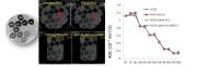
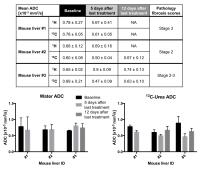
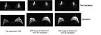
The purpose of this study is to characterize Hematopoietic Acute Radiation Syndrome (hARS) and assess the effect of total body irradiation in a Göttingen minipig model using quantitative diffusion MRI. The minipigs were irradiated at a total dose of either 1.65 Gy (LD30/45 days) or 1.90 Gy (LD70/45 days). The animals underwent diffusion MRI scans prior to and again at 8, 15, or 22 days following irradiation.  The consistent diffusion value prior to irradiation and the significant changes post-irradiation, both observed in this study, suggest that quantitative diffusion MR can be a viable marker for studying the effect of total body irradiation.


3481.   
78 
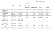
White matter microstructure among perinatally HIV-infected youth: A diffusion tensor imaging study
Manoj Kumar Sarma1, Margaret Keller2, Rajakumar Nagarajan1, David E Michalik3, Judy Hayes2, Karin Nielsen-Saines4, Jaime Deville4, Joseph A Church5, Irwin Walot6, and M. Albert Thomas1
1Radiological Sciences, UCLA School of Medicine, Los angeles, Los Angeles, CA, United States, 2Pediatrics, Harbor-UCLA Medical Center, Torrance, CA, United States, 3Infectious disease-Pediatrics, Miller’s Children’s Hospital of Long Beach, Long Beach, CA, United States, 4Pediatrics, UCLA School of Medicine, Los angeles, Los Angeles, CA, United States, 5Pediatrics, Children’s Hospital Los Angeles, Los Angeles, CA, United States,6Radiology, Harbor-UCLA Medical Center, Torrance, CA, United States
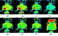
DTI was used to derive in vivo tissue status measurements of subcortical brain regions that are vulnerable to injury in perinatally HIV-infected youths. Quantitative measurements, including the mean diffusivity (MD), fractional anisotropy (FA), axial diffusivity (AD) and radial diffusivity (RD) were determined in of the whole brain in 12 well-characterized HIV youths and in 12 healthy control subjects. We observed widespread brain regions with increased AD values in perinatally HIV-infected youths compared to healthy controls, indicating axonal changes. We also observed increased FA, MD and RD. To confirm these findings a correlation study with neurodevelopement and neurocognitive changes as well as ART effect is needed. Understanding the impact of HIV disease severity on white matter integrity provides potentially useful clinical tools for evaluating ART efficacy during a dynamic period of brain development.


3482.   
79 
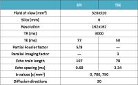
Intravoxel incoherent motion diffusion weighted imaging in evaluating the radio-sensitivity of nasopharyngeal carcinoma xenografts
Youping Xiao1, Yunbin Chen1, Jianji Pan2, Dechun Zheng1, Xiang Zheng1, and Ying Chen1
1Radiology, Fujian Provincial Cancer Hospital, Fuzhou, China, People's Republic of, 2Radiation Oncology, Fujian Provincial Cancer Hospital, Fuzhou, China, People's Republic of
In this present study, by applying the special mouse coil(4 channel), IVIM-DWI with 14 b-factors(0~1000s/mm2) was successfully conducted on nude mice with different radio-sensitive NPC xenografts(CNE-1 and CNE-2) during the course of fractional radiations. The IVIM-DWI parameters of xenografts were found to change characteristically after fractional radiations and were significantly different between different radio-sensitive NPC xenografts, and their corresponding changes also behaved significant correlations with the pathological features of NPC xenografts. Thus, it is suggested IVIM-DWI parameters be valuable in evaluating the micro-structures and radio-sensitivity of NPC xenografts.


3483.   
80 
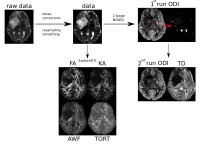
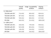
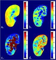
Combining Intravoxel Incoherent Motion Model and Reduced FOV for Evaluation of Single Renal Diffusion and Perfusion
CY Wang1, R Zhang2, L Jiang3, R Wang4, XD Zhang4, H Wang3, K Zhao4, LX Jin3, J Zhang1,2, XY Wang1,4, and J Fang1,2
1Academy for Advanced Interdisciplinary Studies, Peking University, Beijing, China, People's Republic of, 2College of Engineering, Peking University, Beijing, China, People's Republic of, 3Philips Healthcare, Suzhou, China, People's Republic of, 4Department of Radiology, Peking University First Hospital, Beijing, China, People's Republic of
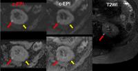
The sequence most commonly used in renal DWI is based on single-shot echo-planar imaging (SS-EPI), which is prone to artifacts and distortions related to susceptibility and eddy currents. Reducing these artifacts and distortions in SS-EPI generally requires a reduced field of view (rFOV) in the phase-encoding direction and/or reduced spatial resolution. To resolve these problems, a 2D rFOV-DWI sequence was introduced for imaging and further IVIM modeling. Compared with conventional full-FOV single-shot DWI techniques, rFOV-DWI methods generally produced images of superior quality. With the application of IVIM model, it is possible to evaluate single renal diffusion and perfusion simultaneously.


3484.   
81 
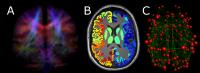
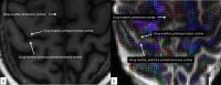
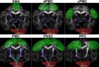
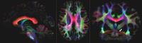
Super-Resolution Track Density Imaging of 1.4 mm isotropic 7T Whole-Brain Diffusion Magnetic Resonance Images
Ralf Lützkendorf1, Robin M. Heidemann2, Thorsten Feiweier2, Michael Luchtmann3, Sebastian Baecke1, Joern Kaufmann4, Joerg Stadler5, Eike Budinger5, and Johannes Bernarding1
1Biometry and Medical Informatics, University of Magdeburg, Magdeburg, Germany, 2Siemens Healthcare GmbH, Erlangen, Germany, 3Department of Neurosurgery, University of Magdeburg, Magdeburg, Germany, 4Department of Neurology, University of Magdeburg, Magdeburg, Germany, 5Leibniz Institute for Neurobiology, Magdeburg, Germany
Track-density imaging (TDI) is a method to generate super-resolution images from fiber-tracking data (1). Here, we applied this technique to 1.4 mm isotropic 7T whole brain diffusion MR imaging data (dMRI). Besides the well-known large and medium-sized fiber tracts the high resolution of the data allowed visualizing the complex interwoven courses of fiber tracts in the cerebellar-pontine angle as well as showing parts of the trigeminus nerve. Combining TDI with high-resolved diffusion data has a great potential for analyzing the anatomy in vivo of brain structures across different scales as well as the neuronal connectome throughout the whole brain.


3485.   
82 Multi-Center Validation of an Acetone-D2O Quantitative Diffusion Phantom
Xiaoke Wang1, Samir D Sharma2, Mustafa R Bashir3, Jean H Brittain2, Jean Shaffer3, Takeshi Yokoo4, Qing Yuan4, Scott B Reeder1,2,5,6,7, and Diego Hernando2
1Biomedical Engineering, University of Wisconsin-Madison, Madison, WI, United States, 2Radiology, University of Wisconsin-Madison, Madison, WI, United States, 3Radiology, Duke University, Durham, NC, United States, 4Radiology, University of Texas Southwestern Medical Center, Dallas, TX, United States, 5Medical Physics, University of Wisconsin-Madison, Madison, WI, United States, 6Medicine, University of Wisconsin-Madison, Madison, WI, United States, 7Emergency Medicine, University of Wisconsin-Madison, Madison, WI, United States
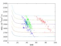
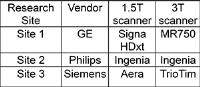
A recently proposed acetone-D2O phantom, which has ADC tunable over the entire physiological range, has shown promise for the development and quality assurance of quantitative diffusion MRI. In this study, this phantom was shipped between three sites with different MRI vendors to demonstrate consistent diffusion quantification across imaging protocols, platforms, and field strengths. The results demonstrated consistent ADC measurements across sites/vendors, field strength, choice of b-values, intra-exam and inter-exam repetition. In conclusion, the acetone-D2O phantom is a promising tool for future multi-center validation and quality assurance of quantitative diffusion MRI techniques.


3486.   
83 
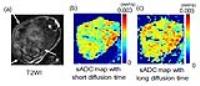
Measuring diffusion time dependence of pseudo-diffusion using flow compensated pulsed and oscillating gradient sequences
Dan Wu1 and Jiangyang Zhang1,2
1Radiology, Johns Hopkins University School of Medicine, BALTIMORE, MD, United States, 2Radiology, New York University School of Medicine, New Yourk, NY, United States
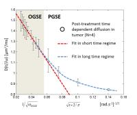
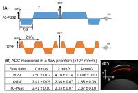
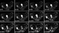
Intravoxel incoherent motion (IVIM) in the capillaries reflects capillary geometry and flow velocity, which may be probed by diffusion MRI measured at varying diffusion times. In this study, we employed flow-compensated pulsed and oscillating gradient sequences to investigate the diffusion time dependence of pseudo-diffusion in the mouse brain with diffusion times ranging from 2.5 ms to 40 ms. We used a simplified IVIM model to characterize the pseudo-diffusion compartment and flow compartment based on the relation between capillary segments and diffusion time/distance. Our results clearly demonstrated diffusion time dependence and suggested that the pseudo-diffusion fraction increased with increasing diffusion time.


3487.   
84 
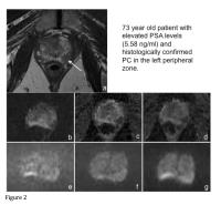
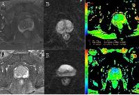
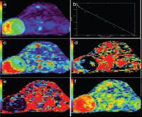
Diffusion weighted imaging of prostate cancer xenografts: comparison of bayesian modeling and independent least squares fitting - Permission Withheld
Parisa Movahedi1, Hanne Hakkarainen2, Harri Merisaari1, Heidi Liljenbäck1, Helena Virtanen1, Hannu Juhani Aronen1, Heikki Minn1, Matti Poutanen1, Anne Roivainen1, Timo Liimatainen2, and Ivan Jambor1
1University of Turku, Turku, Finland, 2A.I. Virtanen Institute for Molecular Sciences, Kuopio, Finland
Tumor growth in mice preclinical prostate cancer model (human prostate cancer cells, PC-3) was followed for 4 weeks by weekly DWI  in control group (n=10) and treatment group (n=9) receiving Docetaxel.  DWI data sets were acquired using 15 b-values in the range of 0-500s/mm2 and 12 b-values the range of 0-2000 s/mm2. The DWI signal decays were fitted using monoexponential, biexponential, kurtosis and stretched exponential models/functions. Bayesian shrinkage prior method and independent least squares fitting have been applied and fitting quality evaluated by corrected Akaike Information Criteria. Bayesian modeling improved quality of DWI parametric maps derived using high b-value DWI data sets. Our result does not support the use of biexponential, kurtosis and stretched exponential models/functions for low b value DWI data sets of PC-3 mice preclinical prostate cancer model.


3488.   
85 Intravoxel Incoherent Motion Diffusion-weighted Magnetic Resonance Imaging for Monitoring the Early Response to ZD6474 from Nasopharyngeal Carcinoma in Nude Mouse - Video Not Available
Yong Zhang1 and Yanfen Cui2
1GE Healthcare China, Shanghai, China, Shanghai, China, People's Republic of, 2Department of Radiology, Shanghai Jiao Tong University School of Medicine, Shanghai, China, People's Republic of
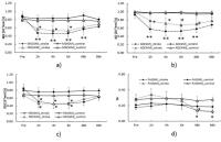
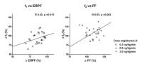
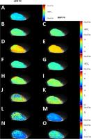
This study was to investigate the feasibility of IVIM DWI to evaluate the early therapeutic effects of ZD6474 upon human NPC xenografts in nude mouse. NPC mice underwent IVIM DWI at baseline and after 1, 3, and 7 days of treatment. In the treated group, the f and D* decreased significantly on day 1 while the ADC and D were significantly higher from day 3 compared with the control group, demonstrating that IVIM DWI is sensitive to detect the ZD6474-induced changes in human NPC nude mouse, and the D* and f parameters could predict early response to anti-angiogenic treatment.  


3489.   
86 Comparison of IVIM and BOLD MR imaging in Functional Evaluation of Diabetic nephropathy
Lihua Chen1, Tao Ren1, Yu Zhang2, Chenglong Wen1, and Wen Shen1
1Tianjin First Center Hospital, Tianjin, China, People's Republic of, 2Philips healthcare, Beijing, China, People's Republic of
To detect the changes of kidney diseases, magnetic resonance imaging(MRI) as a noninvasive approach has been proved to be more suitable for detecting and monitoring diabetic nephropathy(DN). Intravoxel incoherent motion (IVIM) and blood oxygenation level dependent (BOLD) MR imaging have been confirmed their high potential in detecting changes of renal function in patients with chronic renal diseases and transplanted kidneys. We compared the parameters of IVIM and BOLD in patients with DN and heathy controls. The results demonstrated the capacity of IVIM and BOLD for reflecting renal perfusion, diffusion and oxygenation changes in patients with DN. 


3490.   
87 Diffusion Kurtosis Metrics as Biomarker of Fibre Maturity
Grinberg Farida1,2, Ivan I. Maximov1,3, Ezequiel Farrher1, Irene Neuner1,4,5, Eileen Oberwelland6,7, Kerstin Konrad5,6,8, and N. Jon Shah1,2,5
1Institute of Neuroscience and Medicine - 4, Forschungszentrum Juelich GmbH, Juelich, Germany, 2Department of Neurology, Faculty of Medicine, RWTH Aachen University, Aachen, Germany, 3Experimental Physics III, TU Dortmund University, Dortmund, Germany, 4Department of Psychiatry, Psychotherapy and Psychosomatics, RWTH Aachen University, Aachen, Germany, 5JARA - BRAIN - Translational Medicine, Aachen, Germany, 6Institute of Neuroscience and Medicine – 3, Forschungszentrum Jülich GmbH, Juelich, Germany, 7Translational Brain Research in Psychiatry and Neurology, Department of Child and Adolescent Psychiatry, Psychosomatics and Psychotherapy, RWTH Aachen University, Aachen, Germany, 8Child Neuropsychology Section, Department of Child and Adolescent Psychiatry and Psychotherapy, RWTH Aachen University, Aachen, Germany
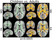
Diffusion tensor imaging has enabled the examination of white matter connectivity and microstructural changes across the lifespan. However, the detection of subtle microstructural changes during typical brain maturation still remains challenging. Recently, diffusion kurtosis imaging has attracted much attention as an efficient method for characterising non-Gaussian water diffusion in brain tissue. Here, we tested whether diffusion kurtosis imaging can extend our knowledge of changes in brain tissue microstructure related to normal brain development. We showed that diffusion kurtosis imaging provides useful biomarkers sensitive to the level of maturity in association, projection and commissural fibres. 


3491.   
88 Using intravoxel incoherent motion MR imaging to measure renal diffusion and perfusion in contrast-induced acute kidney injury - Video Not Available
Bin Zhang1, Long Liang1, Yuhao Dong1, Kannie W.Y. Chan2, Guanshu Liu2, Changhong Liang1, and Shuixing Zhang1
1Department of Radiology, Guangdong Academy of Medical Sciences/Guangdong General Hospital, Guangzhou, China, People's Republic of, 2Russell H. Morgan Department of Radiology and Radiological Sciences, Division of MR Research, The Johns Hopkins University School of Medicine, Baltimore 21287, USA, Baltimore, AL, United States
Contrast-induced acute kidney injury (CI-AKI) is a common iatrogenic event caused by the injection of iodinated contrast agent, and remains the third major source of in-hospital acquired acute renal failure.The objective of our study is to examine the feasibility of using Intravoxel Incoherent Motion (IVIM) MRI to simultaneously measure the pathological changes in kidney diffusion and perfusion in the course of CI-AKI.  Our results showed that the kidney perfusion and diffusion as measured by IVIM are well-correlated with those measured using conventional methods, indicating IVIM MRI can be used as an effective tool for the diagnosis and staging of CI-AKI. 


3492.   
89 
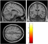
Assessment of brain structural network alterations in major depressive disorder using generalized q-sampling imaging and connectome analysis
Chao-Yu Shen1,2,3, Zhen-Hui Li1, Vincent Chin-Hung Chen4, Ming-Chou Ho5, Yeu-Sheng Tyan1,2, and Jun-Cheng Weng1,2
1Department of Medical Imaging and Radiological Sciences, Chung Shan Medical University, Taichung, Taiwan, 2Department of Medical Imaging, Chung Shan Medical University Hospital, Taichung, Taiwan,3Institute of Medicine, Chung Shan Medical University, Taichung, Taiwan, 4Department of Psychiatry, Chang Gung Memorial Hospital, Chiayi, Taiwan, 5Department of Psychology, Chung Shan Medical University, Taichung, Taiwan
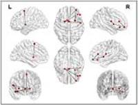
Major depressive disorder (MDD) is the most common mood disorder in the world and the most important precursor of suicide. Despite decades of research, the pathophysiology of MDD remains not well understood. Recently, several MRI studies have focused on structural and functional connectivity evaluation and suggested that alterations of some specific regions of the brain, in both gray and white matter structures and some specific cortical–subcortical neuronal circuits, may play important roles of MDD. Generalized q-sampling imaging (GQI) is a more accurate and sophisticated diffusion MR approach compared to diffusion tensor imaging (DTI), which can extract additional information about the altered diffusion environments to resolve the complicated neural structure changes of neural disease. In this study, we used GQI and graph theoretical analysis to evaluate brain structure and connectivity change of MDD compared to healthy controls and correlation with symptom severity. Our results indicated  GQI indices can help to detect structural and connective abnormalities of MDD patients and these alterations are correlated with depressive severity. 


3493.   
90 
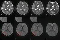
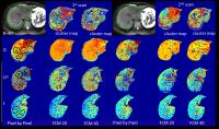
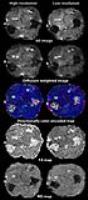
Evaluation of Acute Cerebral Infarction Using a Fast Kurtosis Diffusion imaging Protocol
Chengxu Li1, Tianyi Qian2, Jinsuh Kim3, Philip Zhe Sun4, Jie Lu1, and Kuncheng Li1
1Department of Radiology, Xuanwu Hospital, Capital Medical University, Beijing, China, People's Republic of, 2MR Collaborations NE Asia, Siemens Healthcare, Beijing, China, People's Republic of, 3Department of Radiology, University of Illinois at Chicago, Chicago, IL, United States, 4Martinos Center for Biomedical Imaging, Department of Radiology, Massachusetts General Hospital and Harvard Medical School, Boston, MA, United States
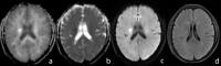
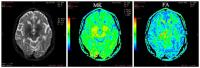
Conventional DKI is limited in use for detecting acute stroke because of its relatively long scan time and current need of offline processing. Here we used a 13 directions protocol combined with simultaneous multi-slice technique with inline reconstruction to test the diffusion pattern at different time points. The results shows the lesion size observed within 24hr on MK matched with the lesion size on T2-FLAIR after one month, which was better than lesion size observed within 24hr on DWI. Early application of the fast DKI to detect the anomalous range of MK was beneficial to predict the range of the eventual infarction for clinical treatment.


3494.   
91 High resolution tract density tract-based spatial statistics and automating fiber-tract quantification analysis in patients suffering from major depressive disorder
Stefan Sommer1,2, Nadja Doerig3,4, Janis Brakowski2, Martin grosse Holtforth5, Sebastian Kozerke1, Erich Seifritz2,4, Simona Spinelli2,4, and Philipp Stämpfli2
1Institute for Biomedical Engineering, ETH and University of Zurich, Zurich, Switzerland, 2Department of Psychiatry, Psychotherapy and Psychosomatics Psychiatric Hospital, University of Zurich, Zurich, Switzerland,3Division Neuropsychology, Departement of Psychology, University of Zurich, Zurich, Switzerland, 4Neuroscience Center, University and ETH Zurich, Zurich, Switzerland, 5Department of Psychology, University of Bern, Bern, Switzerland
In the last few years, tract base spatial statistics (TBSS) and automating fiber-tract quantification (AFQ) have become prominent tools for analyzing diffusion data in group studies. In this study, we introduce optimized high-resolution tract density (optTD) images and analyze these maps using TBSS and AFQ in patients with major depressive disorders. We show a higher sensitivity in the newly introduced optTD compared to traditional FA analyses. Significant group differences were found using both methods indicating robust findings. High resolution optTD maps derived from optimized tractograms provide a promising tool for investigating white-matter abnormalities in mental disorders.


3495.   
92 Diffusional Kurtosis Imaging Study Of Parkinson Disease
Xilun Ma1, Jitian Guan1, Zhiyan Zhang1, Miaomiao Chen1, Yanzi Chen1, Zhiwei Shen1, and Renhua Wu1
1Department of Medical Imaging, the 2nd Affiliated Hospital, Medical College of Shantou University, Shantou 515041, China, Shantou, China, People's Republic of
Eighteen Parkinson Disease’s (PD) patients and four healthy controls(HC) underwent the diffusional kurtosis imaging (DKI) and then we tested the PD’s patients with Hoehn-Yahr scale and Unified Parkinson Disease Rating Scale(UPDRS). As a result, we found a significant decrease of Mean kurtosis (MK) values in the left substantia nigra between PD’s patients and healthy controls. Moreover, the kurtosis fractional anisotropy (KFA) values in right red nucleus of the PD’s patients were positively associated with the UPDRS scores in our study.


3496.   
93 
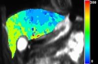
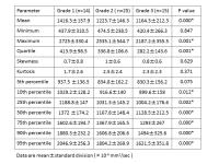
Characterization of Clear Cell Renal Cell Carcinoma with Diffusion Kurtosis Imaging: Correlation between Diffusion Kurtosis Parameters and Tumor Cellularity
Guangyu Wu1 and Yongming Dai2
1renji hospital, shangha, China, People's Republic of, 2MR, Philips Healthcare, Shanghai, China, People's Republic of
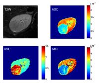
A wide spectrum of the use of DKI to characterize non-Gaussian diffusion pattern in microstructural tumor tissue has been developed involving a variety of tumors. However, the feasibility of DKI in kidney has been assessed in healthy volunteers only. The study was assigned to assess the quantitative DKI in grading of clear cell renal carcinoma (ccRCC) and to compare the correlation between DKI parameters and tumor cellularity and found that DKI could not only quantitatively characterize ccRCC with different grades but also provide valuable information on the diffusion properties related to tumor microenvironment changes or tissue complexity in tumor.


3497.   
94 Diffusional kurtosis imaging study in idiopathic normal pressure hydrocephalus patients, before and after shunt placement surgery analysis
Chanon Ngamsombat 1, Zhe Zhang2, Hua Gua2, Theerapol Witthiwej3, Weerasak Muangpaisan4, Sith Sathornsumetee5, Suwit Charoensak6, Panida Charnchaowanish1, and Orasa Chawalparit1
1Department of Radiology, Faculty of Medicine Siriraj Hospital, Mahidol University, Bangkok, Thailand, Bangkok, Thailand, 2Center for Biomedical Imaging Research, Department of Biomedical Engineering, School of Medicine, Tsinghua University, Beijing, China, Beijing, China, People's Republic of, 3Department of Surgery, Faculty of Medicine Siriraj Hospital, Mahidol University, Bangkok, Thailand, Bangkok, Thailand,4Department of Preventive and Social Medicine, Faculty of Medicine Siriraj Hospital, Mahidol University, Bangkok, Thailand, Bangkok, Thailand, 5Departments of Medicine, Faculty of Medicine Siriraj Hospital, Mahidol University, Bangkok, Thailand, Bangkok, Thailand, 6Department of Psychiatry, Faculty of Medicine Siriraj Hospital, Mahidol University, Bangkok, Thailand, Bangkok, Thailand
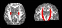
Idiopathic normal pressure hydrocephalus(iNPH) is important reversible cause of dementia and gait abnormality in elderly patients. Diffusional kurtosis imaging(DKI) moreover explains the complexity of white matter abnormality with inclusion of non-Gaussian effects. We aim to identify difference of complexity of white matter alteration in iNPH patients before and after shunt placement surgery by using high resolution DKI. We report significant increase of mean diffusional kurtosis(Kmean), mean diffusivity(MD) and decrease of radial diffusional kurtosis(Krad) , fractional ansiotropy(FA) after shunt placement surgery. High resolution DKI can be used for monitoring and detection complexity of white matter alteration in iNPH patients.


3498.   
95 The Investigation of Cerebral Microstructure Changes of Pediatric Patients  With Type ? Gaucher Disease Using Diffusion Kurtosis Imaging
Huiying Kang1, Ningning Zhang, Kaining Shi2, Yanqiu Lv1, Di Hu1, and Yun Peng1
1Imaging center, Beijing Children’s Hospital, Capital Medical University, Beijing, China, People's Republic of, 2Imaging Systems Clinical Science, Philips Healthcare, Beijing, China, People's Republic of
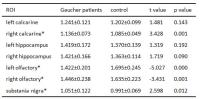
Increasing clinical studies suggested that there are some type ? Gaucher disease (GD?) patients suffering neurological symptom, which was originally defined as non-neuropathlogical involved. This study recruited 38 patients and 32 normal children to investigate morphological changes of brain in GD? patients by using Diffusion Kurtosis Imaging. Our results showed significant decreased MD in bilateral olfactory gyrus, increased MD in right calcarine and substantia nigra and significant increased MK in right olfactory gyrus. Our study suggests a necessity of adjusting the opinion regarding the CNS-involvement of GD?, and DKI analysis is a potential imaging marker in clinical studies of GD?.


3499.   
96 Differentiating Minimal Fat Angiomyolipoma from clear cell Renal Cell Carcinoma: comparison of monoexponential, biexponential, and stretched exponential Diffusion-weighted imaging - Video Not Available
Haojie Li1, Lili Liang1, Anqin Li1, Qiong Li1, Yao Hu1, Hui Lin2, Daoyu Hu1, and Zhen Li1
1Departments of Radiology, Tongji Hospital, Tongji Medical College, WU HAN, China, People's Republic of, 2GE Healthcare,MR Research China,WU HAN, WU HAN, China, People's Republic of
Since DWI with different models may demonstrate different aspects of tissue properties, it should be valuable to compare and explore their roles in renal tumors. To our knowledge, however, no comparison of these different diffusion imaging approaches for the differential diagnosis in renal tumors has been investigated so far. The purpose of this study was to quantitatively compare and evaluate the potential clinical value of various diffusion parameters obtained from monoexponential, biexponential, and stretched exponential DWI models for differentiation of MFAML from ccRCC.
 

The International Society for Magnetic Resonance in Medicine is accredited by the Accreditation Council for
Continuing Medical Education to provide continuing medical education for physicians.