|
Exhibition Hall 10:00 - 11:00 |
|
|
|
Computer # |
|
3692.
 |
25 |
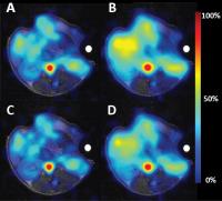
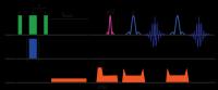
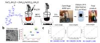
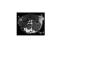
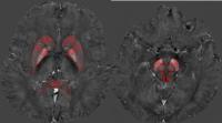
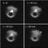
Imaging in vitro and in vivo pH with ioversol by CEST MRI 
Miaomiao Chen1, Xiaolei Zhang1, Yanzi
Chen1, Zhiwei Shen1, Wei Hu1,
Xilun Ma1, and Renhua Wu1
1Radiology Department, Second Affiliated
Hospital, Shantou University Medical College, Shantou,
China, People's Republic of
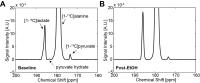
We have developed a CEST MRI method that can measure pH
using ioversol, a contrast agent that is clinically approved
for X-ray imaging and has been repurposed for CEST MRI
studies. Using ioversol as a CEST agent, we have measured pH
over a range of 6.0 - 7.8 pH units by a novel ratiometric pH
MRI method, in a concentration-independent manner. We also
have used this agent and CEST MRI method to measure the
extracellular pH (pHe) within the liver of healthy SD rats.
|
|
3693.
 |
26 |
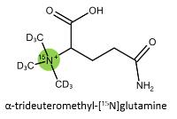
15N Heteronuclear Chemical Exchange Saturation Transfer MRI
Imaging
Haifeng Zeng1,2, Jiadi Xu1,2, Nirbhay
N Yadav1,2, Michael T McMahon1,2,
Bradley Harden3, Dominique Frueh3, and
Peter C.M van Zijl1,2
1Russell H. Morgan Department of Radiology and
Radiological Science, Johns Hopkins University School of
Medicine, Baltimore, MD, United States, 2F.M.
Kirby Research Center for Functional Brain Imaging, Kennedy
Krieger Research Institute, Baltimore, MD, United States, 3Department
of Biophysics and Biophysical Chemistry, Johns Hopkins
University School of Medicine, Baltimore, MD, United States
A two-step heteronuclear enhancement approach to magnify 15N
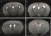
MRI signal through indirect detection of water is described.
Chemical Exchange Saturation Transfer (CEST) works by
continuously perturbation of the spin magnetization of the
exchangeable spins, and then through chemical exchange to
accumulate this perturbation on water proton for signal
magnification. This perturbation is mainly limited to
saturation or excitation pulse on the exchangeable protons.
In this work, the signal of 15N
is detected indirectly through the water signal by first
inverting selectively protons scalar-coupled to 15N
in the urea molecule, followed by chemical exchange of the
amide proton to bulk water.
|
|
3694.
 |
27 |
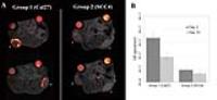
Quantification of macrophage recruitment in double and single
hit head and neck cancer using fluorine-19 MRI
Aman Khurana1, Fanny Chapelin1,
Hongyan Xu1, Partick McConville2,
Quyen Nguyen3, and Eric Ahrens1
1Radiology, University of California, San Diego,
San Diego, CA, United States, 2Molecular
Imaging Inc. La Jolla, San Diego, CA, United States, 3Head
& Neck Surgery, University of California, San Diego, San
Diego, CA, United States
Head and neck carcinoma is a source of significant mortality
worldwide, two prevalent subtypes include double and single
hit tumors. We aimed to evaluate the role of infiltrating
immune cells in worse clinical outcomes with double-hit
tumor patients. A novel 19-Fluorine containing
perfluorocarbon (PFC) emulsion was used to tag macrophages
with high specificity and sensitivity and no background. The
average number of 19F spins within the double hit tumors
were approximately double compared to the single hit group.
This quantifies the tumor associated macrophage burden of
head and neck cancer using a PFC emulsion and proton/19F
MRI.
|
|
3695.
 |
28 |
Simultaneously Trace Blood Perfusion and Glymphatic Passage by
Analyzing Deuterium Oxide Perfusion Imaging with a
Two-Compartment Parallel Model
Cheng-He Li1, Zi-Min Lei1, Sheng-Min
Huang1, Chin-Tien Lu1, Kung-Chu Ho2,
and Fu-Nien Wang1
1Biomedical Engineering and Environmental
Sciences, National Tsing Hua University, Hsinchu, Taiwan, 2Nuclear
Medicine, Chang Gung Memorial Hospital, Linkou, Taoyuan,
Taiwan
This study tried to simultaneously trace blood perfusion and
glymphatic passage by applying two-compartment parallel
model (2CPM) on D2O perfusion using the new
imaging strategy. Six rats were injected with D2O,
and both F1 and
F2 were
quantified from 2CPM. The results show that F1 is
highly coordinated with cerebral blood flow, while F2 is
much irrelevant. Only regions near several arteries show
significant F2 values,
which is speculated as the paravascular pathway of CSF
regulated by glymphatic system. Therefore, using 2CPM for
tracing D2O might noninvasively reveal the
information of both blood and CSF dynamics.
|
|
3696.
 |
29 |
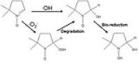
Cellulose-triacetate Nanoparticles as Smart Contrast Agents for
Single Cell Detection.
Laura Szkolar-Sienkiewicz1, Christiane Mallett2,
and Erik M Shapiro1
1Radiology, Michigan State University, East
lansing, MI, United States, 2Michigan
State University, East lansing, MI, United States
It was demonstrated that enzymatically degradable cellulose
triacetate particles offer a novel approach to contrast
modulation. The use of such materials as smart contrast
agents has been discussed and evidence of cellular uptake
and relaxivity modification shown.
|
|
3697.
 |
30 |
Development of intravascular SPION with tunable pharmacokinetics
and relaxivity for preclinical fMRI and micro-MRA
Manasmita Das1,2, Esteban Oyarzabal1,
Heather Decot1, Xiopeng Zong1, Neal
Shah1, Sung Ho Lee1, Jonathan Edward
Frank1, Nazar A Filnov3, and Yen-Yu
Ian Shih1,2
1Biomedical Research Imaging Center, UNC Chapel
Hill, Chapel Hill, NC, United States, 2Department
of Neurology, University of North Carolina Chapel Hill,
Chapel Hill, NC, United States, 3SOP-CNDD,
UNC Chapel Hill, Chapel Hill, NC, United States
We have developed a simple, inexpensive method to synthesize
high-performance intravascular SPION in house. We were able
to tune the relaxivity and PK profile of our home-made SPION
via careful surface functionalization control and came up
with an optimal formulation that offered very robust and
stable contrast for high resolution cerebromicroangiography
and steady state CBV functional imaging. Our future studies
will focus on developing novel MR-detectable inflammation
markers using our home-made iron oxide as the platform
material.
|
|
3698.
 |
31 |
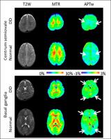
Assessment of brain development in children with developmental
delay using amide proton transfer weighted (APTw) MRI -
Video Not Available
Xiaolu Tang1, Hong Zhang1, Xuna Zhao2,3,
Jinyuan Zhou2, and Yun Peng1
1Imaging Center, Beijing Children’s Hospital,
Capital Medical University, Beijing, China, People's
Republic of, 2Neurosection,
Division of MR Research, Department of Radiology, Johns
Hopkins University, Baltimore, MD, United States, 3Philips
Healthcare, Beijing, China, People's Republic of
Amide proton transfer weighted (APTw) imaging is a novel
molecular MRI technique that can noninvasively detect
cytosolic endogenous mobile proteins and peptides in
myelination process. However it is not well known to brain
developments in children with developmental delay using APTw
MRI. The aim of our work is to explore the brain development
in children with developmental delay (DD) using APTw MRI.
The final conclusion shows that APTw MRI is a promising
technique to assess children with DD at a molecular level.
|
|
3699.
 |
32 |
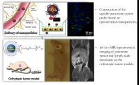
Magnetic/Upconversion Fluorescent Nanoparticle-Based Dual-Modal
Molecular Probes for Imaging of Lymph Node Metastasis From
Pancreatic Cancer in vivo
Kai Cao1, Ruirui Qiao2, and Huimin Wei3
1Radiology, Changhai Hospital, Shanghai, China,
People's Republic of, 2Chemistry,
Chinese Academy of Sciences, Beijing, China, People's
Republic of, 3Clinic
and Translational Medicine Center, Changhai Hospital,
Shanghai, China, People's Republic of
Construction of the specific pancreatic cancer probe based
on upconversion nanoparticles. The probes taking
biocompatibility upconversion nanoparticles with unique
magnetic properties as a carrier, conjugated with ATF
peptide specifically targeting the uPAR. Then, its effect of
detection of tumor and lymph node metastasis would be
further validated in an orthotopic human pancreatic cancer
xenograft model by pathology and dual-modal imaging.
|
|
3700.
 |
33 |
Dynamic PET and cortical thickness comparison between healthy
controls and epilepsy patients using simultaneous PET/MR
Yu-Shin Ding1,2, Shaunak Ohri1, Jean
Logan1, Thomas Koesters1, James Babb1,
and Orrin Devinsky3
1Radiology, NYU School of Medicine, New York, NY,
United States, 2Psychiatry,
NYU School of Medicine, New York, NY, United States, 3Neurology,
NYU School of Medicine, New York, NY, United States
Our results suggest that 1) simultaneous PET/MR imaging
provides a useful imaging tool to identify regional
abnormalities; 2) more information can be rendered from
dynamic PET data; 3) SUVmean_late and cortical thickness are
independent biomarkers for epilepsy. In general, Freesurfer
and SPM are more robust in orientation and segmentation than
FSL.
|
|
3701.
 |
34 |
MRI-Guided Thermosensitive Liposomal Drug Delivery for Cancer
Therapy
Po-Wah So1, Maral Amrahli2, Michael
Wright2, Miguel Centelles2, Wladyslaw
Gedroyc3, Andrew Miller2, and Maya
Thanou2
1Neuroimaging, King's College London, London,
United Kingdom, 2Institute
of Pharmaceutical Sciences, King's College London, London,
United Kingdom, 3Experimental
Medicine, Imperial College London, london, United Kingdom
We have designed and synthesised a novel liposome
formulation, capable of releasing encapsulated drug on
targeting/heating by focussed ultrasound (FUS) for cancer
therapy and being visualised by MRI due to the incorporation
of gadolinium chelates. We show the novel liposomes are
targeted to tumours by FUS and MRI-visible in
vivo and
thus, suitable for theranostic applications in cancer.
|
|
3702.
 |
35 |
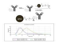
Towards Early Detection of Pancreatic Cancers by Active Feedback
MR Molecular Imaging - Permission
Withheld
Chaohsiung Hsu1, Zhao Li1, Ryan Quiroz1,
Raymond Ngo1, and Yung-Ya Lin1
1Department of Chemistry and Biochemistry, UCLA,
Los Angeles, CA, United States
Early detection of pancreatic cancers using enhanced MRI
techniques increases not only the treatment options
available, but also the patients’ survival rate. This can be
achieved with antibody-conjugated superparamagnetic iron
oxide (SPIO) nanoparticles capable of binding to early stage
pancreatic cancer cells to improve imaging specificity and
innovation methods that can sensitively detect SPIO to
improve imaging sensitivity. The enhanced contrast from SPIO
can then be used to visually assess the distribution and
magnitude of SPIO-targeted tumor cells. In vivo subcutaneous
and orthotopic xenografts mouse models validated the
superior contrast/sensitivity and robustness of this
approach towards early pancreatic cancers detection.
|
|
3703.
 |
36 |
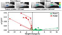
Real Time Detection of Pancreatic Cancer-induced Cachexia using
a Fluorescent Myoblast Reporter and 1H MRS Metabolic Analysis
Paul Thomas Winnard Jr1, Santosh Bharti1,
Marie-France Penet1, Radharani Marik1,
Yelena Mironchik1, Flonne Wildes1,
Anirban Maitra2, and Zaver M Bhujwalla1
1Russell H. Morgan Department of Radiology and
Radiological Science, Johns Hopkins University School of
Medicine, Baltimore, MD, United States, 2MD
Anderson Cancer Center, Houston, TX, United States
Therapeutic options for cancer-induced cachexia are limited
and therefore, efforts to identify signs of precachexia in
cancer patients are necessary for early intervention. Here,
we generated a myoblast cell line expressing a dual
dTomato:GFP construct that was grafted onto the muscle of
mice bearing human pancreatic cancer xenografts to provide
noninvasive live imaging of events associated with
cancer-induced cachexia (i.e., weight loss). 1H
MRS revealed that weight loss in cachectic animals was
associated with a depletion of plasma lipid, cholesterol,
and valine, and decreased skeletal muscle alanine levels,
which may provide informative biomarkers of cachexia.
|
|
3704.
 |
37 |
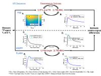
The effect of SNR optimization on cell quantification accuracy
for fluorine-19 MRI sequences: bSSFP, FSE, and FLASH
Kai D. Ludwig1, Erin B. Adamson1,
Christian M. Capitini2,3, and Sean B. Fain1,4,5
1Medical Physics, University of
Wisconsin-Madison, Madison, WI, United States, 2Pediatrics,
University of Wisconsin-Madison, Madison, WI, United States, 3Carbone
Cancer Center, University of Wisconsin-Madison, Madison, WI,
United States, 4Radiology,
University of Wisconsin-Madison, Madison, WI, United States, 5Biomedical
Engineering, University of Wisconsin-Madison, Madison, WI,
United States
Several MRI data acquisition methods have been used for
fluorine-19 (19F) MRI cell tracking and optimizing the image
SNR helps mitigate low sensitivity. An optimization workflow
is presented for three 19F pulse sequences based upon
relaxation parameters measured in a 19F reference phantom.
Bloch simulations reveal signal differences between the
reference phantom and pure 19F cellular label for
SNR-optimized bSSFP, FSE, and FLASH. The simulated relative
errors in 19F signal suggest SNR optimization can compromise
signal quantification and thus in vivo cell quantification
but could provide insight for improved methods to balance
the degree of spin-density weighting and SNR.
|
|
3705.
 |
38 |
Effect of exposure in hypoxia environment caused by high
altitude on magnetic susceptibility in human brain assessed
by quantitative susceptibility mapping
Dandan Zheng1, Wenjia Liu2, Bing Wu1,
and Lin Ma2
1MR Research China, GE Healthcare, Beijing,
China, People's Republic of, 2Radiology
Department, Beijing Military General Hospital, Beijing,
China, People's Republic of
Cerebrospinal fluid fraction, CBF and T2 decay have been
reported to be related with hypoxia caused by high altitude
in previous studies. All these biomarkers maybe associate
with tissue homogeneous magnetism changes, which may result
in the magnetic susceptibility changes. Quantitative
susceptibility mapping (QSM) is a novel technique that
allows mapping of tissue magnetic susceptibility. It has the
potential to be more sensitive with respect to magnetic
tissue properties than conventional magnitude-based
techniques such as transverse relaxation rates. This study
was designed to reveal the effect of exposure in hypoxia
environment on magnetic susceptibility in human brain
assessed by QSM.
|
|
3706.
 |
39 |
Iron accumulation in rat brain with frequent USPIO
administration
Kofi Deh1, Andrew Gorman2, Caspar
Schwiedrzik3, Pascal Spincemaille1,
Martin Prince1, and Yi Wang1
1Weill Cornell Medical College, New York, NY,
United States, 2Tri-Institutional
Training Program in Laboratory Animal Medicine and Science,
New York, NY, United States, 3The
Rockefeller University, New York, NY, United States
An observation of signal loss on functional MRI images of
primates following repeated administration of ultra-small
super-paramagnetic iron oxide (USPIO) particles, led us to
test a hypothesis of iron accumulation in the brain by
performing weekly quantitative susceptibility mapping (QSM)
of rats receiving daily USPIO injections for 9 weeks. We
observed rapid increase in the susceptibility values in
brain ventricles and choroid plexus confirmed by serum iron
measurements and histology. In light of a similar report in
the literature, we recommend monitoring patients receiving
iron therapy with a susceptibility imaging technique such as
QSM.
|
|
3707.
 |
40 |
Comparison Study between T2*, Quantitative Susceptibility
Mapping (QSM), and Histology for Postmortem Human Substantia
Nigra
Jae-Hyeok Lee1, Sun-Yong Baek2,
YoungKyu Song3, Sujeong Lim3, Hansol
Lee3, Minh Phuong Nguyen4, Eun-Joo Kim5,
Gi Yeong Huh6, Se Young Chun4, and
HyungJoon Cho3
1Department of Neurology, Research Institute for
Convergence of Biomedical Science and Technology, Pusan
National University Yangsan Hospital, Yangsan, Korea,
Republic of, 2Department
of Anatomy, Pusan National University School of Medicine,
Yangsan, Korea, Republic of, 3Department
of Biomedical Engineering, Ulsan National Institute of
Science and Technology, Ulsan, Korea, Republic of,4Department
of Electrical and Computer Engineering, Ulsan National
Institute of Science and Technology, Ulsan, Korea, Republic
of, 5Department
of Neurology, Pusan National University Hospital, Busan,
Korea, Republic of, 6Department
of Forensic Medicine, Pusan National University School of
Medicine, Yangsan, Korea, Republic of
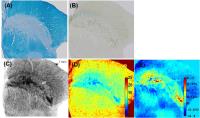
Selective iron deposition in the substantia nigra (SN)
along with the gradual loss of neuromelanin cell (NMC) is
known to be associated with neurodegenerative diseases, such
as Parkinson's disease. Postmortem 40-year-old male and
70-year-old female SN tissues were scanned at various
spatial resolutions with 7T MRI. The association of T2* and
QSM-derived susceptibility values with quantitative NMC and
iron from Perl's Prussian blue staining were investigated
with precise co-registration of MRI and histology. We
identified that T2* and
susceptibility values for NMC and iron regions, which were
segmented from histology were significantly different from
corresponding values of background tissue area.
|
|
3708.
 |
41 |
Arterial spin labeling can detect discreet changes of renal
perfusion after oral application of the pain medication
diclofenac in healthy subjects
Susanne Tewes1, Marcel Gutberlet1, Van
Dai Vo Chieu1, Dagmar Hartung1,
Sebastian Rauhut2, Matti Peperhove1,
Frank Wacker1, Faikah Gueler2, and
Katja Hueper1
1Institute for Diagnostic and Interventional
Radiology, Hannover Medical School, Hannover, Germany, 2Clinic
for Nephrology, Hannover Medical School, Hannover, Germany
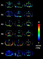
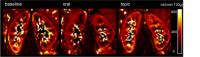
Diclofenac is a nonsteroidal anti-inflammatory drug that is
frequently prescribed to reduce inflammation and pain. It
reduces the prostaglandin-synthesis and may consequently
have an effect on renal perfusion. We investigated whether
arterial spin labeling can detect reduction of renal
perfusion after oral and topical application of diclofenac
compared to baseline measurements. Ten healthy subjects
underwent functional MRI of the kidney. After oral
application of diclofenac renal perfusion was reduced
compared to baseline measurements (321±13 vs. 345±16
ml/(min*100g), p<0.01). No significant changes were found
after topical application. In conclusion, ASL can detect
discreet changes of renal perfusion that occur after
application of diclofenac.
|
|
3709.
 |
42 |
Drug Distribution Kinetics in the Eye assessed by 1H-MRI and
19F-MRS
Christina R Haeuser1, Alfred Ross1,
Markus von Kienlin1, and Basil Künnecke1
1Roche Pharma Research and Early Development, F.
Hoffmann-La Roche Ltd, Basel, Switzerland
Drug treatment of vision impairing diseases often involves
intraocular drug injection into the vitreous humour, a
highly viscous gel-like matter. Drug transport within the
vitreous humour has remained rather elusive although
transport processes are acknowledged to play a pivotal role
for treatment efficacy and adverse effects in the target
tissue. Here, we devised a potentially translational
approach based on contrast-enhanced 1H-MRI and 19F-MRS to
quantitatively ascertain intravitreal drug distribution
kinetics at the macro-scale. We provided proof-of-concept
for the small drug-like molecule trifluoroethanol in
isolated porcine eyes.
|
|
3710.
 |
43 |
Ultra-high field MRI enables the in vivo quantification of the
efficacy of candidate promyelinating molecules in the cuprizone
mouse model
Isaac Mawusi Adanyeguh1, Emilie Poirion1,
Daniel García-Lorenzo2, Marie-Stephane Aigrot1,
Brahim Nait-Oumesmar1, Boris Zalc1,
Alexandra Petiet1,2, and Bruno Stankoff1,3
1Inserm U 1127, CNRS UMR 7225, Sorbonne
Universités, UPMC Univ Paris 06 UMR S 1127, Brain and Spine
Institute, ICM, F-75013, Paris, France, Paris, France, 2Center
for NeuroImaging Research (CENIR), Brain and Spine
Institute, 75013 Paris, France, Paris, France, 3AP-HP,
Saint Antoine Hospital, Department of Neurology, 184 bd
Faubourg Saint Antoine, 75012 Paris, Paris, France
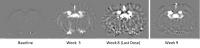
Endogenous remyelination can potentially restore rapid
axonal-conduction and confer neuroprotection in chronic
demyelinating diseases such as multiple sclerosis. We used T2 mapping
to evaluate the ability of two candidate pharmacological
agents to promote remyelination in cuprizone-demyelinated
mice. Demyelination was associated with increase in signal
intensity and T2 values
in the corpus callosum and external capsules. T2 values
showed spontaneous recovery after discontinuation of
cuprizone treatment, an effect accelerated following
administration of the two compounds tested. This study
confirms that in
vivo MRI can
be used to select pharmacological agents for their
therapeutic potential on remyelination.
|
|
3711.
 |
44 |
MRI Assessment of Acute Pathologic Process after Myocardial
Infarction: Role of Magnetic Nanoparticle-based MRI -
Permission Withheld
Cheongsoo Park1,2, Eun-Hye Park3,
Jongeun Kang1,4, Kiyuk Chang3, and
Kwan Soo Hong1,4,5
1Korea Basic Science Institute, Cheongju, Korea,
Republic of, 2The
Catholic University of Korea, Seoul, Korea, Republic of, 3Seoul
St. Mary’s Hospital and College of Medicine, Seoul, Korea,
Republic of,4Chungnam National University,
Daejeon, Korea, Republic of, 5University
of Science and Technology, Daejeon, Korea, Republic of
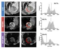
Myocardial infarction (MI) is the major cause of sudden
death in most industrialized society. Imaging of early
disease progression and investigation of relationship
between myocardial necrosis and successive inflammatory
response are needed for optimal treatment of MI. We
conducted cardiac MR imaging of disease progression in acute
MI by using three different MRI methods of Gd (LGE), Mn
(ME), and iron oxide nanoparticles (MNP)-based MRI for
estimation of infarcted and inflammatory regions.
|
|
3712.
 |
45 |
APT MRI of Intracranial Mass Lesions at 3T and Comparison with
DCE Perfusion Parameters
Ayan Debnath1, Prativa Sahoo2, Pradeep
Gupta3, Rakesh Gupta3, and Anup Singh1
1Centre for Bio-Medical Engineering, Indian
Institute of Technolgy Delhi, Delhi, India, 2Philips
India Limited, New Delhi, India, 3Radiology,
Fortis Memorial Research Institute, New Delhi, India
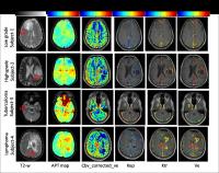
In the current study, Amide Proton Transfer (APT) and
T1-weighted DCE perfusion MRI was performed on patients
with intra-cranial mass lesions(low and high grade tumors,
CNS tuberculoma, CNS lymphoma) at 3T MRI. APT maps provided
a significant difference between lesion and its
contra-lateral side. From the preliminary study it was
observed that APT contrast was low in infection lesion
followed by tumor and lymphoma. APT values showed a
significant (P<0.01) difference between low and high grade
tumors. A weak Inter-class correlation was observed between
APT and perfusion parameters (like cbf, cbv, Ktr, Kep, ve).
Therefore, APT mapping might improve diagnostic value either
alone or in combination with other MRI parameters.
|
|
3713.
 |
46 |
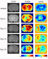
Nuclear Overhauser Enhancement (NOE) mediated Chemical Exchange
Saturation Transfer (CEST) imaging in glioma with different
progression at 7T
Tang Xiangyong1, Dai Zhuozhi1, Shen
Yuanyu1, Hu Wei1, Zhang Zhiyan1,
and Wu Renhua1
12nd Affilicated Hospital, Shantou University
Medical College, Shantou, China, People's Republic of
Nuclear Overhauser Enhancement (NOE) mediated Chemical
Exchange Saturation Transfer (CEST) imaging in glioma with
different progression at 7T
|
|
3714.
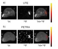 |
47 |
A Comparison between the UTE and PETRA Pulse Sequences for
Fluorine-19 MRI at 3 Tesla
Roberto Colotti1, Jean Delacoste1,
Giulia Ginami1, Maxime Pellegrin2,
Tobias Kober1,3,4, Yutaka Natsuaki5,
David Grodzki6, Ulrich Flögel7,
Matthias Stuber1,8, and Ruud B. van Heeswijk1
1Department of Radiology, University Hospital
(CHUV) and University of Lausanne (UNIL), Lausanne,
Switzerland, 2Division
of Angiology, University Hospital of Lausanne (CHUV),
Lausanne, Switzerland,3Advanced Clinical Imaging
Technology, Siemens Healthcare IM BM PI, Lausanne,
Switzerland, 4LTS5,
École Polytechnique Fédérale de Lausanne, Lausanne,
Switzerland, 5Siemens
Medical Solutions, NAM USA DI MR COLLAB WE, Los Angeles, CA,
United States, 6Siemens
Healthcare GmbH, HC DI MR R&D PLH, Erlangen, Germany, 7Department
of Cardiovascular Physiology, Heinrich Heine University,
Düsseldorf, Germany, 8Center
for Biomedical Imaging (CIBM), Lausanne, Switzerland
Fluorine-19 (19F) MRI of perfluorocarbon
emulsions (PFCs) with multiresonant spectra is challenging
due to destructive phase interference that leads to short T2 relaxation
times (<10 ms). Pulse sequences with very short echo times
(≤ 100 µs) can be used to overcome this challenge. In this
study, in vitro and in vivo 19F
MRI obtained with both UTE and PETRA were acquired and
quantitatively compared.
|
|
3715.
 |
48 |
On the Feasibility of Quantitative Dynamic Whole Body PET/MR
Imaging
Hyungseok Jang1,2, Hyung-Jun Im1,
Arman Rahmim3, Steve Y Cho1, and Alan
B McMillan1
1Department of Radiology, University of
Wisconsin, Madison, WI, United States, 2Department
of Electrical and Computer Engineering, University of
Wisconsin, Madison, WI, United States, 3Department
of Radiology, Johns Hopkins University, Baltimore, MD,
United States
In this study we investigate the feasibility of FDG PET/MR
as a platform for whole body dynamic quantitative PET
imaging. The ability of PET/MR systems to provide truly
simultaneous imaging is advantageous compared to PET/CT for
serial whole body PET acquisitions in that simultaneously
acquired MR images can provide additional information to PET
data, such as the application of motion parameters estimated
from MR images to PET images to correct for misregistration
which is not possible with PET/CT. Further improvements in
workflow can allow integration of multiple MR contrasts,
making dynamic whole body PET/MR a highly feasible and
compelling methodology.
|
|