ISMRM 24th Annual Meeting & Exhibition • 07-13 May 2016 • Singapore

Electronic Poster Session: fMRI

3716 -3739 fMRI: Acquisition
3740 -3763 Brain Connectivity with fMRI
3764 -3787 fMRI Multimodal
3788 -3811 fMRI: Application
3812 -3835 fMRI: Analysis & Models

Exhibition Hall 

10:00 - 11:00

    Computer #

 
3716.   
49 Combined T2*-Weighted Measurements of the Human Brain and Cervical Spinal Cord with Partial Multi-Band Acceleration
Jürgen Finsterbusch1
1Systems Neuroscience, University Medical Center Hamburg-Eppendorf, Hamburg, Germany
Recently, an echo-planar imaging approach has been presented that covers the human brain and cervical spinal cord in the same acquisition in order to investigate the functional connectivity of the two regions. However, the repetition time of this approach (TR) usually exceeds 3 s which is not optimal for a connectivity analysis. Multi-band acceleration is a promising technique to speed up acquisitions, but most neck coil geometries limit its applicability for the cervical spinal cord. In this study, multi-band acceleration is applied to the brain slices only yielding a significantly reduced TR while retaining a good image quality in the cervical spinal cord.


3717.   
50 Comparison of Physiological Noise in Multiband-EPI and Regular EPI fMRI
Zahra Faraji-Dana1,2, Ali Golestani3, Yasha Khatamian3, Simon Graham1,2, and J. Jean Chen1,3
1Department of Medical Biophysics, University of Toronto, Toronto, ON, Canada, 2Sunnybrook Research Institute, Sunnybrook Health Science Centre, Toronto, ON, Canada, 3Rotman Research Institute, Baycrest Health Science Centre, Toronto, ON, Canada
Simultaneous multi-slice echo-planer imaging (EPI) or otherwise known as multiband (MB) EPI provides high temporal and/or spatial resolution in resting-state fMRI (rs-fMRI) studies by modulating and simultaneously imaging multiple slices. However, the effect of slice acceleration on "physiological noise" induced by respiration, cardiac pulsation, variations in respiratory-volume (RVT) and cardiac-rate (CRV) is still unknown. Similar to conventional parallel imaging techniques, residual aliasing occur in MB slice acceleration that could introduce spurious signals into fMRI data. We hypothesize that since a given group of simultaneously acquired slices samples the physiological noise at the same time, the effect of physiological noise may be amplified in MB-EPI. In this study we experimentally verify this hypothesis, and identify the physiological-correction strategy that best corrects this effect.


3718.   
51 Feasibility of line scanning BOLD fMRI on human subjects
Daniel Spitzer1, Jochen Bauer1, and Cornelius Faber1
1Department of Clinical Radiology, Muenster, Germany
Line scanning fMRI is a novel technique to probe the BOLD signal with high temporal resolution, which has been previously demonstrated in small animals. Here, we implement line scanning fMRI on a clinical 3 T scanner and probe the BOLD response in human brain with 100 ms temporal resolution. From our data the hemodynamic response can be derived, perfectly matching the response function as observed with conventional fMRI detection methods.


3719.   
52 High resolution gradient-echo EPI using a shim insert coil at 7T: Implication for BOLD fMRI.
Tae Kim1, Yoojin Lee1, Tiejun Zhao2, Hoby Hetherington1, and Jullie Pan1
1University of Pittsburgh, Pittsburgh, PA, United States, 2Siemens Medical Solution USA, INC, Pittsburgh, PA, United States
High degree/order shimming was applied to improve field homogeneity thereby reducing susceptibility-induced distortion in high resolution gradient-echo EPI using a shim insert coil at 7T. Use of the shim insert improved the overall homogeneity across the entire brain by 30% in comparison to conventional 1st&2nd degree/order shimming. The susceptibility-induced displacement improved by more than 2cm and the number of activated pixels increased by 150% with higher degree shimming in regions such as the anterior temporal and frontal lobes. Our study demonstrates that the use of higher order/degree shims improves GE-EPI BOLD signal at high field.


3720.   
53 DANTE-EPI for CSF Suppression in Cervical Spinal Cord BOLD fMRI at 7T
Alan Charles Seifert1,2, Hadrien Dyvorne1,2, Joo-won Kim1,2, Bei Zhang3, and Junqian Xu1,2,4
1Translational and Molecular Imaging Institute, Icahn School of Medicine at Mount Sinai, New York, NY, United States, 2Department of Radiology, Icahn School of Medicine at Mount Sinai, New York, NY, United States, 3Department of Radiology, NYU Langone Medical Center, New York, NY, United States, 4Department of Neuroscience, Icahn School of Medicine at Mount Sinai, New York, NY, United States
A major challenge in spinal cord fMRI is physiological noise due to pulsating CSF, which confounds detection of BOLD signal.  In this work, we adapt DANTE to reduce CSF signal contamination in spinal cord BOLD fMRI at 7T.  Absolute signal intensity, temporal SNR, and temporal cross-correlation between left and right spinal cord gray matter were measured in DANTE-EPI images at multiple pulse-train lengths and flip angles.  Aggressive DANTE preparation suppresses CSF signal, but also significantly reduces spinal cord signal.  More conservative DANTE preparation yields an optimal tradeoff between adequate CSF attenuation and preservation of spinal cord signal.


3721.   
54 Optimization of EPI protocols for maximum BOLD sensitivity through numerical simulations - Permission Withheld
Steffen Volz1, Martina F. Callaghan1, Oliver Josephs1, and Nikolaus Weiskopf1,2
1Wellcome Trust Centre for Neuroimaging, UCL Institute of Neurology, UCL, London, United Kingdom, 2Department of Neurophysics, Max Planck Institute for Human Cognitive and Brain Sciences, Leipzig, Germany
fMRI studies can suffer substantially from BOLD sensitivity loss due to susceptibility-related magnetic field inhomogeneities. We developed an automated algorithm for optimising arbitrary EPI protocols with respect to BOLD sensitivity based on numerical simulations and a multi-subject field map database, saving time and expensive measurements. In contrast to previous experimental optimization approaches that were limited e.g. to z-shim, gradient polarity and slice tilt for a specific EPI protocol, this algorithm optimizes on a larger parameter space including resolution, echo time and slice orientation. Results were compared to earlier experimental approaches and verified by BOLD sensitivity measurements in healthy volunteers.


3722.   
55 Improved detection of fMRI activity in ventromedial prefrontal cortex using multi-echo EPI
Brice Fernandez1, Laura Leuchs2, Phillip G. Sämann 2, Michael Czisch2, and Victor I. Spoormaker2
1Applications and Workflow, GE Healthcare, Munich, Germany, 2Neuroimaging Unit, Max Planck Institute of Psychiatry, Munich, Germany
Standard fMRI suffers from signal loss in the ventromedial and orbital prefrontal cortex, a region of special interest in affective neuroimaging. Multi-echo EPI (MEPI) is known to have several advantages over EPI. In this work, we test if MEPI is able to reach better performance in detecting task-induced activation in the ventromedial prefrontal cortex (vmPFC) during fear conditioning, known for eliciting activity in this area. We demonstrate that MEPI (by means of the weighted sum combination approach) outperforms standard EPI in vmPFC, which is highly relevant for affective neuroscience and psychiatry given its critical role in emotion regulation.


3723.   
56 Submillimeter spatial and 1 second temporal resolution in fMRI using 3D-EPI-CAIPI with cylindrical excitation
Wietske van der Zwaag1,2, Mayur Narsude3, Olivier Reynaud2, Dan Gallichan2, and José P. Marques4
1Spinoza Centre for Neuroimaging, Amsterdam, Netherlands, 2EPFL, Lausanne, Switzerland, 3Lausanne, Switzerland, 4Donders Institute for Brain, Cognition and Behaviour, Nijmegen, Netherlands
3D-EPI-CAIPI was combined with a cylindrical excitation profile to reduce the brain area from which signal is generated, and hence, the parallel imaging undersampling artefacts and signal loss. This 3D-EPI-CAIPI with cylindrical excitation can be used to acquire fMRI data with submillimetre spatial resolution, 1-second temporal resolution and very high BOLD sensitivity.


3724.   
57 Single vs Multi Echo EPI: a Head-to-Head, Within-Session Cross-Over Comparison for Task Evoked and Seed Based Connectivity Analysis
Jed Wingrove1, Stephen J Wastling1, Prantik Kundu2, Gareth Barker1, Donal Hill1, Owen O'Daly1, and Fernando Zelaya1
1Department of Neuroimaging, King's College London, London, United Kingdom, 2Brain Imaging Center, Mount Sinai Hospital, New York, NY, United States
Resting state fMRI data is highly susceptible to low frequency noise fluctuations from motion and pulsatile physiological movement leading to inaccuracies in observed connectivity. In this study we directly compare conventional single echo EPI against a multi echo EPI (ME-EPI) acquisition and analysis method which denoises and cleans acquired fMRI time series. Task evoked and rs-fMRI data was acquired in 8 volunteers and showed improved spatial localisation and discrete clustering in ME-EPI analysis in comparison to single echo data. Furthermore, this work demonstrates the benefits of using ME-EPI for both functional activation and resting state connectivity investigations.   


3725.   
58 Echo-Time Optimization in Spin Echo EPI using Hypercapnic Manipulation at 3T
Don Marcial Ragot1,2 and Jean Chen1,2
1Medical Biophysics, University of Toronto, Toronto, ON, Canada, 2Roman Research Institute, Toronto, ON, Canada
The optimal echo time (TE) for spin-echo EPI (SE-EPI) BOLD is assumed to be near the tissue T2 (65–100ms at 3 T), but this was never experimentally tested. In this study, we use a hypercapnia paradigm with SE-EPI at different TEs to identify the TE that maximizes BOLD detection at 3 T. Based on the normalized percent change in BOLD signal (?BOLD%/mmHg), BOLD contrast-to-noise ratio (CNR/mmHg) and temporal signal-to-noise ratio (tSNR), we concluded that the optimal SE-EPI TE may be much shorter than the tissue T2, and the optimal TE at 3 T is near 50ms.


3726.   
59 A comparison of time-series SNR and Nyquist ghosting with different parallel imaging autocalibration acquisition schemes in 7 T fMRI with a chin task
Pedro Lima Cardoso1, Jonathan R. Polimeni2, Benedikt Poser3, Markus Barth4, Siegfried Trattnig1, and Simon Daniel Robinson1
1High Field MR Center, Department of Biomedical Imaging and Image-guided Therapy, Medical University of Vienna, Vienna, Austria, 2Athinoula A. Martinos Center for Biomedical Imaging, Department of Radiology, Harvard Medical School, Massachusetts General Hospital, Charlestown, MA, United States, 3Maastricht Brain Imaging Center, Maastricht University, Maastricht, Netherlands, 4Center for Advanced Imaging, University of Queensland, Brisbane, Australia
The present study assesses time-series SNR and residual aliasing (ghosting) levels in accelerated EPI acquisitions at 7 T using single-shot (SS), multi-shot (MS), FLASH and FLEET autocalibration signal acquisition schemes for GRAPPA reconstruction in resting-state and in a chin task (commonly used in fMRI for presurgical planning), which elicits head motion and, consequently, changes in ΔB0. FLASH and FLEET acquisitions yielded significantly higher average tSNR values compared to SS and MS in the chin task and no significant residual aliasing enhancement in both resting-state and chin tasks.


3727.   
60 The dependence of the BOLD response transients on stimulus type and echo time
Martin Havlicek1, Dimo Ivanov1, Benedikt A Poser1, and Kamil Uludag1
1Cognitive Neuroscience, Maastricht University, Maastricht, Netherlands
Adaptation and post-stimulus undershoot observed in the BOLD signal can originate from both active neuronal and passive vascular mechanisms. Using a multi-echo GE-EPI functional MRI sequence with two distinct visual stimuli, we are able to show that not only the BOLD response magnitude but also its shape is affected by the echo-time (TE). As pure oxygenation changes predict approximately a linear scaling of the BOLD signal with TE, this finding indicates that the composition of the underlying physiological mechanisms contributing to the fMRI signal (i.e. CBF, CBV, and CMRO2) varies over time.


3728.   
61 Multiband integrated-SSFP for functional imaging at 7T with reduced susceptibility artifacts
Kaibao Sun1,2, Zhentao Zuo1, Hanyu Shao1, Zhongwei Chen1,2, Bo Wang1, Thomas Martin3, Yi Wang3, Peng Zhang1, Rong Xue1, and Danny JJ Wang3
1State Key Laboratory of Brain and Cognitive Science, Beijing MRI Center for Brain Research, Institute of Biophysics, Chinese Academy of Sciences, Beijing, China, People's Republic of, 2Graduate School, University of Chinese Academy of Sciences, Beijing, China, People's Republic of, 3Laboratory of FMRI Technology (LOFT), Department of Neurology, UCLA, Los Angeles, CA, United States
BOLD fMRI based on echo-planar imaging (EPI) suffers from susceptibility artifacts that impair imaging in specific brain regions, especially severe at ultrahigh fields. Integrated-SSFP (iSSFP), which is modified from balanced SSFP (bSSFP), shows constant magnitude regardless of frequency shift and is proposed to overcome the obstacle. In our 1st experiment, iSSFP achieved better image quality without banding artifacts and more stable signal changes in visual cortex than bSSFP. In our 2nd experiment, using semantic processing task, more tissue signal and greater activations in the inferior portion of the anterior temporal lobe were detected by multiband iSSFP compared to EPI fMRI.


3729.   
62 Calibrated BOLD fMRI with an Optimized ASL-BOLD Dual-Acquisition Sequence
Maria A. Fernandez-Seara1, Zachary B. Rodgers2, Erin K. Englund2, Hee-Kwon Song2, John A. Detre3, Michael C. Langham2, and Felix W. Wehrli2
1Radiology, University of Navarra, Pamplona, Spain, 2Radiology, University of Pennsylvania, Philadelphia, PA, United States, 3Neurology, University of Pennsylvania, Philadelphia, PA, United States
Calibrated fMRI techniques estimate task-induced changes in the cerebral metabolic rate of oxygen (CMRO2) based on simultaneous measurements of cerebral blood flow (CBF) and blood-oxygen-level-dependent (BOLD) signal changes evoked by stimulation. To determine the calibration factor M (corresponding to the maximum possible BOLD signal increase), BOLD signal and CBF are measured in response to a gas breathing challenge (CO2, O2). Here we describe an ASL dual-acquisition sequence that combines a background-suppressed 3D readout with 2D multi-slice EPI. In five subjects we found an average gray matter M-value of 8.71±1.03 and fractional changes of CMRO2 of 12.5±5% in response to a bilateral motor task.


3730.   
63 Comparison of IVIM and pCASL in the brain
Hannah Hare1 and Daniel Bulte1,2
1FMRIB, NDCN, University of Oxford, Oxford, United Kingdom, 2Department of Oncology, University of Oxford, Oxford, United Kingdom
IVIM has been proposed as a method of imaging blood volume fraction and perfusion in the brain. In this study we directly compared both the images and the values measured with IVIM to a multi post label delay pCASL sequence in the same imaging session. Although the units are not directly comparable, the images can be visually compared, and it was hypothesised that inter-subject variability in resting CBF in grey matter across subjects would result in a positive correlation between measures from the 2 modalities. Although visually similar, no correlation was observed in the quantitative data.


3731.   
 
64 The effect of diffusion on quantitative BOLD parameter estimates acquired with the Asymmetric Spin Echo technique
Nicholas P Blockley1, Naomi C Holland1, and Alan J Stone1
1FMRIB Centre, Nuffield Department of Clinical Neurosciences, University of Oxford, Oxford, United Kingdom
The results of a recently described streamlined quantitative BOLD (qBOLD) technique suggest that this method may overestimate the deoxygenated blood volume (DBV), leading to an underestimate of the oxygen extraction fraction (OEF). We hypothesise that this is due to the effect of diffusion, which is assumed to be zero in the analytical qBOLD model. In this study we performed Monte Carlo simulations to investigate this hypothesis and found that DBV and OEF measurements were vessel size dependent. However, the R2’ measurements which underlie qBOLD were found to be a reliable measure of deoxyhaemoglobin content above a vessel radius of 10μm.


3732.   
65 Submillimeter resolution fMRI in the midbrain: measuring T2* changes to a stop-task
Gilles de Hollander1, Robert Trampel2, Birte Forstmann1, and Wietske van der Zwaag3
1Universiteit van Amsterdam, Amsterdam, Netherlands, 2Max Planck Institute for Human Cognitive and Brain sciences, Leipzig, Germany, 3Spinoza Centre for Neuroimaging, Amsterdam, Netherlands
Multi-echo GRE was used to visualise BOLD signal changes in the midbrain, specifically targetting the subthalamic nucleus and substantia nigra, with submillimeter resolution. Midbrain clusters were very small and often only bordered the STN or SN, rather than falling robustly inside it, emphasizing the need for high spatial resolution.


3733.   
66 Characterization of ultra-high resolution Gradient Echo and Spin Echo BOLD fMRI in the human visual cortex at 7 Tesla
Catarina Rua1,2, Mauro Costagli2,3, Mark R Symms4, Laura Biagi3, Mirco Cosottini2,5, Alberto Del Guerra1, and Michela Tosetti2,3
1Department of Physics, University of Pisa, Pisa, Italy, 2Imago7 Research Center, Pisa, Italy, 3IRCCS Stella Maris, Pisa, Italy, 4GE Healthcare, Pisa, Italy, 5Department of Translational Research and New Technologies in Medicine and Surgery, University of Pisa, Pisa, Italy
This study compared GRE-EPI and SE-EPI sequences with different spatial resolutions at 7T for fMRI in the visual cortex. It is demonstrated that SE-EPI yields higher specificity than GRE-EPI. However, the decreasing temporal signal-to-noise at submillimeter acquisitions affected significantly the extension of the activated volume in a SE acquisition. At this level, GRE-based functional maps showed significant increased specificity compared to standard resolutions, and a preserved cluster volume. The reduction of partial volume effects allowed the selection of an activation sub-cluster excluding the highest z-scores, which co-localized preferably in non-gray matter, potentially increasing the performance of UHF high-resolution fMRI.


3734.   
67 Whole brain measurements of the hemodynamic response function variability during a finger tapping task at 7T show regional differences in hrf profiles.
Yohan Boillat1, Rolf Gruetter1,2,3, and Wietske van der Zwaag1,4
1Ecole Polytechnique Fédérale de Lausanne, Lausanne, Switzerland, 2Universite de Lausanne, Lausanne, Switzerland, 3Universite de Geneve, Geneva, Switzerland, 4Spinoza Centre for Neuroimaging, Amsterdam, Switzerland
The temporal properties of the BOLD response, including inter-trial variability, were studied using a 3D-EPI-CAIPI acquisition at 7T with TR=400ms. The HRFs of six different brain regions in the motor network were characterized, showing a reduced post-stimulus undershoot in the cerebellar regions of interest, as well as differences in peak height, with higher response amplitude in M1 than any other region and onset time, with the cerebellar lobule VIII response starting later than the other ROIs. Trial-to-trial variability was highest in CVIII and lowest in SI.


3735.   
68 The impact of through-slice acceleration on the optimum TE in BOLD-based fMRI: a simulation study
Wietske van der Zwaag1, David G. Norris2, and José P. Marques2
1Spinoza Centre for Neuroimaging, Amsterdam, Netherlands, 2Donders Institute for Brain, Cognition and Behaviour, Nijmegen, Netherlands
In this simulation study we evaluate the TE that offers the optimum BOLD contrast per unit time for a given field strength (taking into account the field-specific T1, T2* and ΔT2* values) as a function of the number of slice excitations to acquire the volume (minimum TR achievable), using the Ernst angle for excitation. Generally, optimum BOLD sensitivity is found at TEs closer to T2* when shorter TRs (higher multiband acceleration factors) are employed. At longer TRs efficiency constraints tend to make the optimum TE shorter with this effect being more pronounced at lower static fields.


3736.   
69 Evaluation of PCASL Imaging and T2* Mapping for the Assessment of Cerebrovascular Reactivity in the Hippocampus
Xiufeng Li1, Nicholas Evanoff2, Lynn E. Eberly 3, Anne M. Murray4, Gregory J. Metzger1, and Donald R. Dengel2
1Center for Magnetic Resonance Research, School of Medicine, University of Minnesota, Minneapolis, MN, United States, 2Laboratory of Integrative Human Physiology, School of Kinesiology, University of Minnesota, Minneapolis, MN, United States, 3Division of Biostatistics, School of Public Health, University of Minnesota, Minneapolis, MN, United States, 4Berman Center for Clinical Research, Hennepin County Medical Center, Minneapolis, MN, United States
        The hippocampus is significantly affected in cognitive impairment, including Alzheimer’s disease. Cerebrovascular endothelial dysfunction (CeV-ED) plays an essential role in the initiation and progression of cerebrovascular disease (CeV-D) and cognitive decline. CeV-ED can be assessed with the evaluation of cerebrovascular reactivity (CeV-R) by performing MRI studies with a respiratory challenge, such as the manipulation of end-tidal partial pressure of CO2 (PetCO2) and O2 (PetO2). In the presented studies, ASL imaging and T2* mapping were evaluated for the assessment of the CeV-R in the hippocampus to determine the benefits and disadvantages of each imaging method and to facilitate the imaging method selection for future application studies. 


3737.   
70 Functional Resolution of Gradient Echo, Asymmetric Spin Echo, and Spin Echo in Functional MRI
Eun Soo Choi1 and Gary Glover2
1Electrical Engineering, Stanford University, Stanford, CA, United States, 2Radiology, Stanford University, Stanford, CA, United States
In BOLD fMRI, gradient-echo and spin-echo pulse sequences have been widely used due to their higher functional sensitivity and spatial specificity, however, fair comparison between SE and GRE methods has often not been made properly. In this study, we defined "functional resolution" as a quantitative metric to evaluate spatial specificity of BOLD contrast fMRI and measured it in SE, asymmetric spin-echo, and GRE. Then we conducted spatial smoothing to equalize their functional resolution and compare their functional sensitivity under the equivalent conditions. 


3738.   
71 High resolution IVIM in brain using readout-segmented EPI
Hannah Hare1, Robert Frost1, and Daniel Bulte1,2
1FMRIB, NDCN, University of Oxford, Oxford, United Kingdom, 2Department of Oncology, University of Oxford, Oxford, United Kingdom
1mm isotropic diffusion-weighted images were acquired with readout-segmented EPI at 3T to investigate the effects of partial voluming on the IVIM-derived measures of perfusion and blood volume. The maps produced showed no contrast between white and grey matter, and very high signal from CSF. Both the grey and white matter curves were adequately fitted using a monoexponential model, and only the CSF required a biexponential to fit the data. This suggests that the biexponential signal behaviour typically observed at lower resolutions may arise primarily from the CSF rather than the blood compartment.


3739.   
72 Reduction of run-to-run variability of temporal SNR in accelerated EPI time-series data through FLEET-based robust autocalibration
Anna I. Blazejewska1, Himanshu Bhat2, Lawrence L. Wald1,3, and Jonathan R. Polimeni1
1Athinoula A. Martinos Center for Biomedical Imaging, Department of Radiology, Harvard Medical School, Massachusetts General Hospital, Charlestown, MA, United States, 2Siemens Medical Solutions USA Inc., Charlestown, MA, United States, 3Harvard-MIT Division of Health Sciences and Technology, Massachusetts Institute of Technology, Cambridge, MA, United States
Temporal signal-to-noise ratio (tSNR) provides a crucial metricdetermining sensitivity of the acquisition to BOLD fMRI measurements. Ithas been shown that tSNR may vary dramatically between multiple runs ofaccelerated single-shot EPI acquisitions using single- or multi-shot EPIto acquire autocalibration or ACS data. We applied noise-to-noise ratio(NNR) measure to map run-to-run variability of acquisitions usingconventional multi-shot EPI ACS data as well as recently proposed Fastlow-angle excitation echo-planar technique (FLEET) ACS. tSNR variabilitybetween multiple runs improved in acquisitions using FLEET-ACS, providing the potential to increase sensitivity of BOLD fMRI experiments.
Exhibition Hall 

10:00 - 11:00

    Computer #

3740.   
73 
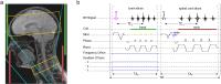
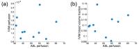
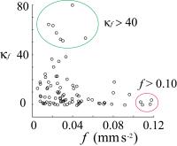
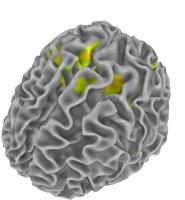
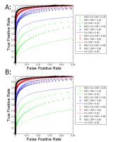
Identifying Foci of Brain Disorders from Effective Connectivity Networks
D Rangaprakash1, Gopikrishna Deshpande1,2,3, Archana Venkataraman4, Jeffrey S Katz1,2,3, Thomas S Denney1,2,3, and Michael N Dretsch5,6
1AU MRI Research Center, Department of Electrical and Computer Engineering, Auburn University, Auburn, AL, United States, 2Department of Psychology, Auburn University, Auburn, AL, United States,3Alabama Advanced Imaging Consortium, Auburn University and University of Alabama Birmingham, Birmingham, AL, United States, 4Department of Diagnostic Radiology, Yale University, New Haven, CT, United States, 5U.S. Army Aeromedical Research Laboratory, Fort Rucker, AL, United States, 6Human Dimension Division, HQ TRADOC, Fort Eustis, VA, United States
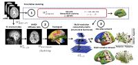
Brain connectivity studies report statistical differences in pairwise connection strengths. While informative, such results are difficult to interpret, since our understanding of the brain relies on region information, rather than connections. Given that large effects in natural systems are likely caused by few pivotal sources, we employed a novel framework to identify sources of disruption from directional connectivity. Using resting-state fMRI, we employed static and time-varying effective connectivities in a probabilistic framework to identify affected foci and associated affected 
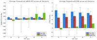
connections. We illustrate its utility in identifying disrupted foci in Soldiers with post-traumatic stress disorder and mild traumatic brain injury.


3741.   
74 
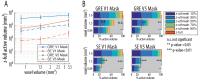
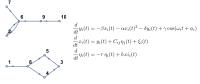
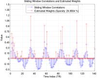
Brain Connectivity Network Dynamics Are Correlated with Cognitive Performance in Multiple Sclerosis
Sue-Jin Lin1,2, Aiping Liu3, Alex MacKay4,5, Brenda Kosaka6, Samantha Beveridge7, Irene Vavasour5, Anthony Traboulsee8, and Martin J McKeown1,2,8
1Graduate Program in Neuroscience, University of British Columbia, Vancouver, BC, Canada, 2Pacific Parkinson’s Research Centre, University of British Columbia Hospital, Vancouver, BC, Canada,3Department of Electrical and Computer Engineering Program, University of British Columbia, Vancouver, BC, Canada, 4Department of Physics and Astronomy, University of British Columbia, Vancouver, BC, Canada, 5Department of Radiology, University of British Columbia Hospital, Vancouver, BC, Canada, 6Department of Psychiatry, University of British Columbia Hospital, Vancouver, BC, Canada,7Graduate Program in Counselling Psychology, University of British Columbia, Vancouver, BC, Canada, 8Neurology, Faculty of Medicine, University of British Columbia, Vancouver, BC, Canada
Brain connectivity networks are usually estimated with the assumption that neural networks do not change over time. However, functional connectivity is inherently non-stationary, changing across time from seconds to minutes. In healthy subjects, dynamic reconfiguration of functional connectivity assessed by fMRI has been estimated and has been linked to cognitive tests, indicating that flexibility of connectivity normally contributes to cognitive performance. In this study, we applied a novel time-varying analysis to study network dynamics in healthy controls and subjects with Multiple Sclerosis (MS).  
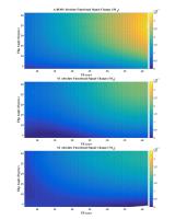


3742.   
75 Parcellation-based connectome assessment by using structural and functional connectivity
Ying-Chia Lin1, Tommaso Gili2,3, Sotirios A. Tsaftaris 1,4, Andrea Gabrielli5, Mariangela Iorio3, Gianfranco Spalletta3, and Guido Caldarelli1
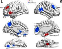
1IMT Institute for Advanced Studies Lucca, Lucca, Italy, 2Enrico Fermi Centre, Rome, Italy, 3IRCCS 
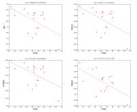
Fondazione Santa Lucia, Rome, Italy, 4Institute of Digital Communications, School of Engineering, The 
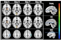
University of Edinburgh, Edinburgh, United Kingdom, 5ISC-CNR, UOS Sapienza, Dipartimento di Fisica, Universita Sapienza, Rome, Italy
Connectome analysis of the human brain structural and functional architecture provides a unique opportunity to understand the organization of brain networks. In this work, we investigate a novel large scale parcellation-based connectome, merging together information coming from resting state fMRI (rs-fMRI) data and diffusion tensor imaging (DTI) measurements.


3743.   
76 
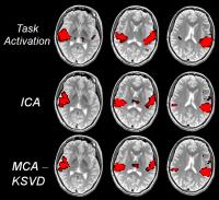
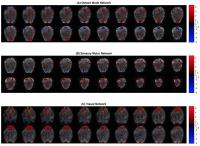
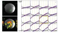
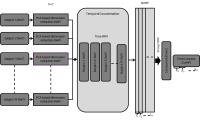
Group NMF Analysis for Resting State fMRI
Bhushan Patil1, Mahesh Panicker1, Radhika Madhavan1, and Suresh Joel1
1Global Research, General Electric Global Research, Bangalore, India
Clustering of resting state fMRI signals for extraction of functional brain networks has been showed to provide value in recent times. Independent component analysis (ICA) is the most commonly used technique to extract functional brain networks. More recently non-negative matrix factorization (NMF) has been successfully utilized for identification of brain functional networks in single-subject resting state fMRI data. NMF may provide complementary information for analyzing resting state fMRI data. However, the technique has not been extended to provide group inferences. This is non-trivial, since the components obtained from single subject NMF is not ordered. Using temporal concatenation, similar to group ICA, we introduce a new framework for back reconstruction of individual subject from group analysis using NMF. This framework will make comparisons between groups possible for NMF. 


3744.   
77 Mixed ICA and Clustering Method Introduced to Study the Life Span Changes in the Within-Network Functional Connectivity of the Default Mode Network
Isa Costantini1, Ottavia Dipasquale1,2, Laura Pelizzari1,2, Maria Marcella Laganà2, Francesca Baglio2, and Giuseppe Baselli1
1Department of Electronics, Information and Bioengineering, Politecnico di Milano, Milan, Italy, 2IRCCS, Don Gnocchi Foundation, Milan, Italy
This study combines the independent component analysis and a local clustering method in order to study the within-network functional connectivity of the default mode network (DMN). Our results strongly support the hypothesis that the long-range FC between anterior and posterior DMN increases from the childhood to the young adulthood and slowly decreases with aging. This joint approach allowed us to obtain more detailed information about within-network FC changes among the DMN sub-regions.


3745.   
78 
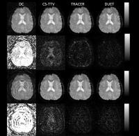
A novel sparse partial correlation method for simultaneous estimation of functional networks in group comparisons
Xiaoyun Liang1, David Vaughan2,3, Alan Connelly1,4, and Fernando Calamante1,4
1Imaging Division, Florey Institute of Neuroscience and Mental Health, Melbourne, Australia, 2Epilepsy Division, Florey Institute of Neuroscience and Mental Health, Melbourne, Australia, 3Department of Neurology, Austin Health, Melbourne, Australia, 4Department of Medicine, University of Melbourne, Melbourne, Australia
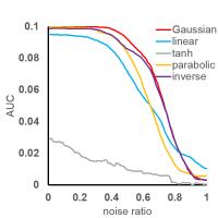
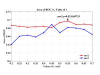
We propose a novel approach, Graphical-LAsso with Stability-Selection (GM-GLASS), by employing sparse group penalties for simultaneously estimating networks from healthy control and patient groups. Simulations demonstrate that both GM-GLASS and JGMSS outperform Fisher Z-transform. Our in vivo results further show that GM-GLASS yields highest contrast of network metrics between groups, demonstrating the superiority of GM-GLASS in detecting significance group differences over JGMSS and Fisher Z-transform. Overall, by controlling confounding variations between subjects, and therefore enhancing the statistical power, our simulated and in vivo results demonstrate that GM-GLASS provides a robust approach for conducting group comparison studies.


3746.   
79 Characterizing cross session coherence in the resting-state human brain
Shuqin Zhou1, Xiaopeng Song1, Yue Cai1, Xuemei Fu1, and Jiahong Gao2
1Department of Biomedical Engineering, Peking University, Beijing, China, People's Republic of, 2Center for MRI Research and Beijing City Key Lab for Medical Physics and Engineering, Peking University, Beijing, China, People's Republic of
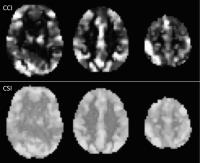
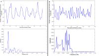
Previous studies suggested that the BOLD signal might be a mixture of different frequency components, but the neurophysiological basis of these components is still unclear. In this study, we attempted to quantify the similarity of the frequency profiles of the resting-state BOLD signals in different sessions by computing the cross session coherence (CSC) of these components. Our results suggested that different frequency components of BOLD signal in the brain might be associated with distinct intrinsic neuronal oscillations rather than random noise.


3747.   
80 
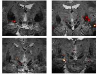
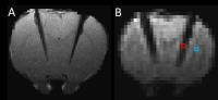
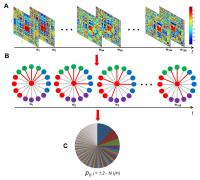
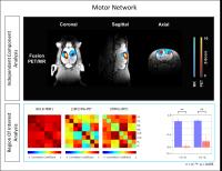
Dynamic Reconfiguration of Intrinsic Functional Connectivity: A Probabilistic Framework - Permission Withheld
Dazhi Yin1, Kristina Zeljic1, Zhiwei Wang1, Qian Lv1, and Zheng Wang1
1Institute of Neuroscience, Chinese Academy of Sciences, Shanghai, China, People's Republic of
Neural basis enabling flexible behavior remains largely unknown. Based on the spatiotemporal dynamics of intrinsic functional connectivity, we proposed a probabilistic modeling framework to quantify the functional flexibility and integration of different brain regions. We then applied this framework to investigate the functional representation of hand preference. Our findings revealed higher functional flexibility and integration for the preferred hand that controls cognitive-motor requirements for skilled movements.


3748.   
81 
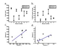
The long-term reproducibility of stimulation and resting state fMRI in the mouse
Yi-Ching Lynn Ho1,2, Fiftarina Puspitasari1, and Kai-Hsiang Chuang1
1Singapore Bioimaging Consortium, Agency for Science, Technology & Research (A*STAR), Singapore, Singapore, 2Interdisciplinary Institute of Neuroscience & Technology (ZIINT), Zhejiang University, Hangzhou, China, People's Republic of
There is a need to evaluate the long-term reproducibility of stimulation and resting state fMRI in the mouse, given the technical challenges of mouse fMRI. We compared 2 sessions of scans done between 3-9 weeks apart on 7 C57BL/6 mice. The intraclass correlation (ICC) indicated significant absolute agreement for forepaw stimulation fMRI results. Interhemispheric functional connectivity scores for a large subcortical area like the CPu were also found to be reproducible, but in a small cortical area like the S1FL, reproducibility did not reach significance. Possible reasons include data coregistration mismatches due to distortion and susceptibility artifacts.


3749.   
82 
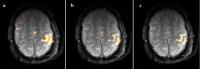
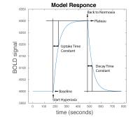
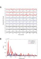
Effects of Anesthesia on Functional Connectivity in Primary Somatosensory Cortex in Monkeys
Tung-Lin Wu1,2, Arabinda Mishra1, Feng Wang1,3, Li Min Chen1,3, and John C. Gore1,2,3
1Vanderbilt University Institute of Imaging Science, Nashville, TN, United States, 2Biomedical Engineering, Vanderbilt University, Nashville, TN, United States, 3Radiology and Radiological Sciences, Vanderbilt University, Nashville, TN, United States
Low-frequency fluctuation of resting state functional MRI (rsfMRI) signals have been linked to changes in the spontaneous neuronal activity, but their relationships have not been established. Anesthesia is known to suppress neuronal activity. Thus, by examining the effects of different levels of anesthesia on changes in inter-regional functional connectivity and the power spectra, we will be able to assess the neuronal origins of the rsfMRI signals. We carried out live anesthetized squirrel monkey experiments that measure how low frequency fluctuations and inter-regional functional connectivity within a small local network (primary somatosensory cortex) vary as isoflurane levels are altered in a small range.


3750.   
83 
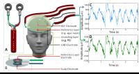
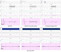
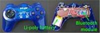
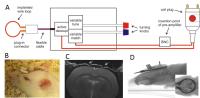
Chronic RF-coil and electrode implantation approach for long-term EEG-fMRI studies in rodents
Tiina Pirttimäki1, Artem Shatillo1, Mikko Kettunen1, Jaakko Paasonen1, Raimo Salo1, Alejandra Sierra Lopez 1, Kimmo Jokivarsi1, Ville Leinonen2, Simon Quittek3, Asla Pitkänen1, and Olli Gröhn1
1Neurobiology, A.I.Virtanen Institute for Molecular Medicine, University of Eastern Finland, Kuopio, Finland, 2Institute of Clinical Medicine - Neurosurgery, University of Eastern Finland and Neurosurgery of NeuroCenter, Kuopio University Hospital, Kuopio, Finland, 3RAPID Biomedical GmbH Technologiepark Wuerzburg-Rimpar, Rimpar, Germany
Simultaneous EEG-fMRI is routinely used in clinical settings as it provides better temporal and spatial information for example when locating seizure onset zones. In pre-clinical research with small rodents, obtaining simultaneous EEG-fMRI in longitudinal studies has been challenged by a number problems including issues related to magnetic susceptibility artifacts. Here, we demonstrate a modified method for permanent MRI coil and EEG electrode implantation that is suitable for long-term chronic follow-up studies on epileptogenesis with improved data consistency across imaging and video-EEG monitoring sessions.


3751.   
84 Longitudinal resting-state fMRI and 1H-MRS characterization in the mouse brain during development of a chronic pain state
David Bühlmann1,2, Joanes Grandjean1, Giovanna Diletta Ielacqua1, Jael Xandry3, and Markus Rudin1,3
1Institute for Biomedical Engineering, ETH and University of Zurich, Zurich, Switzerland, 2Neuroscience Center Zurich, Zurich, Switzerland, 3Institute of Pharmacology & Toxicology, University of Zurich, Zurich, Switzerland
We performed longitudinal resting-state fMRI and single voxel 1H-MRS in a mouse model of chronic pain derived from bone cancer. Linear mixed model analysis of independent components revealed significant functional changes mostly in limbic but also cortical networks. These findings were reproducible across strains and mirror findings from clinical studies on chronic back pain patients. 1H-MRS in the affected ventral hippocampus yielded significant decreases in glutamate, myo-inositol and glycerylophosphorylcholine concentrations in tumor-animals as well as increased glutamine levels. Given the translatability, these readouts could potentially be used to evaluate novel treatments specifically for chronic pain.  


3752.   
85 
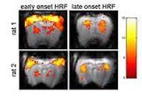
Monitoring longitudinal functional reorganization of a capsular infarct rat model using resting-state fMRI - Video Not Available
Chun-Qiang Lu1 and Shenghong Ju1,2
1Southeast University, Nanjing, China, People's Republic of, 2ZhongDa Hospital, Nanjing, China, People's Republic of
  Many resting-state fMRI studies in stroke patients claim that rs-fMRI measurements is behaviorally relevant. However, most of these studies enroll stoke patients with high heterogeneity. In this study, twenty-three rats underwent photothrombotic stroke lesioning in the PLIC with minimal affect to the nearby area. We monitor longitudinal resting-state brain activity and behavior change in this highly homogeneous white matter infarct rat model by using fcMRI and rat behavior test and try to find out the most behaviorally relevant fcMRI measurements. This project is still going on.


3753.   
86 
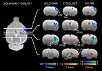
Metabolic and functional connectivity of the rat brain during resting state assessed by simultaneous [18F]FDG-PET/MR
Andre Thielcke1, Mario Amend1, Suril Gohel2, Bharat Biswal2, Bernd J. Pichler1, and Hans F. Wehrl1
1Department of Preclinical Imaging and Radiopharmacy, Werner Siemens Imaging Center, Eberhard Karls University of Tuebingen, Tuebingen, Germany, 2Department of Biomedical Engineering, New Jersey Institute of Technology, Newark, NJ, United States
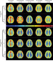
Recent advancement in hardware and software has enabled researchers to study systems level neuroscience using simultaneous PET/MRI. In this study, simultaneous PET/MR was used to investigate resting state networks (RSN) in rats, comparing [18F]FDG PET vs. BOLD-fMRI. RSNs such as default mode network (DMN) have been shown to be disrupted in clinical populations. ICA and ROI-analysis was used to elucidate the complementary nature between PET/MR and visualize brain connectivity. ICA PET and MR data showed prominent RSNs. However, ROI-analysis illustrated different connectivity between network-involved areas. This work suggests the complimentary nature of metabolic connectivity mapping (Cometomics). 


3754.   
87 
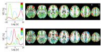
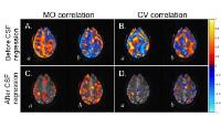
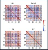
Decoupling flow effects on functional connectivity using R2* resting-sate fMRI - Permission Withheld
Venkata Veerendranadh Chebrolu1, Brice Fernandez2, Suresh E Joel1, Bharath Sundar1, Luca Marinelli3, Rakesh Mullick1, Victor I Spoormaker4, Michael Czisch4, and Thomas K Foo3
1GE Global Research, Bangalore, India, 2GE Healthcare, Munich, Germany, 3GE Global Research, Niskayuna, NY, United States, 4Max Planck Institute of Psychiatry, Munich, Germany
In this work we compare whole brain functional connectivity (FC) estimates from R2* resting-sate fMRI (rs-fMRI) with BOLD rs-fMRI. Thirty-two healthy subjects were imaged using three-echo multi-echo echo-planar-imaging (MEPI) under institutional guidelines. FC matrices based on structural and functional brain parcellation schemes were computed for individual BOLD echoes, R2* and M (initial magnetization approximated by BOLD signal at TE=0). Results tend to show that M might be helpful to decouple flow effects. Positive between network connectivity was observed in BOLD, M and R2* derived matrices. Anti-correlations observed between networks in BOLD and M were significantly lesser in R2* derived matrices.


3755.   
88 Evaluation of Resting State Network by Pupil Diameter Monitoring during fMRI Measurements – The Relationship between the Stability of the Pupil Diameter and the Activation in the Posterior Cingulate
Toshiharu Nakai1, Keiji Matsuda2, Sachiko Kiyama1, and Ichiro Takashima2
1NeuroImaging & Informatics, NCGG, Ohbu, Japan, 2Human Informatics Research Institute, AIST, Tsukuba, Japan
The status of the resting state performance was evaluated by using pupil diameter monitoring during fMRI sessions. The activation in the posterior cingulate was higher in the subjects who kept constant pupil diameters than those with time decay, suggesting that attempts to keep eyes open and fix them to the cross mark target may demand higher consciousness level. In other resting state networks (RSNs), no significant effect was confirmed suggesting that the RSNs are robust and not strongly affected by the tension or eye closing for a short time.   


3756.   
 
89 
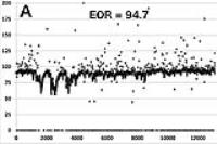
Resting-state fMRI fails to detect disease progression in a multicenter randomized clinical trial of Alzheimer's disease
Coimbra Alexandre1, Farshid Faraji1, Alexander de Crespigny1, Lee Honigberg1, Robert Paul1, and David Clayton1
1Research and Early Development, Genentech, South San Francisco, CA, United States
RS-fMRI was implemented in two multicenter clinical trials of a novel therapeutic for AD.  Although data of good quality were acquired, none of three functional connectivity metrics (FCMs) showed significant progression associated with disease in placebo-treated patients: changes in connectivity in this mild-to-moderate AD population were less than the measurement precision. Significant cognitive decline and brain atrophy were observed. Test-retest precision was similar to other single-center studies. Operational and acquisition improvements could increase data quality (though difficult in multicenter trials), but more sensitive analysis will be needed for RS-fMRI to be a useful tool for the development of AD therapeutics.


3757.   
90 
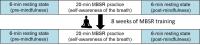
Increased functional connectivity associates with the improved emotion regulation after 8-week mindfulness-based stress reduction (MBSR) training using resting-state fMRI analysis
Yao-Chia Shih1,2, Chang-Le Chen2,3, Shih-Chin Fang4, Tzung-Kuen Wen5, Da-Lun Tang6, Si-Chen Lee7, and Wen-Yih Issac Tseng2,3,8
1Institute of Biomedical Engineering, National Taiwan University, Taipei, Taiwan, 2Institute of Medical Device and Imaging, National Taiwan University College of Medicine, Taipei, Taiwan, 3Graduate Institute of Brain and Mind Sciences, College of Medicine, National Taiwan University, Taipei, Taiwan, 4Department of Neurology, Cardinal Tien Hospital Yonghe Branch, New Taipei City, Taiwan,5Department of Buddhist Studies, Dharma Drum Institute of Liberal Arts, New Taipei City, Taiwan, 6Department of Mass Communication, Tamkang University, Taipei, Taiwan, 7Department of Electrical Engineering, National Taiwan University, Taipei, Taiwan, 8Molecular Imaging Center, National Taiwan University, Taipei, Taiwan
Mindfulness-based stress reduction (MBSR) is modified from Buddhist traditions and aims to improve self-regulation. In this study, we employed the resting-state functional MRI to investigate changes of functional connectivity (FC) before and after MBSR practice, and before and after 8-week MBSR training. We hypothesized that changes in FC may reflect improvements of self-regulation after MBSR training. We found MBSR strengthened FC couplings of right subgenual anterior cingulate cortex and lateral middle orbitofrontal cortex with posterior cingulate cortex in the beginners after 8-week MBSR training. Our findings reveal an underlying neural mechanism of positive effects of MBSR practice on emotional regulation.


3758.   
91 
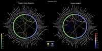
Pattern classification reveals functional connectivity differences in expert and novice meditators
Roberto Guidotti1,2, Mauro Gianni Perrucci1,2, Cosimo Del Gratta1,2, Antonino Raffone3, and Gian Luca Romani1,2
1Neuroscience, Imaging, and Clinical Sciences, Gabriele D'annunzio University Chieti-Pescara, Chieti, Italy, 2Institute for Advanced Biomedical Technologies, Gabriele D'Annunzio University Chieti-Pescara, Chieti, Italy, 3Psychology, La Sapienza University Rome, Rome, Italy
In this work we explored how experience modulates ROI-based fMRI functional connectivity patterns in two different meditators groups: experts and novices. We recorded fMRI data during two styles of meditation (focused attention (Samatha), and open monitoring (Vipassana)), in two groups of subjects (Buddist Theravada Monks, and novices), and we calculated the connectivity pattern between ROIs from the AAL90 atlas. We then used a pattern classification approach to discriminate these groups and find which connections and nodes are important to classify subject experience. Regions having a role in decoding were those implicated in self-awareness and attention control.


3759.   
 
92 Long-term and acute cannabis effects on brain networks - Permission Withheld
Isabelle Berger1,2,3, Philippe Maeder1, Jean-Marie Annoni4, Haithem Chtioui5, Christian Giroud6, Bernard Favrat7, Kim Dao5, Marie Fabritius6, Jean-Frédéric Mall8, Giovanni Battistella1,9, Reto Meuli1, and Eleonora Fornari1,2
1Department of Radiology, Centre Hospitalier Universitaire Vaudois (CHUV), and University of Lausanne, Lausanne, Switzerland, 2CIBM (Centre d'Imagerie Biomédicale), Centre Hospitalier Universitaire Vaudois (CHUV) unit, Lausanne, Switzerland, 3Department of Neurology, Besancon University Hospital, Besançon, France, 4Neurology Units, Department of Medicine, University of Fribourg, Fribourg, Switzerland, 5Department of Clinical Pharmacology and Toxicology, Centre Hospitalier Universitaire Vaudois CHUV, Lausanne, Switzerland, 6CURML (University Center of Legal Medicine), UTCF (Forensic Toxicology and Chemistry Unit), Lausanne, Switzerland, 7CURML (University Center of Legal Medicine), UMPT (Unit of Psychology and Traffic Medicine), Lausanne, Switzerland, 8Department of Psychiatry, SUPAA (Service Universitaire de Psychiatrie de l'Age Avancé), CHUV, Lausanne, Switzerland, 9Department of Neurology, Icahn School of Médicine at Mount Sinai, New York, NY, United States
The purpose of our study was to reveal the changes in functional networks due to chronic and acute cannabis use, and to highlight the anterior insula specific involvement. We explored changes in functional connectivity by means of ICA and seed-based methods. Long-term cannabis use leads to an attenuation of the engagement of the Salience Network regions. The further decrease of activity after acute consumption can reflect the decrease of subject awareness in their performances, or a modulation of networks interplay. Modifications revealed by seed-based connectivity analysis support and clarify the insular role in cannabis addiction.


3760.   
93 Wavelet variance analysis of brain resting state temporal dynamics reveals role of precuneus to reach and sustain abnormal default-mode network activity in major depressive disorder
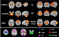
Masaya Misaki1, Hideo Suzuki1, Jonathan Savitz1,2, Brett McKinney3, and Jerzy Bodurka1,4
1Laureate Institute for Brain Research, Tulsa, OK, United States, 2Dept. of Medicine, Tulsa School of Community Medicine, University of Tulsa, Tulsa, OK, United States, 3Tandy School of Computer Science, Dept. of Mathematics, University of Tulsa, Tulsa, OK, United States, 4College of Engineering, University of Oklahoma, Tulsa, OK, United States
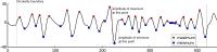
We investigated temporal dynamics of resting-state brain activation in BOLD resting-state networks (RSNs) in patients with major depressive disorder (MDD) and healthy controls (HC). The wavelet variance analysis was applied to the RSNs time courses to assess frequency specific temporal fluctuations. Comparing to HC, MDD subjects had significantly lower fluctuation in the default-mode network (DMN) and the high-visual network in 0.031-0.125Hz and higher fluctuation in the language/auditory and the cerebellum networks in 0.125-0.25Hz and 0.0156-0.031Hz. The low DMN fluctuation in MDD was associated with high precuneus activity that triggered increase of DMN activity.


3761.   
94 
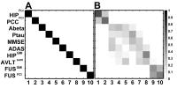
Staging Alzheimer’s Disease Risk by Sequencing Brain Function and Structure, Cerebrospinal Fluid, and Cognition Biomarkers
Guangyu Chen1, Hao Shu1, Gang Chen1, Barney Douglas Ward1, Piero G Antuono2, and Shi-Jiang Li1
1biophysics, medical college of wisconsin, milwaukee, WI, United States, 2Neurology, medical college of wisconsin, milwaukee, WI, United States
A robust temporal ordering sequence of biomarkers for staging the Alzheimer’s disease (AD) progression risk is revealed by integrating brain function and structure, cerebrospinal fluid (CSF), and cognition biomarkers into an event-based model. In this study, we found that functional abnormality in the hippocampus and posterior cingulate cortex networks is the earliest event in the preclinical phase of AD, even antedating the detectable CSF Aβ and p-tau abnormalities; this sheds light on the link between preclinical AD status and its symptomatic onset for accurately identifying progressive AD trajectories along the disease course, given the condition that disease onset is insidious.


3762.   
95 The effect of preterm birth on the thalamocortical development during the neonatal stage: A resting-state fMRI study - Video Not Available
Yue Cai1, Xiushuang Wu2, Yuan Shi3, Lizhi Xie4, and Jiahong Gao5
1Biomedical Engineering, Peking University, Beijing, China, People's Republic of, 2Department of Pediatrics, Daping Hospital, Third Military Medical University, Chong Qing, China, People's Republic of,3Department of Pediatrics, Daping Hospital, Third Military Medical University, Chongqing, China, Chong Qing, China, People's Republic of, 4GE Healthcare, MR Research China, Beijing, Beijing, China, People's Republic of, 5Center for MRI Research and Beijing City Key Lab for Medical Physics and Engineering, Peking University, Beijing, China, People's Republic of
Preterm birth is a leading cause of cognitive impairment in childhood and is associated with cerebral gray and white matter abnormalities. Using the resting-state fMRI imaging analysis, we tested the hypothesis that preterm birth might to some extent affect the thalamo-cortical connections particularly in the thalamo-SM and thalamo-SA projections. Reduced thalamo-SM and increased thalamo-SA connectivity were found in the preterm newborns, and preterm with punctate white matter lesions (PWMLs) exhibited a more sever trend in the thalamo-SA projection.


3763.   
96 Aberrant functional connectivity of resting state networks in subclinical hypothyroidism
Mukesh Kumar1, Ritu Tyagi1, Prabhjot Kaur1, Subash Khushu1, Maria M D'souza1, Tarun Sekhri2, Ratnesh Kanwar2, and Poonam Rana1
1NMR Research Center, Institute of Nuclear Medicine and Allied Science, Delhi, India, 2Thyroid research centre, Institute of Nuclear Medicine and Allied Science, Delhi, India
Cognitive deficit in Subclinical hypothyroidism (SCH) patient is still a topic to research work upon. The present study was conducted to examine resting state networks (RSNs) in SCH using rsfMRI. SCH patients showed significantly decreased functional connectivity in right fronto-parietal network and anterior default mode network (DMN) as compared with control subjects. Our finding suggests cognitive impairment in resting state networks related to attention and emotional processing in SCH patients
Exhibition Hall 

11:00 - 12:00

    Computer #

 
3764.   
1 Altered gray matter volume, cerebral blood flow and functional connectivity in chronic stroke patients: a multi-modal MRI study - Video Not Availble
Peifang Miao1, Caihong Wang1, Peng Li1, Jingliang Cheng1, Dandan Zheng2, and Zhenyu Zhou2
1MRI, The First Affiliated Hospital of Zhengzhou University, Zhengzhou, China, People's Republic of, 2GE Healthcare MR Research, Beijing, China, People's Republic of
In order to investigate the cerebral plasticity in chronic stroke patients well-recovered in global motor function, 29 patients and 30 healthy subjects were recruited to undergo multi-modal MRI techniques. Group comparisons in gray matter volume(GMV), cerebral blood flow(CBF) and resting-state functional connectivity(rsFC) were assessed. Compared with healthy controls, patients exhibited increased GMV in contralesional supplementary motor area, increased CBFs in contralesional superior frontal gyrus and supramarginal gyrus, and increased rsFC in contralesional middle temporal gyrus. The results suggested cerebral structure plasticity, perfusion aberrant and functional reorganization coexist in well-recovered subcortical stroke patients, which may underlie functional recovery of stroke patients.


3765.   
2 Machine learning approach to classify Alzheimer disease and Vascular Dementia using MRI quantitative metrics
Elia Tagliani1, Gloria Castellazzi1,2, Andrea De Rinaldis1,2, Fulvia Palesi2,3, Letizia Casiraghi2,4, Elena Sinforiani5, Paolo Vitali6, Nicoletta Anzalone7, Giovanni Magenes1, Claudia AM Gandini Wheeler-Kingshott2,8, Giuseppe Micieli9, and Egidio D'Angelo2,4
1Department of Electrical, Computer and Biomedical Engineering, University of Pavia, Pavia, Italy, 2Brain Connectivity Center, C. Mondino National Neurological Institute, Pavia, Italy, 3Department of Physics, University of Pavia, Pavia, Italy, 4Department of Brain and Behavioral Sciences, University of Pavia, Pavia, Italy, 5Neurology Unit, C. Mondino National Neurological Institute, Pavia, Italy, 6Brain MRI 3T research center, C. Mondino National Neurological Institute, Pavia, Italy, 7Scientific Institute H. S. Raffaele, Milan, Italy, 8NMR Research Unit, Queen Square MS Centre Department of Neuroinflammation, UCL Institute of Neurology, London, United Kingdom, 9Department of Emergency Neurology, C. Mondino National Neurological Institute, Pavia, Italy
Despite the large number of studies on dementia, efforts to define a clear profile of cognitive impairment for Vascular Dementia (VaD),as well as its differentiation from Alzheimer Disease (AD), are still poor. In this study we tested the power of imaging metrics and adopted a data mining approach, based on Diffusion Tensor Imaging and resting state fMRI, to assess the reliability of machine learning approaches for the automated diagnosis of AD and VaD. Our results show that machine learning algorithms are able to discriminate VaD from AD, representing a suitable approach to build an automated diagnostic system for dementia-like diseases.


3766.   
3 A Preliminary Study of Major Depressive Disorder Using Simultaneous PET/fMRI with Two MID Tasks in a Single Scan
Fuyixue Wang1, Paul Hamilton2, Brian Knutson2, Ian Gotlib2, Matthew Sacchet2, Hershel Mehta2, Christina Schreiner2, Dawn Holley3, Fred Chin3, Bin Shen3, Greg Zaharchuk3, Mehdi Khalighi4, and Gary Glover3
1Department of Biomedical Engineering, Tsinghua University, Beijing, China, People's Republic of, 2Department of Psychology, Stanford University, Stanford, CA, United States, 3Department of Radiology, Stanford University, Stanford, CA, United States, 4Applied Science Lab, GE Healthcare, Menlo Park, CA, United States
The release of dopamine during reward tasks is modulated by major depressive disorder (MDD). In this study of MDD, we used simultaneous PET/fMRI to detect the neurochemical changes of dopamine and neurovascular activity through BOLD contrast during two sequential monetary incentive delay tasks in a single scan. Several modeling methods were proposed and evaluated for dynamic PET data. Six participants with MDD were studied. The results of the group analysis of PET and fMRI show significant effects of dopamine release in ventral striatum bilaterally, close to the nucleus accumbens,  and significant BOLD signals in putamen bilaterally during reward tasks.


3767.   
4 EEG-fMRI at 7T using simultaneous multislice 2D-EPI: safety and functional sensitivity at the single-subject level
João Jorge1,2, Frédéric Grouiller3, Patricia Cotic4, Wietske van der Zwaag5, Patrícia Figueiredo2, and Rolf Gruetter1,6,7
1École Polytechnique Fédérale de Lausanne, Lausanne, Switzerland, 2ISR-Lisboa/LARSyS/Department of Bioengineering, Instituto Superior Técnico, Lisbon, Portugal, 3Biomedical Imaging Research Center, University of Geneva, Geneva, Switzerland, 4Institute of Mathematics, Physics and Mechanics, Ljubljana, Slovenia, 5Spinoza Centre for Neuroimaging, Amsterdam, Netherlands, 6Department of Radiology, University of Lausanne, Lausanne, Switzerland, 7Department of Radiology, University of Geneva, Geneva, Switzerland
The enhanced BOLD sensitivity available at 7T can bring significant advantages for EEG-fMRI studies, and the use of accelerated fMRI sequences such as SMS-EPI could further boost sensitivity. This work investigated whether SMS-EPI can be safely acquired with EEG at 7T, and whether the resulting sensitivity is favorable for combined EEG-fMRI approaches. The adopted SMS-EPI sequence (1.8mm isotropic resolution, whole-brain, TRvol=1.57s) produced no temperature increases when combined with EEG. In a human, eyes-open/closed and resting-state activity patterns could be robustly detected in both modalites, and EEG-derived timecourses produced consistent BOLD predictions, even at a single-subject level with minimal spatial smoothing.


3768.   
5 A simultaneous fMRI-EEG acquisition to minimize the MR gradient artifact on human auditory system
Kevin Wen-Kai Tsai1,2, Hsin-Ju Lee2, Ching-Po Lin2, Li-Wei Ko3, Wen-Jui Kuo2, Toni Auranen4, Simo Särkkä5, and Fa-Hsuan Lin6
1Aim for the Top University Project, National Taiwan Normal University, Taipei, Taiwan, 2Institute of Neuroscience, National Yang-Ming University, Taipei, Taiwan, 3Institute of Bioinformatics and Systems Biology, National Chiao Tung University, Hsinchu, Taiwan, 4Advanced Magnetic Imaging Centre, Low Temperature Laboratory, Aalto University, Espoo, Finland, 5Department of Electrical Engineering and Automation, Aalto University, Espoo, Finland, 6Institute of Biomedical Engineering, National Taiwan University, Taipei, Taiwan
Simultaneous fMRI-EEG acquisition provides a good spatial and temporal resolution from MRI and EEG respective to study the human brain function. However, the EEG signal is impaired due to the strong magnetic gradient switching of concurrent MR imaging. A simultaneous interleaved MR InI-EEG recording strategy is proposed to minimize the distortion of the EEG. Our results suggest that the proposed acquisition strategy can reveal similar BOLD contract activation but preserve better auditory evoked potentials than conventional EPI-EEG acquisition.


3769.   
6 Multimodal elucidation of rat brain function using BOLD-fMRI and CMRO2 [15O]O2-PET
Hans F Wehrl1,2, Jun-ichiro Enmi1, Mario Amend2, Kazuhiro Koshino1, Masako Kunimi1, Takashi Temma1, and Hidehiro Iida1
1Department of Investigative Radiology, National Cerebral and Cardiovascular Center Research Institute, Suita City, Osaka, Japan, 2Department of Preclinical Imaging and Radiopharmacy, Werner Siemens Imaging Center, Eberhard Karls University of Tuebingen, Tuebingen, Germany
The complex interplay of CMRO2, CBF and CMRO2 changes that constitute the BOLD effect are not fully unsterstood. Here we study rat brain activation using combined BOLD-fMRI and CMRO2proportional [15O]O2-PET during the same anesthesia session. To our knowledge, such combined measurements have not been obtained before in small animals. We observed a spatial mismatch between the main activation centers as well as additional activated regions in the CMRO2-PET, not present in BOLD-fMRI. However, statistical significances of activation sites were higher in BOLD-fMRI compared to [15O]O2-PET. This work points towards a complementary nature of both methods in certain brain areas.


3770.   
7 A novel, single scanning session approach to image rat brain activity applying simultaneous [18F]FDG-PET and BOLD-fMRI
Mario Amend1, Tadashi Watabe2, André Thielcke1, Bernd J Pichler1, and Hans F Wehrl1
1Department of Preclinical Imaging and Radiopharmacy, Werner Siemens Imaging Center, Eberhard Karls University, Tuebingen, Germany, 2Department of Nuclear Medicine and Tracer Kinetics, Osaka University Graduate School of Medicine, Osaka, Japan
Functional and metabolic processes during brain activity are still not fully understood. We established a novel protocol to image rat brain activation in a single scanning session, enabling optimal comparison of PET/fMRI data. Performing activation and baseline scans in one single scanning session is challenging due to the half-life (here [18F]FDG: 109.8 min) and decay time of PET-tracers. Applying [18F]FDG-PET and BOLD-fMRI, spatial and quantitative correlations but also mismatches between the glucose proportional PET and the fMRI activation data were found. Our results provide the basis for further studies of brain function and point towards the complementary nature of PET/MR.


3771.   
 
8 Simultaneous, trimodal MR-PET-EEG at 3T in humans: Glutamate drives energy consumption in the default mode network
Irene Neuner1,2,3, Jörg Mauler1, Ravichandran Rajkumar1,2,3, Ezequiel Farrher1, Elena Rota Kops1, Lutz Tellmann1, Jürgen Scheins1, Frank Boers1, Karl Josef Langen1,3,4, Hans Herzog1,2,3, and N. Jon Shah1,3,5
1Institute of Neuroscience and Medicine 4 (INM 4), Forschungszentrum Juelich GmbH, Juelich, Germany, 2Department of Psychiatry, Psychotherapy and Psychosomatics, RWTH Aachen University, Aachen, Germany, 3JARA-BRAIN, Translational Medicine, Aachen, Germany, 4Department of Nuclear Medicine, RWTH Aachen University, Aachen, Germany, 5Department of Neurology, RWTH Aachen University, Aachen, Germany
Within the scope of this explorative pilot trial we focus on the role of GABA and glutamate within the default mode network with regard to energy consumption. In one simultaneous session MR, FDG-PET and EEG data were recorded at a 3T hybrid MR-BrainPET scanner (Siemens, Germany) equipped with a 32 channel MR-compatible EEG system (Brain Products, Germany) in 11 healthy volunteers. The Pearson correlation showed a statistically significant positive correlation between glutamate ratio and mean CMRGlu in the DMN (r = 0.678, n = 11, p = 0.022) but none to GABA. 


3772.   
9 Pharmacological Modulation of Static and Dynamic Functional Connectivity: a Simultaneous PET/MRI Study - Permission Withheld
Hsiao-Ying Wey1, R Matthew Hutchison2, Bruce R Rosen1, and Joseph B Mandeville1
1A. A. Martinos Center, Department of Radiology, Massachusetts General Hospital, Harvard Medical School, Charlestown, MA, United States, 2Center for Brain Science, Harvard University, Cambridge, MA, United States
In this study, we present simultaneous PET/MRI study with pharmacological challenges targeting the μ-opioid receptor system in nonhuman primates to determine the effects of opioid drug on static and dynamic functional connectivity. Mu-opioid receptor occupancy (quantified with PET) and CBV-fMRI signals show dose-dependent reductions to opioid antagonist (naloxone) challenges. Using brain regions showing PET signal changes as seeds, static FC analysis shows an increase in local (within the seed region) and distal (motor cortex) connectivity with putamen after naloxone. Dynamic FC patterns were also modulated with naloxone as indicated by weaker pairwise correlations and larger number of dynamic state transitions.


3773.   
10 Real-time ICA-based artifact correction of EEG data recorded during functional MRI
Ahmad Mayeli1,2, Vadim Zotev1, Hazem Refai2, and Jerzy Bodurka1,3
1Laureate Institute for Brain Research, Tulsa, OK, United States, 2Electrical and Computer Engineering, University of Oklahoma, Tulsa, OK, United States, 3College of Engineering, University of Oklahoma, Tulsa, OK, United States
Recording EEG signals during fMRI acquisition result EEG signals contamination from imaging and BCG artifacts in addition to other types of artifacts. In this abstract we introduced a novel method for detecting and reducing artifacts using second order blind separation in real time. The algorithm was tested on EEG signals from 12 subjects and it has proven successful for reducing various types of artifacts. The proposed algorithm can be applied in real time after the average artifact subtraction of imaging and BCG artifacts for various applications that require a real-time EEG & fMRI system.


3774.   
11 Automatic EEG-assisted retrospective fMRI head motions correction improves rs-fMRI connectivity analysis
Chung-Ki Wong1, Vadim Zotev1, Masaya Misaki1, Raquel Phillips1, Qingfei Luo1, and Jerzy Bodurka1,2,3
1Laureate Institute for Brain Research, Tulsa, OK, United States, 2College of Engineering, University of Oklahoma, Norman, OK, United States, 3Center for Biomedical Engineering, University of Oklahoma, Norman, OK, United States
We utilized an automatic EEG-assisted retrospective motion correction (aE-REMCOR) to improve rs-fMRI connectivity analysis. The aE-REMCOR utilizes EEG data to automatically correct for head movements in fMRI on a slice-by-slice basis. We compared the results of seed-based (posterior cingulate cortex) default-mode network (DMN) connectivity analysis performed with and without aE-REMCOR. The aE-REMCOR reduced the motion-induced position-dependent error in the DMN connectivity analysis. The results show the importance of slice-by-slice fMRI motion corrections to improve rs-fMRI connectivity accuracy especially when the entire group of subjects exhibits rapid head motions, and also provide incentive for conducting simultaneous EEG&fMRI.


3775.   
12 Detecting visual evoked responses in simultaneous EEG-fMRI using Reference Layer Artefact Subtraction (RLAS)
Glyn S. Spencer1, Muhammad E. H. Chowdhury1,2, Karen J. Mullinger1,3, and Richard Bowtell1
1Sir Peter Mansfield Imaging Centre, School of Physics and Astronomy, University of Nottingham, Nottingham, United Kingdom, 2Electrical Engineering, Qatar University, Doha, Qatar, 3Birmingham University Imaging Centre, School of Psychology, University of Birmingham, Birmingham, United Kingdom
Simultaneous EEG-fMRI is limited by large artefacts in EEG recordings resulting from time-varying field gradients, cardiac-related motion and head movement within the magnetic field. Reference layer artefact subtraction (RLAS) reduces artefacts at source by subtraction of artefact voltages recorded from a reference layer on which the EEG leads and electrodes are replicated. Since RLAS does not require prior knowledge of the timing of artefact occurrences, it is an ideal method for correcting pulse and movement artefacts. Here, we apply RLAS in EEG-fMRI experiments for the first time, particularly focusing upon recovery of single-trial, low-frequency, visual-evoked responses from artefact-corrupted data.


3776.   
13 Assessing the stability of neurovascular coupling: A combined fMRI/MEG approach
Marek Allen1, Valentina Tomassini2, Kevin Murphy1, Suresh Muthukumaraswamy3, Krish D. Singh1, and Richard G. Wise1
1Cardiff University Brain Research Imaging Centre, Cardiff University, Cardiff, United Kingdom, 22. Institute of Psychological Medicine and Clinical Neurosciences, Cardiff University, Cardiff, United Kingdom, 3Faculty of Medical and Health Sciences, University of Auckland, Auckland, New Zealand
Neurovascular coupling (NVC) is crucial to maintaining the structural and functional integrity of the brain. Using a graded contrast visual stimulus applied in magnetoencephalography (MEG) and functional MRI (fMRI), we assessed the stability of visual evoked fields (VEFs), gamma power and MRI blood flow responses. A power law was used to relate responses to stimulus contrast and describe the relationship between neuronal responses and blood flow. VEF amplitude was temporally unstable across time points. Gamma power and blood flow (ASL) responses remained stable, making gamma power-based NVC and ASL MRI measures a promising method for testing and exploring NVC in disease states.


3777.   
 
14 EEG-fMRI derived DMN-microstate quantifies meditation induced altered consciousness
Rose Dawn Bharath1 and Rajanikant Panda1
1Neuroimaging and Interventional Radiology, National Institute of Mental Health and Neurosciences, Bangalore, India
Fluctuations in the ICA derived resting state networks can be visualized using simultaneous EEG fMRI. This method can thus be used to understand human consciousness which is the result of synchronous oscillation between multiple networks. EEG fMRI derived "DMN microstate" is seen to accurately assess meditation  induced altered consciousness in the current study.


3778.   
15 New perspectives in simultaneous EEG-fMRI using multiband and quiet pulse sequences
Beatriz Dionisio Parra1,2, Nicolas Hehn1, Xin Liu1,2, Matthew Middione3, Anne Menini1, Darius Burschka2, Florian Wiesinger1, and Ana Beatriz Solana1
1GE Global Research, Munich, Germany, 2TUM (Technical University Munich), Munich, Germany, 3GE Healthcare, California, CA, United States
Three novel fMRI pulse sequences are evaluated together with simultaneous EEG acquisition, with the aim to reduce the induced EEG Gradient Artifacts (GA), increase the spatio-temporal resolution of fMRI and reduce the acoustic noise during scanning: sinusoidal GE- EPI, multiband blipped-CAIPI and single shot T2-prep RUFIS. 

Remarkable results were found for the T2-prep RUFIS sequence, with a significantly reduced gradient artifact amplitude, high temporal resolution and low acoustic noise level, providing eminent advantages to this multimodal technique.


3779.   
16 Mapping dynamics of epileptic seizures in GAERS using simultaneous BOLD fMRI and optical Ca2+ recordings
Lydia Wachsmuth1, Florian Schmid1, Franziska Albers1, Annika Lüttjohann2, Thomas Budde2, and Cornelius Faber1
1Department of Clinical Radiology, University of Münster, Münster, Germany, 2Institute of Physiology I, University of Münster, Münster, Germany
Simultaneous BOLD fMRI and optical Ca2+ recordings using the genetically encoded calcium indicator GCaMP were performed in a rat model of absence epilepsy (GAERS) for seizure mapping.  Spike-and-wave discharge onset times and durations were derived from Ca2+ recordings and used for an event-related analysis with different hemodynamic response functions. BOLD maps showed large scale activations in cortical and subcortical areas, with delayed responses in subcortical areas. In contrast to electrophysiological recordings, Ca2+ recordings are not disturbed by MRI. They allow for cell-specific correlation with the hemodynamic response and provide a tool to obtain epileptic seizure maps with higher specificity.


3780.   
17 A novel test bed for non-BOLD functional MRI
Ruiliang Bai1,2, Tim Bellay 3, Andreas Klaus3, Craig Stewart3, Sinisa Pajevic4, Uri Nevo5, Hellmut Merkle6, Dietmar Plenz3, and Peter J Basser1
1Section on Quantitative Imaging and Tissue Science, DIBGI, NICHD, National Institutes of Health, Bethesda, MD, United States, 2Biophysics Program, Institute for Physical Science and Technology, University of Maryland, College Park, MD, United States, 3Section on Critical Brain Dynamics, LSN, NIMH, National Institutes of Health, Bethesda, MD, United States, 4Mathematical and Statistical Computing Laboratory, Division of Computational Bioscience, Center for Information Technology, National Institutes of Health, Bethesda, MD, United States, 5Department of Biomedical Engineering, Tel-Aviv University, Tel-Aviv, Israel, 6Laboratory for Functional and Molecular Imaging, NINDS, National Institutes of Health, Bethesda, MD, United States
Several fMRI contrast mechanisms have been proposed to measure neuronal activity more directly and accurately than BOLD fMRI. Conclusive findings supporting these non-BOLD fMRI methods have been difficult to obtain, mainly because of the dearth of a reliable and robust test system to vet and validate them. Here we describe the development and testing of a test bed for non-BOLD fMRI, in which calcium fluorescence imaging and MR acquisition can be performed simultaneously on the same organotypic cortical cultures. This experimental design makes it possible to directly correlate any candidate fMRI signal to a robust optical indicator of neuronal activity.


3781.   
18 Anesthesia increases water diffusion in wakefulness/sleep brain regions in the rat brain - Permission Withheld
Yoshifumi Abe1, Tomokazu Tsurugizawa1, and Denis Le Bihan1
1NeuroSpin, Commissariat à l’Energie Atomique-Saclay Center, Gif-sur-Yvette, France
Diffusion fMRI (DfMRI) has been shown to reflect neuronal activation more directly than BOLD fMRI, showing neuronal responses even when neurovascular coupling is abolished. We compared resting state ADC and BOLD fMRI time courses under different anesthesia conditions. While BOLD fMRI showed a widespread signal increase with isofluorane and a decrease with medetomidine, the ADC increased significantly with both agents in specific regions, notably in the wakefulness/sleep network. The amount of ADC increase was correlated with the dose of anesthetic agent, suggesting the suitability of DfMRI to investigate brain resting state or pharmacological challenges quantitatively and without vascular confounding effects.   


3782.   
19 Recruitment of Distinct Cortical and Subcortical Activations by Layer and Frequency Specific Optogenetic Stimulation in Primary Visual Cortex
Russell W Chan1,2, Alex TL Leong1,2, Patrick P Gao1,2, Leon C Ho1,2, Kevin K Tsia2, and Ed X Wu1,2
1Laboratory of Biomedical Imaging and Signal Processing, The University of Hong Kong, Hong Kong, China, People's Republic of, 2Electrical and Electronic Engineering, The University of Hong Kong, Hong Kong, China, People's Republic of
Different layers in mammalian cortex have specific projections and circuit dynamics. However, how optogenetic stimulation frequencies at different infragranular layers contribute to widespread and large-scale cortical and subcortical activities remains largely unexplored. In this study, optogenetic fMRI is used to investigate layer and frequency dependent activities by stimulating excitatory neurons in different infragranular layers of visual cortex. Our results showed that layer and frequency specific optogenetic stimulation recruits distinct widespread and large-scale cortical and subcortical activations. Spatiotemporally varying optogenetic stimulation in combination with fMRI presents unique opportunities in studying the underlying mechanisms of long-range neural circuits and brain functional networks.


3783.   
20 Light-induced activation of the visual network by optogenetic fMRI (ofMRI)
Florian Schmid1, Lydia Wachsmuth1, Franziska Albers1, Nathalie Just1, Miriam Schwalm2, Albrecht Stroh2, and Cornelius Faber1
1Department of Clinical Radiology, University of Münster, Münster, Germany, 2Research Group Molecular Imaging and Optogenetics, Johannes Gutenberg-University Mainz, Mainz, Germany
Optogenetic fMRI is a novel tool in neurophysiology and neuroimaging. However, ofMRI is prone to light-induced artifacts. Here, the unspecific activation of the visual pathways in ofMRI in rats was investigated. It was caused by the stimulation light and was also detected in naïve rats without the presence of opsins.  Visual stimulation of the eyes resulted in similar activation. Visual pathway activation by intrabrain illumination could be suppressed by additional low-level constant light applied to the eyes. We provide evidence that the activation of the visual pathways is at least partly caused by light scattered diffusely inside the brain.


3784.   
21 Development of Carbon Nanotube Optrodes to Acquire LFP and BOLD Concurrently with Optogenetic Stimulation
Corey Cruttenden1, Jennifer M. Taylor2, Xiao-Hong Zhu2, Yi Zhang2, Hannes M. Wiesner2, Anders Asp3, Erin Larson3, Wilson Yu3, Rajesh Rajamani1, Mark Thomas3, Esther Krook-Magnuson3, and Wei Chen2
1Mechanical Engineering, University of Minnesota, Minneapolis, MN, United States, 2Center for Magnetic Resonance Research (CMRR), University of Minnesota, Minneapolis, MN, United States,3Neuroscience, University of Minnesota, Minneapolis, MN, United States
Carbon nanotube optrodes are under development for simultaneous optogenetics, neural recording, and fMRI BOLD signal acquisition. First-generation devices demonstrate capability in combining optogenetic stimulation and neural recording in vivo, but in vitro MR-images reveal severe susceptibility artifacts generated by terminal silver leads. New devices utilize carbon fiber wires in place of silver. Initial in vitro images of the second-generation device show a dramatic reduction of image artifacts, indicating that BOLD signal should be obtainable around the optrodes. The new devices will enable the combination of the aforementioned techniques, providing a platform for novel brain investigations.


3785.   
22 Concurrent 32-channel electrophysiological recording and fMRI in bilateral rat striatum - Permission Withheld
Saul Jaime1,2, Hanbing Lu1, Jose Cavazos2,3, and Yihong Yang1
1Neuroimaging Research Branch, National Institute on Drug Abuse, NIH, Baltimore, MD, United States, 2Physiology, Uni. Texas Health Science Center-San Antonio, San Antonio, TX, United States,3Neurology, Uni. Texas Health Science Center-San Antonio, San Antonio, TX, United States
Despite the high clinical and pre-clinical value of fMRI, its success depends on the identification of the underlying neurophysiological basis of the fMRI BOLD signal. In previously reported simultaneous electrophysiology and fMRI studies, experiments were limited by the poor spatial resolution or poor source localization of intra-cranial or surface EEG techniques employed to record neural activity. In order to overcome these limitations, we have developed a method that allows concurrent whole brain fMRI acquisition and 32-channel intra-cerebral electrophysiological recording in the rat brain.     


3786.   
23 MRI-compatible wireless multi-purpose game controller for social neuroscience fMRI experiments
Ying-Hua Chu1, Yi-Cheng Hsu1, Pu-Yeh Wu1, Kevin W.-K. Tsai2, Wen-Jui Kuo2, and Fa-Hsuan Lin1
1Institute of Biomedical Engineering, National Taiwan University, Taipei, Taiwan, 2Institute of Neuroscience, National Yang Ming University, Taipei, Taiwan
We developed a MRI-compatible wireless multi-purpose game controller. With the presence of the game controller, EPI shows minimal distortion and time-domain SNR degradation. Subjects’ responses were successfully recorded from a two-person hyper-scanning experiment.


3787.   
24 A new way of looking at brain connectivity. pH fMRI: myth or reality?
Vitaliy Khlebnikov1, Jeroen CW Siero1, Alex Bhogal1, Peter R Luijten1, Dennis WJ Klomp1, and Hans Hoogduin1
1Radiology, University Medical Center Utrecht, Utrecht, Netherlands
A new way of looking at brain connectivity. pH fMRI: myth or reality?
Exhibition Hall 

11:00 - 12:00

    Computer #

 
3788.   
25 Loss of functional specificity in basal ganglia in Parkinson's disease: a rs-fMRI study
Anne-Charlotte PHILIPPE1, Pierre BERROIR1, Marie VIDAILHET2, and Stéphane LEHERICY1
1Brain and Spine Institute, CENIR, INSERM U1127/CNRS UMR7225, Sorbonne Universités, UPMC, CHU Pitié-Salpêtrière, Paris, France, 2Brain and Spine Institute, INSERM U1127/CNRS UMR7225, Sorbonne Universités, UPMC, CHU Pitié-Salpêtrière, Paris, France
Loss of neuronal specificity in the basal ganglia (BG) has been reported In Parkinson’s disease (PD)[1]. However, loss of specificity has not been characterized using non-invasive methods in PD. We proposed an innovative method to characterize the functional specificity of BG. We tested the hypothesis that loss of specificity may result in larger spatial extent of overlap between anatomo-functional territories in the BG. We performed population statistics to compare the extent of overlap between PD subjects and controls. As expected, the motor territories had larger extent of overlap in PD subject than in controls revealing a loss of specificity of the BG in PD.


3789.   
26 Increased activation of precuneus and posterior cingulate cortex in resting state fMRI in patients with functional movement disorders after undergoing a motor retraining program
Kwan-Jin Jung1, Sarah Mufti2, and Kathrin LaFaver2
1Radiology, University of Louisville, Louisville, KY, United States, 2Neurology, University of Louisville, Louisville, KY, United States
Functional movement disorders (FMD) can be significantly reduced with a one-week motor retraining program. Our study compared resting state fMRI before and after treatment of 6 FMD patients. We found increased activity in the posterior default mode network, specifically the precuneus and posterior cingulate cortex, in 4 out of 6 patients after treatment, which correlated with clinical improvement of motor symptoms. Our findings suggest that restoration of normal movements in FMD patients are accompanied by increased default mode network activation.


3790.   
27 Demonstration of brain tumor-related NVU in both task-based fMRI and resting state fMRI
Shruti Agarwal1, Noushin Yahyavi-Firouz-Abadi1, Haris I. Sair1, Raag Airan1, and Jay J. Pillai1
1Division of Neuroradiology, Russell H. Morgan Department of Radiology and Radiological Science, Johns Hopkins University School of Medicine, Baltimore, MD, United States
The phenomenon of neurovascular uncoupling (NVU) is a limitation of clinical fMRI, particularly in presurgical mapping. A previous study using task-based motor activation (tbfMRI) and breath hold cerebrovascular reactivity (BH CVR) mapping demonstrated that BH CVR was capable of detecting NVU in low-grade perirolandic tumors. In this study we demonstrated the effect of NVU on resting state fMRI (rsfMRI) data within the sensorimotor network through comparison to both BH CVR and task-based fMRI data. 


3791.   
28 Why a clinical sign does not always correlate with lesion location?
Rajanikant Panda1, Rose Dawn Bharath1, Shriram Varadharajan1,2, Sankalp Tikoo1, Sarbesh Tiwari3, Surabhi ramawat1, Shiva Karthik1, Indira Devi Bhagavatula4, and Arun Gupta1
1Department of Neuroimaging & Interventional Radiology, National Institute of Mental Health and Neurosciences (NIMHANS), Bangalore, India, 2Bangalore, India, 3Department of Neuroimaging & Interventional Radiology, National Institute of Mental Health and Neurosciences (NIMHANS), bangalore, India, 4Department of Neurosurgery, National Institute of Mental Health and Neurosciences (NIMHANS), Bangalore, India
Understanding the degree of functional reserve in patient with brain tumor, when lesion is located in the eloquent cortex is important in presurgical evaluation to predict the surgical outcome as well reducing postoperative neurological deficits. For achieving this, a detailed knowledge of the functional topography and connectivity in whole brain level is crucial. In this study, our aim to understand the brain hyper and hypo connectivity in patients with high grade tumor who have deficits and who do not have deficits. 


3792.   
29 Functional brain changes in breachers following a blast-overpressure: an acute longitudinal assessment with MRI
Fatima Nasrallah1,2, Trina Kok1, Mary Stephenson1, Jiesen Wang3, Alexandre Schaefer3, You Jin1, Benjamin Thomas1, Pamela Pun Boon Li4, Melissa Teo Ai Ling4, Julie Yeo Su Li4, Jia Lu4, John Tottman1, and David Townsend5
1Clinical Imaging Research Centre, NUS/A*STAR, Singapore, Singapore, 2Queensland Brain Institute, Queensland, Australia, 3Clinical Imaging Research Centre, NUS, Singapore, Singapore, 4Defense Singapore Organisation, DSO, Singapore, Singapore, 5clinical Imaging Research Centre, NUS/A*STAR, Singapore, Singapore
Blast injury is one of the most common types of mild traumatic brain injury. In this work we have investigated the longitudinal changes induced by a blast-overpressure injury in breacher trainers using resting state functional connectivity MRI. We show that reductions in connectivity in the Thalamic and Cerebellar regions at Day 1 following a blast are regained and increased 1 month post blast. 


3793.   
30 Parkinson’s disease laterality impact onto the default mode network deactivation by the audio-motor transformation
Oleksii Omelchenko1, Zinayida Rozhkova2, and Iryna Karaban3
1Human and Animal Physiology, Taras Shevchenko National University of Kyiv, Kyiv, Ukraine, 2Radiology, Medical Clinic BORIS, Kyiv, Ukraine, 3Department of extrapyramidal disorders, D. F. Chebotarev Institute of Herontology, Kyiv, Ukraine
Asymmetry of motor symptoms is considered a crucial criterion for PD and in?uence hemisphere-associated cognitive functions. DMN was shown to loose it’s functional connectivity in PD. We hypothesized that AMT play important role in switching DMN to task-related deactivation state. AMT exploration for PD laterality specific DMN connectivity analysis during movements was done. AMT and movement execution in PD evokes activation auditory cortex, SMN and pC. In PD patients DMN deactivates exceptionally at the period of motor activity. pC participates as a DMN ‘hub’ and DMN deactivation ‘switching’ trigger during AMT. PD symtoms lateralization play important role in DMN functioning.


3794.   
31 Evaluation in brain function and the correlation with depression and anxiety of obese patients using resting-state fMRI
Cheng-Jui Li1,2, Vincent Chin-Hung Chen3, Hse-Huang Chao4, Ming-Chou Ho5, and Jun-Cheng Weng1,2,6
1Department of Medical Imaging and Radiological Sciences, Chung Shan Medical University, Taichung, Taiwan, 2Department of Biomedical Sciences, Chung Shan Medical University, Taichung, Taiwan,3Department of Psychiatry, Chang Gung Memorial Hospital, Chiayi, Taiwan, 4Tiawan Center for Metabolic and Bariatric Surgery, Jen-Ai Hospital, Taichung, Taiwan, 5Department of Psychology, Chung Shan Medical University, Taichung, Taiwan, 6Department of Medical Imaging, Chung Shan Medical University Hospital, Taichung, Taiwan
Obesity has reached epidemic proportions globally to become a major public health problem. Obesity-related health problems are numerous including strokes, cardiovascular disease, diabetes mellitus, and increased risk for developing cancer. Reward mechanism of obese patients regarding functional connectivity has been declared by several studies, but few studies mentioned about the correlation between functional images and clinical indices. Thus, our study aimed to find out abnormal functional connectivity over obese patients based on amplitude low frequency fluctuation (ALFF) and regional homogeneity (ReHo) using voxel-based analysis, and the correlation between functional images and clinical indices, including body mass index (BMI) and hospital anxiety and depression scale (HADS).   We found the brain functional abnormality in the obese patients compared to healthy controls, and the correlation with depression and anxiety.  The potential functional imaging markers may provide guidance for managing obesity and disordered eating behaviors.


3795.   
32 Local and Distant Functional Connectivity Density Alteration in Primary Open-angle Glaucoma: A Resting-state functional MRI Study
Zhenyu Liu1 and Ting Li2
1Key Laboratory of Molecular Imaging, Institute of Automation, Beijing, China, People's Republic of, 2Beijing Tongren Hospital, Beijing, China, People's Republic of
To measure functional connectivity density maps were applied with resting-state functional MRI. Twenty-one patients and twenty-two age- and gender-matched healthy controls were evaluated by 3T MRI using resting-state blood oxygenation level dependent imaging. POAG patients showed decreased FCD values in visual cortices and increased FCD values of DMN components. This study demonstrates POAG can induce reorganization of brain function in multiple brain regions, including visual cortices and DMN.


3796.   
33 Altered Amplitude of Low-Frequency Fluctuation and functional connectivity in High Myopia: A Resting-State fMRI Study
Xue-wei Zhang1,2, Wei-hong Zhang2, and Qin Long3
1Department of Interventional Radiology, China Meitan General Hospital, Beijing, China, People's Republic of, 2Department of Radiology, Peking Union Medical College Hospital, Beijing, China, People's Republic of, 3Department of Ophthalmology, Peking Union Medical College Hospital, Beijing, China, People's Republic of
High myopia (HM) can result in serious vision problems. To date, the pathophysiologic mechanism remains unknown. The authors tried to explore the potential locations which involved in the brain abnormalities of HM patients, through observing altered ALFF of different bands and functional connectivity of the brain by rs-fMRI. The results showed that, not only visual cortex but also multiple brain regions were noted to have abnormal changes in the brain of HM patients. In conclusion, the findings indicated that high myopia affects many functional networks, and different bands of ALFF may provide a new way to explore the underlying mechanism.


3797.   
34 Accuracy of Functional Localization in Pre-surgical Function MRI
Mu-Lan Jen1, Islam S. Hassan2, Ping Hou1, Guang Li1, Ashok J. Kumar2, Colen R. Rivka2, and Ho-Ling Liu1
1Department of Imaging Physics, The University of Texas M. D. Anderson Cancer Center, Houston, TX, United States, 2Department of Diagnostic Radiology, The University of Texas M. D. Anderson Cancer Center, Houston, TX, United States
This study evaluates the errors associated with the spatial transformation process by using algorithms commonly applied for clinical pre-surgical fMRI. The images from nine right-handed patients with brain tumors were retrospectively analyzed. Significant differences (P<0.05) were found when comparing results from automated registration (AR) vs. coordinate matching (CM)  and AR vs. AR with manual adjustment (AR, adjusted). No statistical significance was found between CM and AR, adjusted. This study established a platform for evaluating the functional localization accuracy in pre-surgical fMRI, and highlighted the necessity of quality control for the AR processing as a clinical routine.


3798.   
35 Action observation network in children with unilateral cerebral palsy: an fMRI study.
Laura Biagi1, Giuseppina Sgandurra1, Leonardo Fogassi2, Andrea Guzzetta1,3, Giovanni Cioni1,3, and Michela Tosetti1
1IRCCS Stella Maris, PISA, Italy, 2Department of Neuroscience, University of Parma, Parma, Italy, 3Department of Clinical and Experimental Medicine, University of Pisa, PISA, Italy
Mirror Neuron System (MNS) activation constitutes a powerful mechanism for recovery of motor deficits after stroke. We studied with fMRI the MNS (re)-organization in children with congenital unilateral cerebral palsy (UCP), using a goal-directed hand action stimulus. With respect to age-matched controls, UCP children present differences, appearing more lateralized to the dominant hemisphere as adults. The subject-specific pattern of lateralization seems related to the type and extension of the lesion and correlates negatively with the severity of the hand impairment. This paradigm might be useful to explore MNS in UCP and to monitor possible motor improvements in response to therapy.  


3799.   
36 Resting-State Functional Connectivity in Patients with Treatment-Resistant Depression
Jia Liu1, Mingrui Xia2, Qiyong Gong1, and Yong He2
1Huaxi MR Research Center (HMRRC), Department of Radiology, West China Hospital of Sichuan University, Chengdu, China, People's Republic of, 2State Key Laboratory of Cognitive Neuroscience and Learning& IDG/McGovern Institute for Brain Research, Beijing Normal University, Beijing, China, People's Republic of
We used a data-driven graph theoretical approach—whole-brain functional connectivity strength (FCS) mapping to investigate functional connectivity patterns in 24 patients with RDD, 40 patients with NDD, and 48 healthy comparison subjects. Our study indicated that when compared with HC, the alteration of FCS in NDD group was mainly located in the medial frontal gyrus while the alterations of RDD group were located in the non-medial frontal areas and NDD demonstrated lower FCS in the limbic regions which was not obvious in the RDD group. Meanwhile, lower FCS in the default mode network and visual recognition network was observed in the group with NDD when compared with the group with RDD.


3800.   
37 When mint smells blue and diagonal: an fMRI study on olfactory synesthesias - Permission Withheld
Helena Melero1, Susana Borromeo1, Alexandra Cristobal-Huerta1, Eva Manzanedo1, and Juan Antonio Hernandez-Tamames1
1Universidad Rey Juan Carlos, Madrid, Spain
Neuroimaging experiments on grapheme-color synesthesia have provided evidence of structural and functional peculiarities in the synesthetic brain and several explanatory models have been proposed. Nevertheless, data from other modalities are needed in order to test their predictions. For the first time, we investigated brain activity in response to olfactory stimuli in multiple synesthetes. Results showed differential activity in areas that participate in high level visual processing, memory, language, lexical meaning and emotion. These findings suggest that the Conceptual Mediation Model and the Emotional Binding Theory may be complementary and reinforce the idea that meaning and emotion are intrinsically related processes. 


3801.   
38 Data-driven model for evaluation of cerebrovascular-reserve measurement with hypercapnia BOLD
Lenka Vondrácková1,2, Pawel Krukowski3, Johannes Gerber3, Jennifer Linn3, Jan Kybic2, and Jan Petr1
1Institute of Radiopharmaceutical Cancer Research, Helmholtz-Zentrum Dresden-Rossendorf, Dresden, Germany, 2Center for Machine Perception, Faculty of Electrical Engineering, Czech Technical University in Prague, Prague, Czech Republic, 3Department, University Hospital Carl Gustav Carus, Dresden, Germany
Hypercapnia BOLD with the breath-holding task is a technically easier and more clinically available alternative to cerebrovascular reserve (CVR) mapping than administration of CO2 enriched air using an air-tight mask. The disadvantage is complicated data evaluation in case the subject does not adhere to the breathing protocol completely. Here, a data-driven approach for evaluation is presented that is more robust to protocol deviations and produces a reasonable CVR map in most cases where the standard model-driven approach fails. This is demonstrated on randomized evaluation of CVR maps of a group of 86 subjects with stenosis or vessel occlusion.


3802.   
39 Multi Voxel Pattern Analysis (MVPA) classification reveals distributed neural presentations of specific finger movement sequences in the human striatum: a task-based functional MRI study.
Kasper Winther Andersen1, Kristoffer H Madsen1, Tim B Dyrby1, and Hartwig R Siebner1,2
1Danish Research Centre for Magnetic Resonance, Centre for Functional and Diagnostic Imaging and Research, Copenhagen University Hospital Hvidovre, Denmark, Copenhagen, Denmark, 2Department of Neurology, Copenhagen University Hospital Bispebjerg, Copenhagen, Denmark
The basal ganglia is a group of subcortical nuclei, which receives connections from almost the entire cerebral cortex. Here, we investigated the involvement of the striatum, which is the input structure of the basal ganglia, in a sequential finger movement task using fMRI. Sixteen healthy controls performed finger sequences with different complexities. Multi Voxel Pattern Analysis was used to discriminate the distributed signal in striatum and cortex. We found that the distributed signals in contra-lateral striatum are discriminative of the finger sequence performed, which points to a significant role of the basal ganglia in the control of finger sequences.


3803.   
40 Heterogeneity of hemodynamic response dynamics across the subcortical-cortical visual pathway
Laura Lewis1,2, Kawin Setsompop2,3, Bruce R Rosen2,3, and Jonathan R Polimeni2,3
1Society of Fellows, Harvard University, Cambridge, MA, United States, 2Athinoula A. Martinos Center for Biomedical Imaging, Massachusetts General Hospital, Boston, MA, United States, 3Department of Radiology, Harvard Medical School, Boston, MA, United States
            The hemodynamic response is nonlinear in response to short duration stimuli, and this nonlinearity varies across cortex. To study the nonlinearities in subcortical vs. cortical structures, we performed high-resolution fMRI at 7 Tesla to characterize responses to short visual stimuli in superior colliculus (SC), lateral geniculate nucleus (LGN) and primary visual cortex (V1). Response nonlinearity was increased in SC, and response timecourses were consistently narrower in LGN than in V1. We conclude that analysis of subcortical activations using fMRI will require flexible models of the hemodynamic response, and suggest future studies to identify the neural and vascular factors contributing to these nonlinearities.


3804.   
41 Morphological component analysis of functional MRI data based on sparse representations and dictionary learning
Hien M. Nguyen1, Jingyuan Chen2, and Gary H. Glover3
1Department of Electrical Engineering & Information Technology, Vietnamese - German University, Binh Duong New City, Vietnam, 2Department of Electrical Engineering, Stanford University, Stanford, CA, United States, 3Department of Radiology, Stanford University, Stanford, CA, United States
A data-driven method for identifying functional connectivity networks utilizing sparse representations is presented. Specifically, fMRI signals are decomposed into morphological components which have sparse spatial overlap. Allowing sparse spatial overlap between components is more physically plausible than the statistical independence assumption of the Independent Component Analysis (ICA) method. The proposed formulation is related to the Morphological Component Analysis (MCA) and uses a K-Singular Value Decomposition (SVD) algorithm for dictionary learning. Experimental results prove that the MCA-KSVD method can identify functional networks in task and resting-state fMRI and thus can be used as an alternative method for investigating brain functional connectivity.


3805.   
42 fMRI activation patterns during Successful Face-Name Recognition
Yunqing Li1, Prasanna Karunanayaka1, and Qing X Yang1,2
1Radiology, Penn State College of Medicine, Hershey, PA, United States, 2Neurosurgery, Penn State College of Medicine, Hershey, PA, United States
The contributions of distinct anatomical brain regions within the medial temporal lobe (MTL) during successful learning and recognition is poorly understood. In this research, we attempt to unravel how the MTL brain structures interact and integrate information during successful memory recall under different conditions during the performance of a face-name fMRI task.


3806.   
43 Long-term intensive training induced cortical thickness alterations in world class gymnasts - Permission Withheld
Meng Li1, Min Lu2, Shumei Li1, Junjing Wang3, Bin Wang3, Guihua Jiang1, Ruibin Zhang3, Xue Wen3, Jun Wang4, and Ruiwang Huang3
1Department of Medical Imaging, Guangdong No. 2 Provincial People's Hospital, Guangzhou, China, People's Republic of, 2Key Laboratory of Mental Health, Institute of Psychology, Chinese Academy of Sciences, Beijing, China, People's Republic of, 3Center for the Study of Applied Psychology, Key Laboratory of Mental Health and Cognitive Science of Guangdong Province, South China Normal University, Guangzhou, China, People's Republic of, 4State Key Laboratory of Cognitive Neuroscience and Learning, Beijing Normal University, Beijing, China, People's Republic of
World class gymnasts are typical elite athletes whose motor skills and experience are much more than non-athletes. Therefore, changes in brain structure may be expected to occur after long-term intensive training. Thus, we utilized the vertex-wise and ROI-wise methods to investigate the alterations in the cortical thickness of gymnasts. We found the increased thickness in some regions of parietal, occipital, and frontal cortex in the gymnasts, and the significant correlation between thickness with years of training in right superior frontal cortex. Our study indicates that in response to long-term training, neuroanatomical adaptations and plastic changes occur in gymnasts’ cortical thickness. 


3807.   
44 Effects of Hyperglycemia and Hyperinsulinemia on Amplitude of Low Frequency Fluctuations in Medial Frontal and Posterior Cingulate Cortices in Healthy Non-Diabetic Subjects
Nicolas R. Bolo1,2, Alan M. Jacobson3, Brandon Hager1, Gail Musen2,4, Matcheri Keshavan1,2, and Donald C. Simonson5,6
1Psychiatry, Beth Israel Deaconess Medical Center, Boston, MA, United States, 2Psychiatry, Harvard Medical School, Boston, MA, United States, 3Research Institute, Winthrop University Hospital, Mineola, NY, United States, 4Research Division, Joslin Diabetes Center, Boston, MA, United States, 5Division of Endocrinology, Brigham and Women's Hospital, Boston, MA, United States, 6Harvard Medical School, Boston, MA, United States
Our goal is to elucidate the independent effects of hyperglycemia and hyperinsulinemia on brain function. We measured whole-brain amplitude of low frequency fluctuations (ALFF) in slow-band 5 (SB5: 0.01-0.027Hz) and slow-band 4 (SB4: 0.027-0.073Hz) in 10 healthy non-diabetic subjects using resting state fMRI during fasting baseline euglycemia (EU), hyperglycemia (HG) and euglycemic hyperinsulinemia (EU-HI).  SB5 fractional ALFF was decreased in the left medial frontal gyrus and right posterior cingulate/cuneus/precuneus cortices during HG, but not during EU-HI, relative to EU. Our findings may help understand brain functional adaptations to chronic hyperglycemia in diabetes, and their implications for comorbid neuropsychiatric complications.


3808.   
45 ECG-derived respiratory signal for physiological noise correction in simultaneous EEG-fMRI for enhanced mapping of epileptic activity
Rodolfo Abreu1, Sandro Nunes1, Alberto Leal2, and Patrícia Figueiredo1
1ISR-Lisboa/LARSyS and Department of Bioengineering, Instituto Superior Técnico, Universidade de Lisboa, Lisboa, Portugal, 2Department of Neurophysiology, Centro Hospitalar Psiquiátrico de Lisboa, Lisboa, Portugal
We propose a physiological noise model where respiratory-induced BOLD signal fluctuations were extracted from a surrogate of the respiratory signal estimated by Empirical Mode Decomposition. We optimized this model on a subject-specific basis, and evaluate its impact EEG-correlated fMRI mapping of epileptic networks, by comparing the epileptic maps obtained with and without physiological noise correction. This assessment was performed in terms of specificity and sensitivity, having been obtained not only substantial improvements for both measures, but also plausible and patients' semiology concordant epileptic networks.


3809.   
46 Pelvic floor muscles contraction assessed by fMRI
Joel Daouk1, Ludovic Viart2, Fabien Saint2, and Olivier Baledent1,3
1Bioflow Image, University of Picardie Jules Verne, Amiens, France, 2Urology, CHU Amiens, Amiens, France, 3Medical image processing, CHU Amiens, Amiens, France


In this work, we compared two fMRI paradigms to assess the volunteer pelvic floor muscles contraction.

Paradigms suited the block-design method and the action phases consisted in contraction of the pelvic floor muscles (continuously in "Continuous" paradigm and repeated in "pulsed" paradigm) and rest phases were complete relaxation.


Results showed that there was no significant difference in cluster size between "continuous" and "pulsed" paradigms. However, Z-max was significantly higher in the “pulsed” paradigm than in the “continuous” one (p <0.001). fMRI, with a pulsed paradigm, is a suitable technique to assess volunteer pelvic floor muscles contraction.


3810.   
47 A Brain Resting State network specific T2* Study in neonatal infants
Maryam Abaei1, Tomoki Arichi1,2, Anthony Price1, Eugene P Duff3, Emer Hughes1, Giulio Ferrazzi1, Jacques-Donald Tournier 1, Jonathan O'Muircheartaigh1,4, Serena Counsell1, A David Edwards1,5, Steve M Smith3, Daniel Rueckert6, and Joseph V Hajnal1,5
1Centre for the Developing Brain, King's College London, London, United Kingdom, 2Department of Bioengineering, Imperial College, London, United Kingdom, 3Department of Clinical Neurosciences, Oxford University, Oxford, United Kingdom, 4Institute of Psychiatry, Kings College London, London, United Kingdom, 5Division of Imaging Sciences and Biomedical, King's College London, London, United Kingdom, 6Biomedical Image Analysis Group, Department of Computing, Imperial College, London, United Kingdom
Previous studies reported that, although the majority of RSNs are present at the time of normal birth, but they appear to mature at different rates with the "higher-order" networks developing later than primary sensory. T2* in neonates are up to 2 times longer than those typically seen in the mature adult brain and decrease with increasing gestational age at scan. In this study we assessed T2* for different RSNs and hypothesized that those which mature earlier on structural MRI would have shorter T2* than networks in cortical regions that develop later in gestation.


3811.   
48 How feedback, verbal instruction and reward influence learning brain self-regulation? A real-time fMRI study.
Pradyumna Sepulveda1,2, Ranganatha Sitaram3,4,5,6, Mohit Rana3, Cristián Montalba1, Cristián Tejos1,2, and Sergio Ruiz3,5
1Biomedical Imaging Center, Pontificia Universidad Católica de Chile, Santiago, Chile, 2Department of Electrical Engineering, Pontificia Universidad Católica de Chile, Santiago, Chile, 3Institute of Medical Psychology and Behavioral Neurobiology, University of Tübingen, Tübingen, Germany, 4Sree Chitra Tirunal Institute of Medical Sciences and Technology, Trivandrum, India, 5Department of Psychiatry, Faculty of Medicine, Interdisciplinary Center for Neuroscience, Pontificia Universidad Católica de Chile, Santiago, Chile, 6Institute for Biological and Medical Engineering, Pontificia Universidad Católica de Chile, Santiago, Chile
Explicitly instructing subjects to use mental imagery and giving monetary reward  are two strategies used to complement contingent neurofeedback (NF) in the process of learning to self-regulate BOLD signal with real-time fMRI NF. However, it is yet to be defined which is the optimal protocol design in rtfMRI-NF studies, critical step for potential clinical applications. The present study compares the influence of these two strategies in NF learning. Results showed a positive effect of monetary reward in BOLD signal change. Mental imagery had no significant impact in rtfMRI learning.  Despite variation of strategies, brain patterns during NF training were similar.
Exhibition Hall 

11:00 - 12:00

    Computer #

 
3812.   
49 Improved Characterization of Low-Frequency Fluctuations in Resting-State fMRI using GLM Correction of Baseline and Physiological Noise
Olivia Viessmann1, Peter Jezzard1, and Harald Moeller2
1Nuffield Department of Clinical Neuroscience, University of Oxford, FMRIB Centre, Oxford, United Kingdom, 2Max-Planck Institut fuer Kognitions-und Neurowissenschaften, Leipzig, Germany
We present a method to correct mutiband rs-fmri frequency spectra for baseline and physiological noise components using a GLM in the frequency domain. We acquired short-TR (328ms) rs-fmri whole-brain data in a group of younger and older subjects to compare the fractional amplitude of low frequency fluctuations (fALFF) between 0.01 and 0.1Hz that is thought to decline with age. We tested the ability of the GLM approach to minimise baseline and physiological noise contributions to the fALFF. GLM post-processing increased the statistical significance of the group fALFF difference. 


3813.   
50 Nonlinear kernel canonical correlation analysis (kCCA) in fMRI
Zhengshi Yang1, Xiaowei Zhuang1, Tim Curran2, and Dietmar Cordes1,3
1Cleveland Clinic Lou Ruvo Center for Brain Health, Las vegas, NV, United States, 2Department of Psychology and Neuroscience, University of Colorado Boulder, Boulder, CO, United States, 3Department of Radiology, University of Colorado-Denver, Denver, CO, United States
Kernel representation is an efficient method to extract nonlinear features without significantly increased computational complexity. Linear kernel CCA has been applied to fMRI data but the performance of nonlinear kernel CCA is still not clear. Here we investigate the accuracy of five types of kernel on simulated fMRI data and then apply Gaussian kernel CCA on real fMRI data. It provides a more sensitive and specific way to detect activation pattern.


3814.   
51 Addressing multi-centre image registration of 3T arterial spin labeling images from the GENetic Frontotemporal dementia Initiative (GENFI)
Henk Mutsaerts1, David Thomas2, Jan Petr3, Enrico de Vita2, David Cash2, Matthias Van Osch4, Paul Groot5, John Van Swieten6, Robert Laforce Jr7, Fabrizio Tagliavini8, Barbara Borroni9, Daniela Galimberti8, James Rowe10, Caroline Graff11, Giovanni Frisoni9, Elizabeth Finger12, Sandro Sorbi13, Alexandre Mendonça14, Martin Rossor2, Jonathan Rohrer2, Mario Masellis1, and Bradley MacIntosh1
1Sunnybrook Research Institute, Toronto, ON, Canada, 2London, United Kingdom, 3Dresden, Germany, 4Leiden, Netherlands, 5Amsterdam, Netherlands, 6Rotterdam, Netherlands, 7Quebec City, QC, Canada, 8Milan, Italy, 9Brescia, Italy, 10Cambridge, United Kingdom, 11Stockholm, Sweden, 12London, ON, Canada, 13Florence, Italy, 14Lisbon, Portugal
One obstacle in multi-centre arterial spin labeling (ASL) studies is the variability attributed to differences between vendor- or site-specific ASL implementations. This multi-centre study compares spatial registration methods from ASL to 3D-T1, to reduce the between-subject variability of cerebral blood flow (CBF) maps. Our results demonstrate that choices of image registration have profound effects on ASL data collected using different pulse sequences and/or sites. A rigid-body registration of CBF images to segmented gray matter images produced the most robust similarity outcome as a standard approach across the different ASL implementations.


3815.   
52 The Novel Anisotropic Filtering Method for Noise Reduction in fMRI Utilizing Phase Information
Vahid Malekian1,2,3, Danny JJ Wang4, Gholam-Ali Hossein-Zadeh2,5, and Abbas Nasiraei Moghaddam1,2
1Biomedical Engineering, Amirkabir University of Technology (Tehran Polytechnic), Tehran, Iran, 2School of Cognitive Sciences, Institute for Research in Fundamental Sciences (IPM), Tehran, Iran,3Donders Institute for Brain, Cognition and Behaviour, Radboud University, Nijmegen, Netherlands, 4Neurology, UCLA, Los Angeles, CA, United States, 5School of Electrical and Computer Engineering, University of Tehran, Tehran, Iran
To minimize noise and artifacts in fMRI studies, authors have mostly focused on magnitude-based filtering methods and have neglected phase data due to its noisy nature. However, fMRI is a complex-valued data with also valuable phase information. Here, we propose a novel spatial weighted averaging method which uses the phase information along with magnitude to create a reference signal and utilize it iteratively to updates weights. We evaluate the method on experimental A-BOSS fMRI dataset and compared with conventional smoothing methods. The results indicate that the approach can suppress noise effectively and preserve the boundaries of active regions.  


3816.   
53 Regional optimization of physiological noise models improves functional connectivity measurements in resting-state fMRI at 7T
Sandro Nunes1, Marta Bianciardi2, Afonso Dias1, Luís M. Silveira3, Lawrence L. Wald2, and Patrícia Figueiredo1
1ISR-Lisboa/LARSyS and Department of Bioengineering, Instituto Superior Técnico – Universidade de Lisboa, Lisbon, Portugal, 2Department of Radiology, A.A. Martinos Center for Biomedical Imaging, MGH and Harvard Medical School, Boston, MA, United States, 3INESC-ID, Instituto Superior Técnico – Universidade de Lisboa, Lisbon, Portugal
We develop physiological noise models based on cardiac/respiratory recordings, with lag optimization at various levels of specificity (group, dataset, regional and voxel), where regional optimization was achieved by clustering the lagged BOLD responses across the brain. We compare these models, both in terms of the spurious variance explained in the data and the specificity and reproducibility of functional connectivity measurements from three well-known resting-state networks in rs-fMRI at 7T. Voxelwise models explain the most variance in the data; however, connectivity strength specificity and test-retest reproducibility indicate that optimization at the regional/cluster level produces the most accurate networks.


3817.   
54 CSF Signal as a Complex-Valued RETROICOR Regressor Removes Unwanted Physiological Signal and Increases the Accuracy of Spatial Correlation in Complex-Valued fMRI
Mary Kociuba1 and Daniel Rowe1,2
1Mathematics, Statistics, and Computer Science, Marquette University, Milwaukee, WI, United States, 2Biophysics, Medical College of Wisconsin, Milwaukee, WI, United States
Discarding the phase component of the time-series removes relevant biological information from a complex-valued signal. Although, commonly implemented retrospective image correction techniques fail to account for physiological artifacts in both the magnitude and phase components of the time-series.  Using the CSF signal, observed during the data acquisition, as a complex-valued regressor increases the statistical power of fMRI analysis, through reducing unwanted physiological variability in the complex-valued signal of interest. The improved performance of implementing the complex-valued image correction methods is demonstrated with a comparison of magnitude-only and complex-valued spatial correlations. 


3818.   
55 An overcomplete and efficient ICA for BOLD-fMRI. - Permission Withheld
Michael Hütel1,2, Andrew Melbourne1, Jonathan Rohrer2, and Sebastien Ourselin1,2
1Translational Imaging Group, University College London, London, United Kingdom, 2Dementia Research Centre, University College London, London, United Kingdom
Independent Component Analysis (ICA) has been proven to produce compact representations of recurrent patterns in BOLD-fMRI imaging data. Most ICA implementations used in BOLD-fMRI, however, optimize for spatial sparse decompositions rather than independent decompositions. We describe a neural-network ICA framework that optimizes directly for sparsity and also allows for overcomplete basis representation.  


3819.   
56 Sample entropy (SampEn) differentiate patients with Major Depressive Disorder (MDD) from healthy controls and explores mechanism of MDD
Kai Wang1, Yunwen Shao1, Tongxin Chen1, Chuangjian Cai1, Yan Zhu2, and Kui Ying3,4
1Department of Biomedical Engineering, Tsinghua University, Beijing, China, People's Republic of, 2Psychiatry Department, Yu Quan Hospital, Tsinghua University, Beijing, China, People's Republic of,3Department of Engineering Physics, Tsinghua University, Beijing, China, People's Republic of, 4Key Laboratory of Particle and Radiation Imaging, Ministry of Education, Beijing, China, People's Republic of
Entropy indicates system irregularity. Sample Entropy (SampEn), an estimation of entropy in a relatively robust way, was used to differentiate patients with Major Depressive Disorder (MDD) from healthy controls and to explore the mechanism of MDD. In this work, we found five clusters with significantly different SampEn in MDD group from that of control group, located in left frontal lobe, left parietal lobe, left temporal lobe, cerebellum and occipital lobe respectively, and the overall accuracy obtained with linear support vector machine formed by SampEn of those regions was 86.7%(p=0.000). 


3820.   
57 Useful Metrics to Facilitate Clinical Application of Resting-state fMRI
Tie-Qiang Li1, Guochun Fu2, and Peter Aspelin3
1Department of Medical Physics, Karolinska University Hospital, Stockholm, Sweden, 2Department of CLINTEC, Karolinska Institute, Stockholm, Sweden, 3Department of Radiology, Karolinska University Hospital, Stockholm, Sweden
Resting-state fMRI is useful for studying functional networks in the human brain and the abnormalities associated with various neuropsychiatric disorders. However, a lack of quantitative metrics closely associated with underlying neurophysiological characteristics has made its translation to clinical settings difficult. In this study, we propose two voxel-based metrics, namely the functional connection counter index (CCI) and connection strength index (CSI). These metrics depicts high contrast between different brain tissues and can be used for quantitative data-driven analysis to detect changes in functional connectivity related to neuropathology. 


3821.   
58 A Novel Model-based Segmentation Approach for Improved Activation Detection in fMRI studies - Permission Withheld
Wei-Chen Chen1 and Ranjan Maitra2
1pbdR Core Team, Silver Spring, MD, United States, 2Statistics, Iowa State University, Ames, IA, United States
Functional Magnetic Resonance Imaging (fMRI) provides a popular approach to imaging cerebral activation in response to stimuli. Reliably detecting activation is, however, not an easy proposition because only a very small proportions of voxels show true activation. These truly activated voxels are known to be spatially localized, yet incorporating this information is challenging to implement practically. We provide a model-based approach that incorporates spatial context in a practical and methodologically sound manner while postulating our a priori expectation that a certain proportion of voxels is truly active. Results on simulation experiments for different noise levels are uniformly encouraging. The methodology is also illustrated on a sports imagination experiment and shows its potential in making possible the adoption of fMRI as a clinical tool to detect awareness and improve treatment in individual patients in persistent  vegetative state, such as traumatic brain injury survivors.  


3822.   
59 Estimating directed functional connectivity through autoregressive models and orthogonal Laguerre basis functions
Andrea Duggento1, Gaetano Valenza2,3, Luca Passamonti4,5, Maria Guerrisi1, Riccardo Barbieri3,6, and Nicola Toschi1,7
1Department of biomedicine and prevention, University of Rome "Tor Vergata", Rome, Italy, 2Department of Information Engineering, and Research Centre “E. Piaggio”, University of Pisa, Pisa, Italy,3Massachusetts General Hospital and Harvard Medical School, Boston, MA, United States, 4Institute of Bioimaging and Molecular Physiology, National Research Council, Catanzaro, Italy, 5Department of Clinical Neurosciences, University of Cambridge, Cambridge, United Kingdom, 6Department of Electronics, Informatics and Bioengineering, Politecnico di Milano, Milano, Italy, 7Department of Radiology, Martinos Center for Biomedical Imaging and Harvard Medical School, Boston, MA, United States
Classical multivariate Granger causality-based approaches to estimating effective functional connectivity are almost exclusively based on linear autoregressive models. In order to better represent the nonlinear, multiple-time scales interactions which concur to the formation of the BOLD signals, we present a novel approach to Granger causality based on a Volterra-Wiener decomposition with use of the discrete-time, orthogonal Laguerre basis. After validation in synthetic noisy oscillator networks, we analyze timeseries data from the "HCP-500-Subjects PTN Release", revealing a clear-cut, directed interactions between components which highlights strong driving roles of the posterior occipital-inferior parietal networks, superior parietal as well as of the novel “cognitive" cerebellar regions.


3823.   
60 A Software as a Service Cloud-based Platform for integrated analysis of T1-based segmentation and structure-based resting-state fMRI Processing
Yue Li1, Hangyi Jiang1,2, Can Ceritoglu3, James Pekar2,4, Michael I Miller1,3, Susumu Mori1,2, and Andreia Vasconcellos Faria2
1AnatomyWorks, LLC, Baltimore, MD, United States, 2Department of Radiology and Radiological Science, Johns Hopkins University, Baltimore, MD, United States, 3Center for Imaging Science, Johns Hopkins University, Baltimore, MD, United States, 4F. M. Kirby Research Center for Functional Brain Imaging, Kennedy Krieger Institute, Baltimore, MD, United States
Brain resting state fMRI (rs-fmri) is a useful tool for research, although its clinical impact is still limited, mainly because data from single subjects is low in power. We have shown that this limitation can be addressed by “borrowing strength” from the population, specifically by eschewing voxel-based analyses in favor of assessing connectivity between atlas parcels [1]. We now present user-friendly software for signal processing, integrated with structural analysis, under a web-based platform "BrainGPS" (https://braingps.mricloud.org). Users can submit data and download results (co-registered structural and functional images, rs-fmri time-courses, and parcel-based correlation matrices) in a few minutes.


3824.   
61 Sparse Estimation of Quasi-periodic Spatiotemporal Components in Resting-State fMRI
Alican Nalci1,2, Bhaskar D. Rao2, and Thomas T. Liu1
1Center for Functional MRI, University of California, San Diego, La Jolla, CA, United States, 2Electrical and Computer Engineering, University of California, San Diego, La Jolla, CA, United States
Recent studies suggest the presence of complex recurrent spatiotemporal patterns in resting-state fMRI.  These patterns may affect the performance of existing preprocessing and analysis approaches, such as global signal regression and ICA.  In this work we present an approach for the sparse estimation of quasi-periodic spatiotemporal components in resting state fMRI. Our algorithm successfully estimates spatiotemporal components in a sample resting-state fMRI dataset and our results suggest that the removal of these components may represent an alternative to global signal regression. 


3825.   
62 Alzheimer’s Disease is Associated with Hypo-Brain Entropy
Zhengjun Li1 and Ze Wang1,2
1Psychiatry, University of Pennsylvania, Philadelphia, PA, United States, 2Center for Cognition and Brain Disorders, Hangzhou Normal University, Hangzhou, China, People's Republic of
Brain entropy (BEN) mapping provides a way to characterize temporal brain dynamics, and thus the health of regional brain functionality. In this study, we aimed to examine its sensitivity for differentiating Alzheimer’s disease (AD) from healthy controls, as well as its relationship to AD severity. We found reduced BEN in the limbic and prefrontal area in AD compared to controls, and a negative correlation between BEN and AD severity in a widespread area of neural cortex. These results suggest BEN as a potential biomarker for AD.


3826.   
63 Task-Driven Functional Connectivity of White Matter in Projection Pathways of the Human Brain
Xi Wu1, Wuzhong Bi1, Stephen K Bailey2, Laurie E Cutting3,4, Jiliu Zhou1, Adam W Anderson4,5,6, John C Gore4,5,6, and Zhaohua Ding5,6,7
1Department of Computer Science, Chengu University of Information Technology, Chengdu, China, People's Republic of, 2Brain Institute, Vanderbilt University, Nashville, TN, United States, 3Kennedy Center, Chengu University of Information Technology, Nashville, TN, United States, 4Department of Radiology and Radiological Sciences, Vanderbilt University, Nashville, TN, United States, 5Institute of Imaging Science, Vanderbilt University, Nashville, TN, United States, 6Department of Biomedical Engineering, Vanderbilt University, Nashville, TN, United States, 7Department of Electrical Engineering and Computer Science, Vanderbilt University, Nashville, TN, United States
Functional MRI has proven to be most effective in detecting neural activity in brain cortices on the basis of hemodynamic responses, but meanwhile has poor sensitivity in detecting neural activity in white matter. In this study, we demonstrate that MRI signals in the projection pathways have significant correlations to the primary motor cortex in finger tapping conditions, and distributions of the correlations bear clear relations with the sidedness of the task. This indicates that MRI signals in white matter may also encode neural activity, which may be detectable with sensitive methods.


3827.   
64 Differences in slow drift among echoes in multiband multiecho EPI data compromise TE-dependent analysis
Kun Lu1, Javier Gonzalez-Castillo 2, Matthew Middione3, Brice Fernandez4, Valur Olafsson5, Prantik Kundu6, Ajit Shankaranarayanan7, and Thomas Liu1
1Center for Functional Magnetic Resonance Imaging, University of California, San Diego, La Jolla, CA, United States, 2Laboratory of Brain and Cognition, Section on Functional Imaging Methods, National Institutes of Health, Bethesda, MD, United States, 3Applied Sciences Laboratory West, GE Healthcare, Menlo Park, CA, United States, 4Applications and Workflow, GE Healthcare, Munich, Germany,5Neuroscience Imaging Center, University of Pittsburgh, Pittsburgh, PA, United States, 6Brain Imaging Center, Icahn Institute of Medicine at Mt. Sinai, New York, NY, United States, 7Applications and Workflow, GE Healthcare, Menlo Park, CA, United States
We recently observed differences in slow signal drift between multiple echoes in multiband multiecho EPI data.  Such differences in drift compromise the TE dependence model and could impact the TE dependence analysis (e.g  Multiecho Independent Component Analysis ME-ICA). We first designed a metric MEQA to quantitatively measure the observed drift differences, then investigated the effects of the drift differences on ME-ICA analysis.


3828.   
65 Reproducibility of low frequency components in scan-rescan resting state fMRI data
Katherine A Koenig1, Wanyong Shin1, Sehong Oh1, and Mark J Lowe1
1Imaging Sciences, The Cleveland Clinic, Cleveland, OH, United States
This work investigates the reproducibility of low frequency fluctuations by assessing the relationship of scan-rescan timeseries data taken during a resting state fMRI. We look at the relationship between various cortical regions and white matter and CSF.


3829.   
66 High Resolution fMRI with Constrained Evolution Reconstruction
Xuesong Li1, Lyu Li1, Xiaodong Ma1, Xue Zhang1, Zhe Zhang1, Bida Zhang2, Sen Song3, and Hua Guo1
1Center for Biomedical Imaging Research, Department of Biomedical Engineering, School of Medicine, Tsinghua University, Beijing, China, People's Republic of, 2Healthcare Department, Philips Research China, Shanghai, China, People's Republic of, 3Department of Biomedical Engineering, School of Medicine, Tsinghua University, Beijing, China, People's Republic of
fMRI with high temporal and/or spatial resolution is beneficial for psychology and neuroscience studies, but is limited by various factors. Compressed Sensing (CS) based methods for accelerating fMRI data acquisition are promising, however, it may be problematic because the over-smoothing effects may contaminate the hemodynamic osculation of fMRI data. This study aimed to develop a new method, Dual Extended TRACER (DUET), based on Temporal Resolution Acceleration with Constrained Evolution Reconstruction (TRACER), for accelerating fMRI acquisitions using golden angle variable density spiral. Results show DUET can recover fMRI hemodynamic signals in even 14 fold under-sampling. Compared with other methods, DUET provides better signal recovery, higher fMRI signal sensitivity and more reliable activity maps.


3830.   
67 Complex-Valued Correlation Increases Sensitivity and Specificity in the Analysis of Low Contrast-to-Noise fMRI Time-Series
Mary Kociuba1 and Daniel Rowe1,2
1Mathematics, Statistics, and Computer Science, Marquette University, Milwaukee, WI, United States, 2Biophysics, Medical College of Wisconsin, Milwaukee, WI, United States
The standard in fMRI is a magnitude-only statistical analysis of the data, despite evidence of task related change in the phase time-series. This study demonstrates the increased sensitivity and specificity  of implementing complex-valued correlation models for low magnitude and phase contrast-to-noise ratio (CNR) values in fMRI data sets.


3831.   
68 Modeling Resting Cerebral Perfusion from BOLD Signal Dynamics During Hyperoxia
M. Ethan MacDonald1, Avery J.L. Berman1,2, Erin L. Mazerolle1, Rebecca J. Williams1, and G. Bruce Pike1
1Departments of Radiology & Clinical Neurosciences, University of Calgary, Hotchkiss Brain Institute, Seaman Family MR Research Centre, Foothills Medical Centre, Calgary, AB, Canada, 2Montreal Neurological Institute, Montreal, QC, Canada
In this work we demonstrate the use of BOLD-fMRI during hyperoxia to obtain perfusion parameters, including CBF, CBV, and MTT. During BOLD imaging, subjects breathing from a respiratory circuit inhaled air whose oxygen content was increased from 21% to 70%. The exhaled oxygen concentration was processed to obtain an arterial input function, and the concentration of bound oxygen in the venous blood was determined by modeling the BOLD time series. Through deconvolution modeling we were able to obtain measurements of CBF, venous CBV, and MTT within expected ranges.


3832.   
69 Mean-Shift Clustering Technique For fMRI Activation Detection - Permission Withheld
Leo Ai1 and Jinhu Xiong2
1University of Minnesota, Minneapolis, MN, United States, 2University of Iowa, Iowa City, IA, United States
It has been shown the application of Mean-Shift Clustering (MSC) to fMRI analysis can increase detection sensitivity in low contrast to noise situations. In this study, MSC was utilized with a feature space containing both temporal and spatial features to further increase its detection power. If successful, the proposed technique can improve detection in techniques that inherently have low CNR, such as non-proton fMRI or non-BOLD fMRI.


3833.   
70 Deterministic Estimation of Spatiotemporal Motifs in Resting-State fMRI
Alican Nalci1,2 and Thomas Liu2
1Electrical and Computer Engineering, University of California San Diego, La Jolla, CA, United States, 2Center for Functional MRI, University of California San Diego, La Jolla, CA, United States
In resting-state fMRI data,  dynamic  quasi-periodic spatio-temporal patterns have  previously  been identified in both animal and humans with potential links to infra slow electrical activity. These prior studies used an iterative pattern-finding algorithm that employed heuristic learning rules for the dynamic adjustment of correlation thresholds.   Here we present a novel deterministic non-iterative approach for estimating spatiotemporal motifs in resting-state fMRI data without the need for heuristic learning rules. 


3834.   
71 A Systematic Investigation on the BOLD Contrast in S1- and S2-SSFP fMRI
Mahdi Khajehim1 and Abbas Nasiraei Moghaddam1,2
1Biomedical Engineering, Amirkabir University of Technology, Tehran, Iran, 2School of Cognitive Sciences, Institute for Research in Fundamental Sciences (IPM), Tehran, Iran
Non-balanced SSFP can provide banding artifact free images, with reduced sensitivity to B1 inhomogeneity and SAR level. Recently, non-balanced SSFP which is known by the S1 and S2 signals, has been utilized for fMRI. In this study to model the BOLD contrast in S1- and S2-SSFP fMRI a Monte Carlo simulation has been performed. It will enable us to accurately investigate the dependence of the BOLD contrast on vessel size and acquisition parameters. Results are in accordance with the reported experimental values and show the increased sensitivity to capillary-sized vessels for only S2 signal.


3835.   
72 Monte Carlo Simulation for A-BOSS fMRI
Mahdi Khajehim1 and Abbas Nasiraei Moghaddam1,2
1Biomedical Engineering, Amirkabir University of Technology, Tehran, Iran, 2School of Cognitive Sciences, Institute for Research in Fundamental Sciences (IPM), Tehran, Iran
Averaged-BOSS fMRI has been proposed as an alternative to BOSS fMRI to eliminate its spatial coverage problem. The feasibility of A-BOSS has been previously investigated. Due to the complexity presented in A-BOSS fMRI, in this work a Monte Carlo simulation has been performed for a comprehensive investigation on its achievable functional contrast and to compare the results with S1 and S2 SSFP fMRI. The obtained results show the superior absolute signal change for A-BOSS and demonstrate the desirable acquisition parameters.
 

The International Society for Magnetic Resonance in Medicine is accredited by the Accreditation Council for
Continuing Medical Education to provide continuing medical education for physicians.