|
Exhibition Hall 14:30 - 15:30 |
|
|
|
Computer # |
|
3980.
 |
49 |
Whole body broadband and uniform 31P
MRSI with sub-second power calibration at 7T. 
Dennis WJ Klomp1, Joost Löring1, Joep
WM van Oorschot1, Peter R Luijten1,
and Wybe JM van der Kemp1
1Radiology, UMC Utrecht, Utrecht, Netherlands
We have integrated a body RF coil tuned at the 31P
frequency in a 7T MR system that includes pick-up probes for
fast and reliable power calibration. With this setup, full
body and broadband 31P
MRSI can be obtained in single breath-holds. When averaged
over less than 5 minutes, excellent 31P
spectra can be shown from liver and heart as demonstrated in
healthy volunteers.
|
|
3981.
 |
50 |
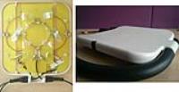
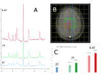
31P MRS Signal-to-Noise Ratio in Human Brain at 3, 7, and 9.4
Tesla Using Dual Tuned Head RF Coils
Marek Chmelik1,2, Diana Bencikova3,
Christian Mirkes4, Christopher T. Rodgers5,
Gunamony Shajan4, Klaus Scheffler4,6,
Siegfried Trattnig1,2, and Wolfgang Bogner1
1High Field MR Centre, Department of Biomedical
Imaging and Image-guided Therapy, Medical University of
Vienna, Vienna, Austria, 2Christian
Doppler Laboratory for Clinical Molecular MR Imaging,
Vienna, Austria, 3Department
of Nuclear Physics and Biophysics, Faculty of Mathematics,
Physics and Informatics, Comenius University, Bratislava,
Slovakia, 4High-Field
MR Center, Max Planck Institute for Biological Cybernetics,
Tuebingen, Germany, 5OCMR,
RDM Cardiovascular Medicine, University of Oxford, Oxford,
United Kingdom, 6Department
for Biomedical Magnetic Resonance, University of Tuebingen,
Tuebingen, Germany
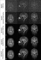
The purpose of this study was to quantitatively compare the
SNR of brain 31P-MRS
coils capable of covering the whole brain at various B0 field
strenghts. SNR was compared using phantoms and in vivo in
clinically acceptable measurement times at 3T (birdcage), 7T
(23ch-array) and 9.4T (27ch-array). Data showed
approximately 3-fold higher SNR at 7T than at 3T. 9.4T
provided an additional -more than linear- increase. WSVD
coil combination outperformed Brown’s coil combination.
Especially with the 7T coil the necessity of advanced coil
combination algorithms is apparent.
|
|
3982.
 |
51 |
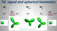
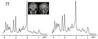
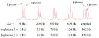
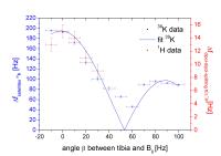
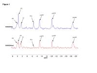
In vivo comparison of quadrupole splitting of potassium
resonance with dipole-dipole splitting of total creatine
resonance in proton MR spectroscopy of human calf muscle
Manuela Barbara Rösler1,2, Nicolas G.R. Behl1,
Nadia Benkhedah1, Armin Michael Nagel1,3,
and Reiner Umathum1
1Medical Physics in Radiology, German Cancer
Research Center, Heidelberg, Germany, 2Institute
for Biomedical Engineering, University and ETH Zurich,
Zurich, Switzerland, 3Diagnostic
and Interventional Radiology, University Medical Center Ulm,
Ulm, Germany
Theory predicts that the residual quadrupole interaction of
spin-3/2 nuclei with electrical field gradients and the
dipole-dipole interaction of coupled spin-1/2 nuclei depend
similarly on the angle between the privileged direction and
the static magnetic field. In this work, we compare the
splitting of the 39K
resonance with the splitting of the total creatine resonance
in 1H
MR spectroscopy in vivo at human calf muscle. We find
similar behavior under variation of the angle between B0 and
tibia. Therefor we conclude that the potassium ions and
creatine are located in an equivalent electromagnetic
environments.
|
|
3983.
 |
52 |
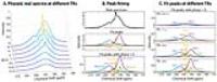
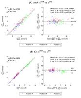
Model-based fitting of in
vivo 129Xe spectra in mice reveals five robust
dissolved-phase peaks
Rohan S. Virgincar1, Scott H. Robertson2,
Simone Degan3, Geoffry Schrank4, Mu He5,
John Nouls4, and Bastiaan Driehuys4
1Biomedical Engineering, Duke University, Durham,
NC, United States, 2Medical
Physics Graduate Program, Duke University, Durham, NC,
United States, 3Center
for Molecular and Biomolecular Imaging, Duke University,
Durham, NC, United States, 4Radiology,
Duke University, Durham, NC, United States, 5Electrical
and Computer Engineering, Duke University, Durham, NC,
United States
Inhaled 129Xe
exhibits chemical shifts which carry useful information
about the underlying physiology. However, their resonant
frequencies have been reported with a variability of 2-3 ppm
likely attributable to using simplistic peak finding methods
and inconsistent reference frequencies. In this work, we use
robust non-linear curve fitting of the complex
dissolved-phase spectrum in mice to identify resonances, and
report shifts relative to an accurate reference frequency.
At short 129Xe
replenishment times curve fitting identified two peaks at
197.4±0.9 and 193.0±0.7 ppm, but as replenishment time was
increased, five distinct peaks became apparent at 198.4±0.4,
195.5±0.4, 193.9±0.2, 191.3±0.2, and 190.7±0.3 ppm.
|
|
3984.
 |
53 |
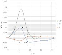
Dynamic 1H MRS study of water T2* and water concentration
contributions to water signal intensity changes in premotor
cortex of the norm and in early stage schizophrenia during
hemodynamic response to a single stimulus.
Svetlana Sergeevna Batova1, Andrei Valerievich
Manzhurtsev2, Maxim Vadimovich Ublinskii2,3,
Irina Sergeevna Lebedeva4, Tolibjon Abdullaevich
Akhadov3, Petr Evgenevich Menshchikov5,
and Natalia Alexandrovna Semenova2,3,5
1Lomonosov Moscow State University, Moscow,
Russian Federation, 2Emanuel
Institute of Biochemical Physics of Russian Academy of
Sciences, Moscow, Russian Federation, 3Radiology,
Scientific Research Institute of Children's Emergent Surgery
and Trauma, Moscow, Russian Federation, 4Scientific
Centre of Mental Health, Moscow, Russian Federation, 5Semenov
Institute of Chemical Physics of Russian Academy of
Sciences, Moscow, Russian Federation
Using dynamic 1H
MRS we have separated T2* and water concentration
contributions to changes of MRS detectable water signal in
motor cortex after activation by a single short stimulus. We
revealed effects of schizophrenia on both parameters in the
period of hemodynamic response to stimulation. Decreased
changes of T2* and water concentration in schizophrenia
might reflect a lower vasodilation caused by a single short
stimulus.
|
|
3985.
 |
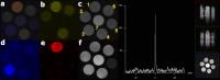
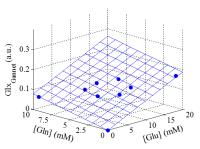
54 |
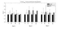
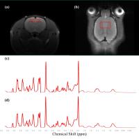
Assessment of the glutamatergic activity changes induced by
Schizophrenia on rat model: A In Vivo Proton Magnetic Resonance
Spectroscopy (¹H MRS) Study at 9.4 T
Chi-Hyeon Yoo1,2, Do-Wan Lee1, Kyu-Ho
Song1, Song-I Lim1,2, Dong-Cheol Woo2,
and Bo-Young Choe1
1Department of Biomedical Engineering, and
Research Institute of Biomedical Engineering, The Catholic
University of Korea College of Medicine, Seoul, Korea,
Republic of, 2Asan
Institute for Life Science, Asan Medical Center, Seoul,
Korea, Republic of
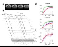
To investigate schizophrenia (SZ)-induced effects in the
glutamatergic activity on prefrontal cortex of rat, we used
proton magnetic resonance spectroscopy (¹H MRS) to estimate
the concentration of glutamate (Glu) and glutamine (Gln).
With a short echo time (TE) and 9.4 T of our study, Glu, Gln
and glutamate-complex (Glx) were reliably quantified with a
low Cramer-Raw low bound (CRLB) value, and Glu, Glx showed
significant increase. As our results the SZ-induced change
in the glutamatergic activity can be reliably detected by ¹H
MRS.
|
|
3986.
 |
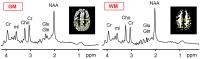
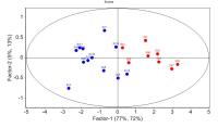
55 |
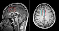
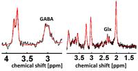
Chronic pain related alterations of regional and interregional
glutamate and GABA associations in the human brain
Alexander Gussew1, Lisa Janetzki2,
Marianne Cleve1, Constanze Borys3, and
Jürgen r Reichenbach1
1Medical Physics Group, Institute of Diagnostic
and Interventional Radiology, Jena University Hospital -
Friedrich Schiller University Jena, Jena, Germany, 2Institute
of Psychosocial Medicine and Psychotherapy, Jena University
Hospital - Friedrich Schiller University Jena, Jena,
Germany, 3Department
of Psychiatry and Psychotherapy, Jena University Hospital -
Friedrich Schiller University Jena, Jena, Germany
1H-PRESS and MEGA-PRESS spectroscopy was
performed in anterior cingulate cortex (aCC), insula (Ins)
and posterior cortex (PC) of 13 matched pairs of chronic low
back pain patients and healthy volunteers to quantify
regional and interregional Glx and GABA associations in the
resting state. Volunteers had negative correlations between
GABA in aCC and Glx in Ins and PC (rho < -0.55) as well as
positive correlations between Glx in aCC, Ins and PC
(rho > 0.65). In contrast, patients had no any comparable
metabolic associations, which may be ascribed to disordered
functional pathways between brain regions due to the
disease.
|
|
3987.
 |
56 |
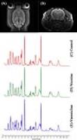
The Influence of Varenicline on Repeated Nicotine-Induced Rats:
In Vivo Proton Magnetic Resonance Spectroscopy at 9.4T
Song-I Lim1,2, Kyu-Ho Song1, Chi-Hyeon
Yoo1,2, Dong-Cheol Woo2, and Bo-Young
Choe1
1Department of Biomedical Engineering, and
Research Institute of Biomedical Engineering, The Catholic
University of Korea College of Medicine, Seoul, Korea,
Republic of, 2Asan
Institute for Life Sciences, Asan Medical Center, Seoul,
Korea, Republic of
Nicotine effects the activation of nicotinic acetylcholine
receptors (nAChRs) in multiple areas of the brain.
Varenicline is a partial agonist acting at the α4β2 nAChRs.
The purpose of the study is to compare the in
vivo effects
of nicotine and varenicline that contribute to the reward
system. The results show the tendency of increased Glu level
in nicotine group. Moreover, GSH and NAA levels tended to
decrease in the nicotine group. It satisfies that high
resolution and short TE component adequately spilt the
overlapped metabolite spectra and quantify the cerebral
neurochemicals. We found that varenicline effectively
inhibits the reward cycle.
|
|
3988.
 |
57 |
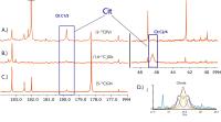
Choline metabolism is reprogrammed differently in mutant IDH1
cells - Permission Withheld
Pavithra Viswanath1, Jose Izquierdo-Garcia1,
Larry Cai1, Joanna Phillips2, Russell
Pieper2, and Sabrina M Ronen1
1Radiology, University of California San
Francisco, San Francisco, CA, United States, 2Neurological
Surgery, University of California San Francisco, San
Francisco, CA, United States
Abnormal choline metabolism with increased levels of
phosphocholine (PC) driven by overexpression of choline
kinase α is considered a hallmark of cancer. For the first
time, we show that glioma cells with the IDH1 mutation
reprogram choline metabolism differently. Using 13C-MRS
to quantify [1,2-13C]-choline flux to PC in IDH1
mutant cells from two genetically engineered glioma models,
we show that reduced PC synthesis is characteristic of
mutant IDH1 cells. Furthermore, reduced PC synthesis is
driven by down-regulated choline kinase α expression. Our
study points to unusual reprogramming of choline metabolism
in IDH1 mutant glioma cells, pointing to novel therapeutic
opportunities.
|
|
3989.
 |
58 |
Reciprocity based metabolite quantification at 3T
Niklaus Zoelch1, Andreas Hock1,2, and
Anke Henning1,3
1Institute for Biomedical Engineering, UZH and
ETH Zurich, Zurich, Switzerland, 2Department
of Psychiatry, Psychotherapy and Psychosomatics, University
of Zurich, Zurich, Switzerland, 3Max
Planck Institute for Biological Cybernetics, Tuebingen,
Germany
At 1.5 T reciprocity principle based quantification
strategies have been successfully used to quantify brain
metabolites. But these methods all rely on the assumption
that the magnitude of the RF transmission field B1+ and
the reception field B1- are
equal at all points in the subject. This is not true at
higher field strengths and for example differences in the
concentrations measured in the left and right hemisphere are
observed when these methods are directly applied at higher
fields. Here a further development is presented, proposing a
correction for deviations of B1+ from
B1- to
allow concentration measurements at 3T and even higher field
strength without the need of assumptions about
concentrations of an internal reference. The obtained
metabolite concentrations in vivo in 31 healthy volunteers
highly agree with values estimated with internal water
referencing, demonstrating the capabilities of this new
method, which might make concentration measurements in
diseased tissue more reliable.
|
|
3990.
 |
59 |
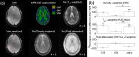
In vivo metabolite quantification using ERETIC with corrections
for changes in the RF transmission and recepetion field
Niklaus Zoelch1, Andreas Hock1,2, and
Anke Henning1,3
1Institute for Biomedical Engineering, UZH and
ETH Zurich, Zurich, Switzerland, 2Department
of Psychiatry, Psychotherapy and Psychosomatics, University
of Zurich, Zurich, Switzerland, 3Max
Planck Institute for Biological Cybernetics, Tuebingen,
Germany
With ERETIC (Electric Reference To access In vivo
Concentrations) metabolite signals measured in vivo are
referenced to a signal measured in a phantom while directly
correcting for differences in the coli loading conditions
between the in vivo and in vitro measurement. This is
beneficial compared to using an internal reference because
no assumption about the concentration or the relaxation rate
of the internal reference is need. However in contrast to
the signal of an internal reference the ERETIC signal
contains no information about B1 during
transmission or reception and changes in B1 between
the in vivo and in vitro measurement or at different
positions are misinterpreted as metabolite concentration
changes. The aim of this work was to tackle this problem by
incorporating reception sensitivity corrections into the
ERETIC method and by using a volume based power optimization
to avoid differences during transmission. As a result, the
obtained metabolite concentrations agree well with the
values obtained with internal water referencing in healthy
volunteers.
|
|
3991.
 |
60 |
Using partially suppressed water signal to improve J-edited
proton MRS - Permission Withheld
Zhengchao Dong1,2, Joshua Kantrowitz1,2,
and Hong Wang1,3
1Columbia University, New York, NY, United
States, 2New
York State Psychiatric Institute, New York, NY, United
States, 3Tianjin
University, Tianjin, China, People's Republic of
Magnetic field drift and subject motion during J-editing
proton MRS scan will not only cause frequency and phase
drifts in the spectrum but also alter the linewidth and
lineshape. When the linewidths/shapes of edit-on and
edit-off spectra do not match, the J-difference spectrum
will have residue of Cr peaks; even when edit-on/off spectra
match each other, the linebroadening and distortion will
deteriorate the quality of the difference spectrum. In this
study, we used the partially suppressed water signals to
match and to transform the edit-on and edit-off spectra so
as to improve the quality of the J-edited spectrum.
|
|
3992.
 |
61 |
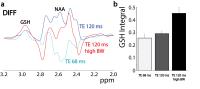
Echo-time optimization for J-difference editing of glutathione
at 3T
Kimberly L Chan1,2,3, Nicolaas AJ Puts2,3,
Karim Snoussi2,3, Ashley D Harris2,3,4,5,6,
Peter B Barker2,3, and Richard AE Edden2,3
1Biomedical Engineering, Johns Hopkins School of
Medicine, Baltimore, MD, United States, 2Radiology
and Radiological Science, Johns Hopkins School of Medicine,
Baltimore, MD, United States, 3F.M.
Kirby Center for Functional Brain Imaging, Kennedy Krieger
Institute, Baltimore, MD, United States, 4Radiology,
University of Calgary, Calgary, AB, Canada, 5Hotchkiss
Brain Institute and Alberta Children's Hospital Research
Institute, University of Calgary, Calgary, AB, Canada, 6CAIR
Program, Alberta Children's Hospital Research Institute,
University of Calgary, Calgary, AB, Canada
Glutathione is involved in maintaining redox balance, and
can be detected in vivo in brain tissue using MEGA-PRESS
editing. In literature to-date, echo times from 68 to 131
ms have been stated as optimal; in this abstract, the
TE-dependence of MEGA-edited GSH signals is investigated
using simulations, and phantom and in vivo experiments. It
is shown that, in vivo, there is a moderate (15%) benefit of
detecting GSH at TE 120 ms over 68 ms. We also demonstrate
that the longer echo time allows the use of
higher-bandwidth, more rectangular slice-selective
refocusing pulses, giving a further 57% gain in signal.
|
|
3993.
 |
62 |
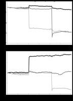
Post acquisition frequency correction in GABA editing
Jan Willem van der Veen1, Stefano Marenco2,
Karen Berman2, and Jun Shen1
1Magnetic Resonance Spectroacopy Core, NIH, NIMH,
Bethesda, MD, United States, 2NIH,NIMH,CTNB,
Bethesda, MD, United States
Patient motion and magnetic field drift may shift the
frequency of the GABA editing pulse relative to that of
metabolites. Using the frequency location of the residual
water we corrected for changes caused by frequency
variations by using averaged reference signals simulated at
specific editing frequency offsets. Our analysis also showed
that GABA editing with a top-hat editing pulse is highly
robust in the presence of frequency variations.
|
|
3994.
 |
63 |
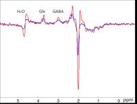
Macromolecule Suppressed GABA Editing using MEGA-SPECIAL
Sequence with Spectral-spatial RF Pulse
Meng Gu1, Adam Kerr2, Ralph Hurd3,
and Daniel Spielman1
1Radiology, Stanford University, Stanford, CA,
United States, 2Electrical
Engineering, Stanford University, Stanford, CA, United
States, 3GE
Healthcare, Menlo Park, CA, United States
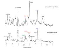
MEGA PRESS has been used to edit the GABA resonance at 3ppm.
Due to the wide transition bandwidth of the editing pulse,
macromolecule resonances are coedited. To suppress
macromolecule signals, a symmetric suppression method has
been proposed resulting in reduced GABA signal. We present a
new editing method by incorporating spatial and spectral
selectivity into the SPECIAL refocusing RF pulses to achieve
both GABA editing and macromolecule suppression. Phantom
studies showed higher edited GABA signal compared with MEGA
PRESS and 90% macromolecule suppression. In-vivo studies
demonstrated significantly higher edited GABA signal
compared with MEGA PRESS.
|
|
3995.
 |
64 |
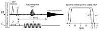
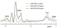
Dual J-difference editing of glutathione and lactate at 3T
Kimberly L Chan1,2,3, Karim Snoussi2,3,
Richard AE Edden2,3, and Peter B Barker2,3
1Biomedical Engineering, Johns Hopkins School of
Medicine, Baltimore, MD, United States, 2Radiology
and Radiological Science, Johns Hopkins School of Medicine,
Baltimore, MD, United States, 3F.M.
Kirby Center for Functional Brain Imaging, Kennedy Krieger
Institute, Baltimore, MD, United States
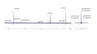
Glutathione (GSH), a redox metabolite, and lactate, a
product of anaerobic energy metabolism, can both be
detected in the human brain using J-difference editing.
Editing each will usually co-edit the other to some degree,
as the GSH editing target spin is at 4.56 ppm and the
lactate spin is at 4.1 ppm. In this abstract, we
investigate optimal simultaneous detection of both
metabolites, using a combination of simulations, and phantom
and in vivo experiments. We demonstrate a new acquisition
protocol applying 10 ms editing pulses at 4.35 ppm, which
successfully edits both GSH and lactate signals with near
maximal efficiency.
|
|
3996.
 |
65 |
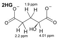
J-Difference Editing of 2-Hydroxyglutarate
Kimberly L Chan1,2,3, Richard AE Edden2,3,
and Peter B Barker2,3
1Biomedical Engineering, Johns Hopkins School of
Medicine, Baltimore, MD, United States, 2Radiology
and Radiological Science, Johns Hopkins School of Medicine,
Baltimore, MD, United States, 3F.M.
Kirby Center for Functional Brain Imaging, Kennedy Krieger
Institute, Baltimore, MD, United States
2-hydroxyglutarate (2HG) is formed in some brain tumors due
to a mutation of isocitrate dehydrogenase (IDH), and is
becoming an important biomarker for tumor
classification. Various approaches have been proposed for
the in vivo measurement of 2HG using MR spectroscopy,
including spectral-editing using the MEGA-PRESS technique.
This abstract investigates 2HG editing at 3T using
density-matrix simulations and phantom experiments. It is
demonstrated that MEGA-PRESS detection of 2HG is best
performed at an echo time of 100 ms, applying editing pulses
to the 1.9 ppm spins and detecting the 4.0 ppm signal, and
employing high-bandwidth refocusing pulses.
|
|
3997.
 |
66 |
Volumetric Navigated MEGA-SPECIAL for real-time zero- and
first-order shim and motion corrected GABA MRS
Muhammad Gulamabbas Saleh1, Jamie Near2,
A Alhamud1, Lindie du Plessis1, André
J.W. van der Kouwe3, and Ernesta M Meintjes1
1Human Biology, MRC/UCT Medical Imaging Research
Unit, University of Cape Town, Cape Town, South Africa, 2Douglas
Mental Health University Institute and Department of
Psychiatry, McGill University, Montreal, QC, Canada, 3Athinoula
A. Martinos Center for Biomedical Imaging, Massachusetts
General Hospital, Charlestown, MA, United States
During macromolecule (MM) suppressed GABA MRS acquisition,
subject motion may cause the spectra to be acquired at an
incorrect region of interest and with suboptimal shim.
Furthermore, effective MM-suppression requires the editing
pulses to be applied consistently at 1.7 ppm, necessitating
real-time frequency updates, which can be exacerbated in the
presence of motion. We demonstrate that a pair of 3D EPI
volumetric navigators acquired once per TR is able to
perform accurate motion and magnetic field inhomogeneity
correction in real time during MM-suppressed MEGA-SPECIAL
GABA MRS.
|
|
3998.
 |
67 |
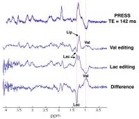
Simultaneous MEGA-PRESS editing of valine and lactate -
Permission Withheld
Thomas Lange1, Cheng-Wen Ko2,
Shang-Yueh Tsai3, Martin Buechert4,
and Ping-Hong Lai5
1Medical Physics, Department of Radiology,
University Medical Center Freiburg, Freiburg, Germany, 2Dept.
of Computer Science and Engineering, National Sun Yat-sen
University, Kaohsiung, Taiwan,3Graduate Institute
of Applied Physics, National Chengchi University, Taipei,
Taiwan, 4Magnetic
Resonance Development and Application Center, University
Medical Center Freiburg, Freiburg, Germany,5Dept.
of Radiology, Veterans General Hospital Kaohsiung,
Kaohsiung, Taiwan
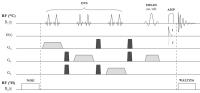
The MEGA-PRESS sequence allows difference editing of
J-coupled metabolites on the basis of their specific
coupling behavior. In this work, we demonstrate MEGA-PRESS
editing of valine and lactate from a large background of
lipid signal. Exploiting the very similar J-coupling
constants of valine and lactate, it is demonstrated that
both metabolites can be edited simultaneously with an echo
time of 142 ms, allowing an editing efficiency of 100% with
negligible lipid co-editing. Simultaneous valine/lactate
editing is validated in vitro and successfully demonstrated
in one brain abscess patient. The method may prove
clinically useful for distinguishing brain abscesses from
brain tumors.
|
|
3999.
 |
68 |
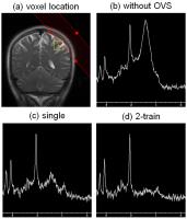
Optimized B1-robust Outer Volume Suppression for MR Spectroscopy
Martin A Janich1, Ralph Noeske2, Timo
Schirmer1, and Rolf F Schulte1
1GE Global Research, Munich, Germany, 2GE
Healthcare, Potsdam, Germany
Outer Volume Suppression (OVS) applied to MR spectroscopy
improves voxel localization and suppresses undesired
signals. Goal of this work was the numerical optimization of
a train of broadband SLR pulses for B1-robustness
and T1 effects
and its application to PRESS in the human brain at 3T. The
technique improved localization and better suppressed
subcutaneous fat at around 1.5ppm in MRS voxels close to the
scalp.
|
|
4000.
 |
69 |
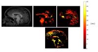
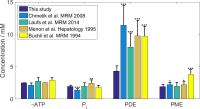
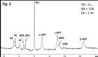
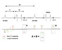
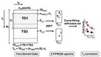
Quantitative Imaging of ATP Production Rates and their
Functional Changes in Healthy Human Brain -
Permission Withheld
Xiao-Hong Zhu1, Byeong-Yeul Lee1, and
Wei Chen1
1CMRR, Radiology Department, University of
Minnesota, Minneapolis, MN, United States
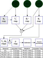
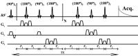
We established a practical protocol for quantitatively
imaging the cerebral metabolic rates of ATP production via
ATPase and creatine kinase (CK) reactions in human brain at
7T using three dimensional (3D) chemical shift imaging (CSI)
and in vivo 31P
MR spectroscopy (MRS) in combine with magnetization transfer
(MT) approach. Subsequently, we applied this 3D 31P-MT
imaging protocol to quantify the regional phosphorous
metabolites concentrations, ATPase and CK reaction rate
constants and fluxes and the intracellular pH in human brain
at rest and during visual stimulation. The results of this
study provide the values of key parameters relevant to ATP
metabolism in absolute scale, which allow quantitative
evaluation of regional cerebral energetics in resting human
brain and its functional changes.
|
|
4001.
 |
70 |
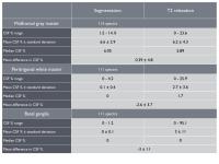
CSF fraction calculation for single voxel spectroscopy:
comparison of water signal T2 biexponential fitting and image
segmentation in a pediatric population
Frances C Robertson1, Martha J Holmes1,
Francesca Little2, Mark F Cotton3, Els
Dobbels3, Andre JW van der Kouwe4,5,
Barbara Laughton3, and Ernesta M Meintjes1
1Department of Human Biology, University of Cape
Town, Cape Town, South Africa, 2Department
of Statistical Sciences, University of Cape Town, Cape Town,
South Africa, 3Department
of Paediatrics & Child Health, Stellenbosch University,
Stellenbosch, South Africa, 4A.A.
Martinos Centre for Biomedical Imaging, Massachusetts
General Hospital, Charlestown, MA, United States, 5Department
of Radiology, Harvard Medical School, Boston, MA, United
States
For partial volume correction in 1H-MRS
the voxel fraction of brain matter (BM) and cerebral spinal
fluid (CSF) can be calculated via biexponential fitting of
T2 relaxation of the unsuppressed water signal or via
segmentation of a high-resolution structural image. We
compared voxel CSF percentages obtained using these two
methods and investigated whether discrepancies could be
explained by head movement between voxel positioning and MRS
acquisition. Subjects with large differences in CSF% between
methods tended to show greater displacement than those with
no difference between methods. Inconsistencies may be due to
segmentation inaccuracy in particular regions or subject
motion.
|
|
4002.
 |
71 |
Rotation optimization for semi-LASER implementation under the
constraint of maximum gradient strength -
Permission Withheld
Ralph Noeske1
1GE Healthcare, Potsdam, Germany
The semi-LASER sequence is less prone to chemical shift
displacement errors and shows higher B1-robustness
compared to PRESS. To achieve short echo times high
amplitude crusher gradients are used to suppress unwanted
coherence signals. Goal of this work was to implement an
algorithm that prevents violation of gradient strength
constraints due to unrestricted rotation of the voxel while
optimizing for shortest possible echo time.
|
|
4003.
 |
72 |
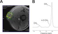
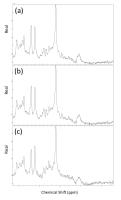
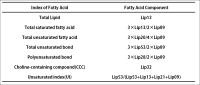
Hepatic lipid metabolite changes in high-fat diet induced liver
steatosis model by in vivo 1H-MRS at 9.4T
Joo-Yeon Kim1, Yeong-Jae Jeon1,2,
Sang-Woo Kim1,2, and Hyeon-Man Baek1,2
1Bioimaging Reseach Team, Korea Basic Science
Institute, Ochang, Korea, Republic of, 2Bio-Analytical
Science, University of Science and Technology, Ochang,
Korea, Republic of
The aim of this study was to characterize hepatic lipid
metabolites changes in high-fat diet induced liver steatosis
model using in vivo 1H-MRS. MR imaging an single-voxel
1H-MRS was performed using a PRESS sequence at 9.4T.
Significant increase in lipid signals at 0.9, 1.3, 2.1, 2.3,
2.8, 4.1, 4.3, and 5.3 ppm was found in mice with high-fat
diet (p<0.001). TL, TUB, UI, and Cho were increased with
high-fat diet.Therefore, 1H-MRS is useful in detecting and
characterizing various hepatic lipid alterations at early
phase in mouse liver steatosis prior to development of
fibrosis.
|
|