14:15
|
|
Muscle Quality and Function 
John Thornton1
1MRC Centre for Neuromuscular Diseases,
University College London, London, United Kingdom
Target audience: This presentation is intended to inform
those interested in the application of quantitative MRI
to probe structure, function or pathology in skeletal
muscle. Objectives: To outline the properties of
skeletal muscle pertinent to quantitative MRI, the
various MRI-accessible quantities that reflect muscle
quality, and how MRI measurements correlate with disease
severity and functional assessment
|
14:45
|
|
Clinical Applications - Permission Withheld
Thomas M Link1
1Department of Radiology and Biomedical
Imaging, UCSF, San Francisco, CA, United States
Over the past decade advanced quantitative MRI
techniques have evolved which allow to characterize bone
and muscle structure and function. Clinically applicable
techniques analyzing bone quality and strength are high
resolution, morphological MRI, UTE and MRS. These
techniques have shown promise in clinical studies,
providing information beyond bone mineral density, the
current standard measurement. Novel technologies
focusing on the assessment of muscle structure and
function are chemical shift-based fat quantification
techniques, MRS, T2 relaxation time measurements and
BOLD MRI, all of which are also clinically applicable
and were used in investigating pain syndromes and
disorders of muscle function.
|
15:15
 |
0177.
 |
Bound- and Pore-Water MRI of Cortical Bone in Osteoporotic
Patients
Mary Kate Manhard1, S Bobo Tanner2,
Daniel F Gochberg3, Jeffry S Nyman4,
and Mark D Does1
1Biomedical Engineering, Vanderbilt
University, Nashville, TN, United States, 2Department
of Medicine, Vanderbilt University, Nashville, TN,
United States, 3Vanderbilt
University Institute of Imaging Science, Vanderbilt
University, Nashville, TN, United States, 4Department
of Orthopaedics & Rehabilitation, Vanderbilt University,
Nashville, TN, United States
Osteoporotic fractures are a growing problem, and X-ray
based methods do not always identify individuals at risk
of a fracture. MRI based methods of bound and pore water
in cortical bone have the potential to offer new
information about fracture resistance. These methods
were implemented on both osteoporotic volunteers and
healthy controls in the tibia. Osteoporotic subjects had
significant decreases in bound water concentration and
slight increases in pore water concentration compared to
healthy subjects. These promising results will allow for
further investigation of changes of bound and pore water
concentrations across diseases and with response to
various treatment methods.
|
15:27
|
0178.
 |
Magnetic resonance elastography characterization of skeletal
muscle stiffness changes resulting from pressure ulcers
Jules Laurent Nelissen1,2, Willeke Traa3,
Larry de Graaf1, Kevin Moerman4,
Cees Oomens3, Aart Nederveen5,
Ralph Sinkus6, Klaas Nicolay1, and
Gustav Strijkers2
1Biomedical NMR, Eindhoven University of
Technology, Eindhoven, Netherlands, 2Preclinical
and Translational MRI, Academic Medical Center,
Amsterdam, Netherlands, 3Biomechanics
of Soft Tissues, Eindhoven University of Technology,
Eindhoven, Netherlands, 4MIT
media lab, Massachusetts Institute of Technology,
Cambridge, MA, United States, 5Radiology,
Academic Medical Center, Amsterdam, Netherlands, 6Imaging
Sciences & Biomedical Engineering, King's College
London, London, United Kingdom
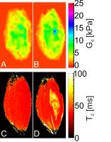
We have investigated the feasibility of using Magnetic
Resonance Elastography (MRE) to quantify muscle-tissue
mechanical properties and changes therein related to the
development of deep tissue injury type of pressure
ulcers. MRE measurements were performed before and after
damage-inducing indentation of the tibialis-anterior
muscle of Sprague Dawley rats. Current study
demonstrates that changes in muscle-tissue mechanical
properties associated with deep tissue injury can be
quantified by MRE. We expect that better knowledge of
changes in soft tissue mechanical properties due to
damage, measured with MRE, will provide new insights in
the aetiology of deep tissue injury and other muscle
pathologies.
|
15:39
|
0179.
 |
Selective in Vivo Bone Imaging with Long-T2 suppressed PETRA
MRI
Cheng Li1, Jeremy F. Magland1, Xia
Zhao1, Alan C. Seifert2, and Felix
W. Wehrli1
1Radiology, University of Pennsylvania,
Philadelphia, PA, United States, 2Translational
and Molecular Imaging Institute, Icahn School of
Medicine at Mount Sinai, New York, NY, United States
An IR-based long-T2 suppressed PETRA sequence was
designed and optimized to image sub-millisecond-T2
tissue components, e.g. collagen-bound bone water. To
minimize scan time signal was sampled repeatedly after
each inversion with individual excitation flip-angle
designed to yield constant short-T2 signal amplitude. A
fast non-iterative reconstruction algorithm combined
with phase-modulated excitation pulse was applied to
minimize image artifacts due to non-uniform excitation
profile, allowing for increased flip-angle and higher
SNR. Optimized long-T2 suppressed PETRA allows imaging
of bone matrix water, opening up new possibilities for
anatomic bone imaging at isotropic resolution and
quantification in clinically practical scan times.
|
15:51
|
0180.
 |
In vivo skeletal muscle fiber length measurements using a
novel MRI diffusion tensor imaging approach: reproducibility
and sensitivity to passive stretch. - Permission Withheld
Jos Oudeman1, Valentina Mazzoli1,2,3,
Marco A Marra2, Klaas Nicolay3,
Mario Maas1, Nico Verdonschot2,
Andre M Sprengers2, Aart J Nederveen1,
Gustav J Strijkers4, and Martijn Froeling5
1Radiology, Academic Medical Center,
Amsterdam, Netherlands, 2Orthopedic
Research Lab, Radboud UMC, Nijmegen, Netherlands, 3Biomedical
NMR, Eindhoven University of Technology, Eindhoven,
Netherlands,4Biomedical Engineering and
Physics, Academic Medical Center, Amsterdam,
Netherlands, 5Radiology,
University Medical Center, Utrecht, Utrecht, Netherlands
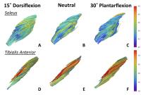
Diffusion Tensor Imaging in combination with
tractography facilitates 3D visualizations of the muscle
architecture, which is described by fiber length and
pennation angle. In order to get accurate fiber length
estimation, tendinous structures need to be separated
from muscles. In this work we propose a new method for
semiautomatic tendon segmentation. The fiber length
obtained after tendon segmentation is seen to be
reproducible. Furthermore the sensitivity of the method
allows for detection of change in fiber length whit
muscle stretch. The observed behavior is in agreement
with the known antagonistic function of muscles.
|
16:03
|
0181.
 |
31P-MRS and MRI of lower leg muscle oxidative metabolism in
heart failure patients
Ding Xia1, Stuart D. Katz2, and
Ravinder R. Regatte1
1Center for Biomedical Imaging, Department of
Radiology, New York University Langone Medical Center,
New York, NY, United States, 2Division
of Cardiology, Department of Medicine, New York
University Langone Medical Center, New York, NY, United
States
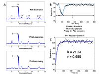
We measured the lower leg muscle oxidative metabolism in
healthy volunteers (n=5) and heart failure patients
(n=6) with quantitative 31P-MRS
and MRI at 3T clinical scanner. The post-exercise rate
of phosphocreatine (PCr) resynthesis was decreased in
heart failure subjects (i.e. delayed PCr recovery time)
compared to healthy volunteers in global calf muscle, as
well as in predominantly fast twitch (type II)
gastrocnemius muscle (medial and lateral, GM and GL) and
predominantly slow twitch (type I) soleus (SOL) muscle.
|
16:15
|
|
Adjournment & Meet the
Teachers |
|