16:30
|
|
Introduction by Moderator |
16:33
|
 |
Arterial Spin Labeled Measurement of Renal Perfusion 
Ananth J Madhuranthakam1
1Radiology, UT Southwestern Medical Center,
Dallas, TX, United States
ASL has become a mainstream application for brain
perfusion, but still has challenges for renal perfusion.
Various improvements including pseudo-continuous
labeling combined with background suppression and
timed-breathing approaches have enabled robust renal
perfusion imaging. This presentation will discuss
different types of arterial spin labeling technique
along with the acquisition methods and strategies for
robust renal perfusion imaging without the
administration of exogeneous contrast agent.
|
16:48
|
0267.
 |
Assessing longitudinal renal blood flow changes in children
following renal replacement therapy using Arterial Spin
Labelling MRI
Fábio Nery1, Enrico De Vita2,3,
Chris A. Clark1, Isky Gordon1, and
David L. Thomas3
1UCL Institute of Child Health, Developmental
Imaging and Biophysics Section, LONDON, United Kingdom, 2National
Hospital for Neurology and Neurosurgery, Lysholm
Department of Neuroradiology, LONDON, United Kingdom, 3UCL
Institute of Neurology, Department of Brain Repair and
Rehabilitation, LONDON, United Kingdom
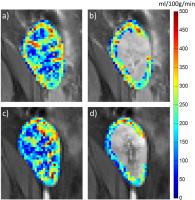
Arterial spin labelling (ASL) is a contrast-free MRI
technique that allows for the quantitative measurement
of organ perfusion. In this study, we non-invasively
evaluated renal perfusion changes in sixteen children
within the first year following renal replacement
therapy using ASL. Each child was scanned in three
occasions : (A) immediately post-transplant; (B) “1
month” post-transplant and (C) “1 year” post-transplant.
The highest renal cortical blood flow was seen on the
first scan in the majority of children while in later
scans equilibrium between child and kidney was reached.
|
17:00
|
0268.
 |
Noninvasive Measurement of Single Renal Oxygen Extraction
Fraction using Focused Asymmetric Spin Echo Approach - a
feasibility study
CY Wang1, R Zhang2, L Jiang3,
R Wang4, XD Zhang4, H Wang3,
K Zhao4, LX Jin3, J Zhang1,2,
XY Wang1,4, and J Fang1,2
1Academy for Advanced Interdisciplinary
Studies, Peking University, Beijing, China, People's
Republic of, 2College
of Engineering, Peking University, Beijing, China,
People's Republic of, 3Philips
Healthcare, Suzhou, China, People's Republic of, 4Department
of Radiology, Peking University First Hospital, Beijing,
China, People's Republic of
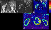
This study demonstrates the feasibility of combining
2D-RF excitation pulse and ASE sequence (focused ASE
sequence, FASE) for single renal OEF measurement.
Comparison between images acquired with full-FOV ASE and
focused ASE was conducted to confirm the advantages of
the focused ASE sequence for single renal imaging. The
new technique could reduce artifacts and distortion
caused by susceptibility differences, and limit spatial
blurring due to T2-decay, which is promising for
diagnosis of some renal diseases.
|
17:12
|
 |
How Bold is BOLD MRI of the Kidney: Detailing Renal Hypoxia
with MRI, Electrochemical Physiological Methods and Optical
Imaging
Thoralf Niendorf1
1Berlin Ultrahigh Field Facility (B.U.F.F.),
Max-Delbrück Center for Molecular Medicine, Berlin
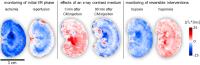
This presentation is designed
to inspire the preclinical and clinical imaging, renal
physiology, and nephrology communities to foster
explorations into the assessment of renal oxygenation
and haemodynamics by exploiting the powers of MRI. For
this purpose the merits
and limitations of renal BOLD-MRI are surveyed together
with their implications. Explorations into detailing the
relation between renal T2* and renal tissue
partial pressure of oxygen (pO2) are
discussed. Multi-modality in
vivo approaches
suitable for detailing the role of the confounding
factors that govern T2* are considered.
Future directions of MRI assessment of renal oxygenation
and perfusion are explored.
|
17:27
|
0269.
 |
Quantitative MRI of Renal Function in a Mouse Model of
Unilateral Ureteral Obstruction
Haiying Tang1, Matthew Fronheiser1,
Guoqiang Zhang2, Adrienne Pena1,
Daniel Kukral1, Cindy Cai2, Rachel
Zebo2, Jeff L L Zhang3, Bradley
Zinker2, Anthony Azzara2, Patrick
Chow1, Feng Luo4, and Wendy Hayes1
1Bristol Myers Squibb, Princeton, NJ, United
States, 2Bristol
Myers Squibb, Hopewell, NJ, United States, 3Radiology,
University of Utah, Salt Lake City, UT, United States, 4Bristol
Myers Squibb, Wallingford, CT, United States
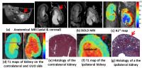
Recent advances in magnetic resonance imaging (MRI)
allow the development of non-invasive and quantitative
tools to assess renal function. DCE-MRI using low dose
Gd-based contrast has been established as a reliable
technique for measuring glomerular filtration rate (GFR)
in individual kidneys. Other promising markers for renal
function include R2* measured with BOLD MRI, and the
longitudinal relaxation time T1. Unilateral ureteral
obstruction (UUO) has been developed in rodents as a
model of renal fibrosis. The purpose of the study is to
evaluate the various MRI techniques in assessing kidney
tissue properties and renal function in the UUO mouse
model.
|
17:39
 |
0270.
 |
Determination of Parameters Variation in DTI, BOLD, and ASL
MRI for Transplanted Kidneys
Maryam Seif1, Laila Yasmin Mani2,
Chris Boesch1, Bruno Vogt2, and
Peter Vermathen1
1Depts. Radiology and Clinical Research,
University of Bern, Bern, Switzerland, 2Dept.
Nephrology, Hypertension and Clinical Pharmacology,
Hospital University of Bern, Bern, Switzerland
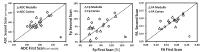
DTI, BOLD and ASL MRI techniques have gained acceptance
to evaluate different physiological aspects of the renal
function both in research and clinics. However, there
are not yet sufficient studies available investigating
the accuracy and repeatability of renal MRI
techniques. The main aim of this study was therefore to
evaluate the reproducibility of DTI, BOLD MRI and ASL
parameters derived from two scans and to investigate
whether there are significant correlations between
renal parameters obtained from these MR techniques in
transplanted kidneys.
|
17:51
|
|
Diffusion
Hersh Chandarana1
1NYU School of Medicine
Conventional methods of measuring renal function
including estimated GFR are insensitive to early renal
dysfunction and cannot assess single kidney
function/dysfunction. Advance MR imaging techniques
including diffusion weighted imaging (DWI) are being
investigated to study renal microstructure and function
in health and disease. Various flavors of diffusion
weighted imaging including intra-voxel incoherent motion
(IVIM) and diffusion tensor imaging (DTI) have shown
considerable promise in evaluation of kidney structure
and function.
|
18:06
|
0271.
 |
Diffusion Tensor Imaging (DTI) of the kidneys incorporating
advanced geometric distortion correction using reversed
phase encoding images.
Jose Teruel1,2, Jeremy C. Lim3,
Eric E. Sigmund4, Elissa Botterill5,
Jas-mine Seah6, Shawna Farquharson7,
Elif E. Ekinci6,8, and Ruth P. Lim5,9
1Department of Circulation and Medical
Imaging, Norwegian University of Science and Technology,
Trondheim, Norway, 2St.
Olavs University Hospital, Trondheim, Norway, 3Department
of Radiology, The Royal Melbourne Hospital, Melbourne,
Australia, 4Department
of Radiology, NYU Langone Medical Center, New York, NY,
United States, 5Department
of Radiology, Austin Health, Melbourne, Australia, 6Department
of Endocrinology, Austin Health, Melbourne, Australia, 7Florey
Neuroscience Institute, Melbourne, Australia, 8Department
of Endocrinology, The University of Melbourne,
Melbourne, Australia, 9Departments
of Radiology and Surgery, The University of Melbourne,
Melbourne, Australia
Diffusion tensor imaging is emerging as a promising
technique for structural and functional evaluation of
the kidneys. However, diffusion sequences employing echo
planar imaging readout are prone to geometric
distortions due to static field inhomogeneities arising
from different magnetic susceptibilities from adjacent
tissues and bowel gas. In this study, we evaluated the
efficacy of distortion correction using a reversed phase
encoding approach for diffusion tensor imaging of
healthy controls and patients with Type 1 diabetes.
|
18:18
|
0272.
 |
Kidney diffusion-weighted imaging based on multi-band
multi-shot DW-EPI acquisition and multi-band multiplexed
sensitivity encoding (MB-MUSE) reconstruction
Hing-Chiu Chang1,2, Arnaud Guidon3,
Mustafa R. Bashir4, Dan Xu5, Lloyd
Estkowski6, Ersin Bayram7, Allen
W. Song2, and Nan-Kuei Chen2
1Department of Diagnostic Radiology, The
University of Hong Kong, Hong Kong, Hong Kong, 2Brain
Imaging and Analysis Center, Duke University Medical
Center, Durham, NC, United States, 3Global
MR Application and Workflow, GE Healthcare, Boston, MA,
United States, 4Department
of Radiology, Duke University Medical Center, Durham,
NC, United States, 5Global
MR Application and Workflow, GE Healthcare, Waukesha,
WI, United States, 6Global
MR Application and Workflow, GE Healthcare, Menlo Park,
CA, United States, 7Global
MR Application and Workflow, GE Healthcare, Houston, TX,
United States
DWI has been shown to be useful in characterizing renal
carcinoma with quantitative measurement of ADC. However,
with echo-planar imaging (EPI) based DWI protocols, the
application of body DWI remains limited due to
suboptimal EPI image quality. The multi-band multi-shot
EPI with multiplexed sensitivity encoding (MB-MUSE) has
been developed and shown to be useful in achieving
high-resolution and high-quality DWI and DTI of brains,
with improved scan throughput. In this study, we propose
to use MB multi-shot EPI to acquire kidney DWI data with
reduced geometric distortion and bilateral coverage,
demonstrating the feasibility of MB multi-shot DWI of
body applications.
|
18:30
|
|
Adjournment & Meet the
Teachers |
|