| |
16:00
|
0990.
 |
Harmonic-Phase versus Sine-Wave Modeling for Measuring Regional
Cardiac Function from Tagged MRI Images 
El-Sayed H. Ibrahim1, Scott Swanson1,
Jadranka Stojanovska1, Claire Duvernoy1,
and Rodica Pop-Busui1
1University of Michigan, Ann Arbor, MI, United
States
MRI tagging is a valuable method for evaluating regional
heart function. This study compares the harmonic-phase
(HARP) and sine-wave modeling (SinMod) tagging analysis
techniques for evaluating myocardial strain and torsion in
healthy controls and type-1-diabetes patients. All SinMod
measurements were significantly larger than those by HARP.
Nevertheless, there existed consistency in the measurements
by each technique, as seen by the good correlation between
the HARP and SinMod measurements in both normals and
patients, except for apical strain (patients and controls)
and mid-ventricular strain in patients. The inter-observer
agreement was better in SinMod than in HARP for both torsion
and strain.
|
| |
16:12
|
0991.
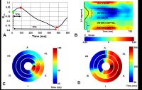 |
Regional cardiac mechanical activation times using cine DENSE
strain imaging strongly predict electrical activation times in
cardiac resynchronization therapy
Daniel A Auger1, Kenneth C Bilchick2,
and Frederick H Epstein1,3
1Department of Biomedical Engineering, University
of Virginia, Charlottesville, VA, United States, 2Department
of Medicine, Cardiovascular Medicine, University of
Virginia, Charlottesville, VA, United States,3Radiology
and Medical Imaging, University of Virginia,
Charlottesville, VA, United States
A widely held goal in cardiac resynchronization therapy
(CRT) is to implant the left-ventricular (LV) pacing lead in
a late-activating region. Time to peak shortening (TPS) has
been used to image mechanical activation; however electrical
activation time is directly related to the time of onset of
contraction rather than TPS. Using cine DENSE in heart
failure patients, we show that the time of onset of
shortening (TOS) shows a strong correlation with electrical
activation time, whereas a lower correlation was found using
TPS. Cine DENSE of TOS is a promising method for the
detection of late-activating segments in CRT patients
|
| |
16:24
|
0992.
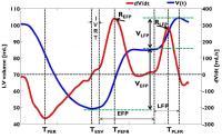 |
Left ventricular (LV) volume and rate of volume change (dV/dt)
during the early and late filling periods evaluated from
respiratory triggered, high frame rate cine SSFP as markers of
LV diastolic function: Direct correlation with Echocardiography
Jiming Zhang1, Benjamin Y Cheong1, Jie
Chen1, Amol Pednekar2, Claudio Arena1,
Melissa L Andrews1, and Raja Muthupillai1
1Diagnostic and Interventional Radiology, CHI St
Luke's Health, Houston, TX, United States, 2Phillips
Healthcare, Cleveland, OH, United States
LV chamber volumes measured using MR cine SSFP imaging and
trans-mitral flow velocities measured with echo are
considered de facto standards for evaluating LV systolic and
diastolic function respectively. Our results show that the
relative change in LV volume as well as peak LV volume-rate
between the early and late filling periods of the cardiac
cycle as measured from high-frame rate cine SSFP imaging,
correlate well with conventional echo-based diastolic
function index (E/A ratio). The results from the study
suggest that a free-breathing, high frame rate MR cine SSFP
imaging approach can evaluate both systolic and diastolic
function from a single LV volume data-set.
|
| |
16:36
|
0993.
 |
An extended 3D whole-heart myocardial first-pass perfusion
sequence: Alternate-cycles interchanging high-resolution and
isotropic imaging
Merlin J Fair1,2, Peter D Gatehouse1,2,
Liyong Chen3,4, Ricardo Wage2, Edward
VR DiBella5, and David N Firmin1,2
1NHLI, Imperial College London, London, United
Kingdom, 2NIHR
Cardiovascular BRU, Royal Brompton Hospital, London, United
Kingdom, 3UC
Berkeley, Berkeley, CA, United States, 4Advanced
MRI Technologies, Sebastopol, CA, United States, 5UCAIR,
University of Utah, Salt Lake City, UT, United States
An alternate-cycle acquisition strategy is proposed for 3D
whole-heart first-pass perfusion, capturing two separate
datasets from the same first-pass, each pushing separate
boundaries of currently achievable parameters whilst
maintaining clinically feasible acquisition times. It is
postulated this approach may also confer an advantage with
regard to artefact detection.
|
| |
16:48
|
0994.
 |
Reduced field-of-view 3D stack-of-spirals perfusion imaging with
high spatiotemporal resolution
Yang Yang1, Li Zhao2, Xiao Chen3,
Kelvin Chow4, Peter W. Shaw4, Jorge A.
Gonzalez4, Frederick H. Epstein1,5,
Craig H. Meyer1,5, Christopher M. Kramer4,5,
and Michael Salerno1,4,5
1Biomedical Engineering, University of Virginia,
Charlottesville, VA, United States, 2Radiology,
Beth Israel Deaconess Medical Center & Harvard Medical,
Boston, MA, United States, 3Medical
Imaging Technologies, Siemens Healthcare, Princeton, NJ,
United States, 4Medicine,
University of Virginia, Charlottesville, VA, United States, 5Radiology,
University of Virginia, Charlottesville, VA, United States
3D CMR perfusion imaging enables whole ventricular coverage
at the same cardiac cycle permitting quantification of
ischemic burden of patients being evaluated for coronary
artery disease. Current 3D techniques have limited
spatial-temporal resolution. We developed an efficient
outer-volume suppressed 3D Stack-of-Spiral perfusion
sequence with motion-guided compressed sensing
reconstruction which can acquire 20 partitions with 2 mm
in-plane and 4 mm through-plane resolution with a temporal
foot print of 180 ms. A pilot study of 10 subjects using
this technique demonstrates clinically acceptable image
quality.
|
| |
17:00
|
0995.
|
Multi-center study of whole-heart dynamic 3-dimensional cardiac
magnetic resonance perfusion imaging for the detection of
coronary artery disease – analysis of diagnostic performance in
women
Sandra Hamada1, Alexander Gotschy2,3,
Lukas Wissmann3, Sebastian Kozerke3,
Cosima Jahnke4, Ingo Paetsch4, Rolf
Gebker5, Nikolaus Marx6, Hatem Alkadhi1,
and Manka Robert1,7
1Institue of Diagnostic and Interventional
Radiology, University Hospital Zurich, Zurich, Switzerland, 2Department
and Policlinic of Internal Medicine, University Hospital
Zurich, Zurich, Switzerland, 3Institue
for Biomecical Engineering, University and ETH Zurich,
Zurich, Switzerland, 4University
Heart Center Leipzig, Leipzig, Germany, 5German
Heart Institute Berlin, Berlin, Germany, 6Department
of Cardiology, Pneumology, Angiology and Intensive Care
Medicine, University Hospital RWTH Aachen, Aachen, Germany, 7Department
of Cardiology, University Heart Center, Universitiy Hospital
Zurich, Zurich, Switzerland
Coronary heart disease accounts for a large amount of
morbidity and mortality in women. To contribute to evidence
for non-invasive testing in women, this study compares
diagnostic performance of whole heart dynamic 3D myocardial
first-pass perfusion stress imaging in female and male with
findings in coronary angiography. 61 female and 139 male
with suspected and known coronary artery disease were
enrolled and a whole heart dynamic 3D-CMR first-pass
perfusion imaging was performed at rest and at stress. Whole
heart dynamic 3D-CMR perfusion imaging shows high
sensitivity, specificity and diagnostic accuracy
in women and men and therefore seems to be a suitable
testing tool for myocardial ischemia in women and men.
|
| |
17:12
|
0996.
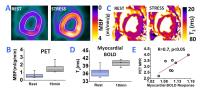 |
Towards Reliable Non-Contrast Enhanced MR-based Myocardial
Perfusion Imaging: Myocardial BOLD MRI Using Late Effects of
Regadenoson with Simultaneous 13N-ammonia PET Validation in a
Whole-body Hybrid PET/MR System
Hsin-Jung Yang1, Damini Dey1, Jane
Sykes2, John Butler2, Xiaoming Bi3,
Behzad Sharif1, Sotirios Tsaftaris4,
Debiao Li1, Piotr Slomka1, Frank Prato2,
and Rohan Dharmakumar1
1Cedars Sinai Medical Center, Los Angeles, CA,
United States, 2Lawson
Health Research Institute, london, ON, Canada, 3Siemens
Healthcare, Los Angeles, CA, United States, 4IMT
Institute for Advanced Studies Lucca, Lucca, Italy
Over the past two decades myocardial BOLD MRI has seen major
technical advancements and a number of clinical validation
studies. However, the reliability of BOLD MRI still remains
a key weakness for its widespread adoption for routine
clinical use due to the unpredictable motions during stress
tests. We investigated whether the unique pharmocokinetics
of regadenoson, a new coronary vasodilator that is rapidly
becoming the agent of choice for cardiac stress testing, can
be used to markedly improve the reliability of myocardial
BOLD MRI. Studies were performed in a canine model and
validated in a clinical PET/MR system.
|
| |
17:24
|
0997.
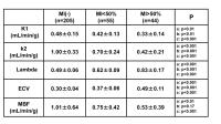 |
Comprehensive assessments of myocardial tissue kinetic
parameters of K1, k2, MBF, lambda and ECV by using a synergistic
quantitative analysis of first-pass myocardial perfusion MRI and
pre-and post-contrast T1 mapping in patients with myocardial
infarction.
Akimasa Yamada1, Masaki Ishida1,
Takashi Ichihara2, Takahiro Natsume2,
Yoshitaka Goto1, Mio Uno1, Motonori
Nagata1, Yasutaka Ichikawa1, Kakuya
Kitagawa1, and Hajime Sakuma1
1Radiology, Mie University Hospital, Tsu-Mie,
Japan, 2Faculty
of Radiological Technology, Fujita Health University School
of Health Science, Toyoake-Aichi, Japan
In this study, we proposed a new method that synergistically
analyzes quantitative perfusion MRI and T1-mapping for
quantifying k2, as well as K1, myocardial blood flow, lambda
and extracellular volume fraction. Nineteen patients with
previous myocardial infarction (MI) were studied. Myocardial
segments were categorized into 3 groups by presence or
absence as well as severity of MI in each segment.
Quantitative measurement was successful in all segments with
significant difference among the 3 groups of myocardial
segments for all tissue kinetic parameters including k2.
Synergistic assessment of quantitative perfusion MRI and
T1-mapping is promising for more detailed myocardial tissue
characterization.
|
| |
17:36
|
0998.
 |
Bayesian Intravoxel Incoherent Motion Imaging to Map Perfusion
in the Human Heart
Georg Spinner1, Constantin von Deuster1,2,
Christian Torben Stoeck1, and Sebastian Kozerke1
1Institute for Biomedical Engineering, ETH
Zurich, Zurich, Switzerland, 2Division
of Imaging Sciences and Biomedical Engineering, King's
College London, London, United Kingdom
In vivo cardiac Intravoxel Incoherent Motion Imaging (IVIM)
is particularly challenging due to low signal-to-noise
ratio, cardiac and respiratory motion. To address the
limitation, a spin-echo (SE) based sequence employing
motion-compensated diffusion gradients during cardiac
contraction was used in combination with Bayesian Shrinkage
Prior (BSP) inference. In this work, parameter maps of four
volunteers (two slices) are compared to standard segmented
least squares (LSQ) regression. Bayesian inferred IVIM
parameter maps showed reduced intra-subject variation
relative to LSQ. It is concluded that the proposed method is
a promising alternative to map myocardial perfusion without
the need for contrast agent administration.
|
| |
17:48
|
0999.
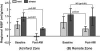 |
Non-contrast Vasodilatory Response Assessment in a porcine model
of Acute Myocardial Infarction using Arterial Spin Labeled CMR
Hung Phi Do1, Venkat Ramanan2, Graham
A Wright2,3, Nilesh R Ghugre2,3, and
Krishna S Nayak4
1Department of Physics and Astronomy, University
of Southern California, Los Angeles, CA, United States, 2Physical
Sciences Platform, Sunnybrook Research Institute, Toronto,
ON, Canada, 3Department
of Medical Biophysics, University of Toronto, Toronto, ON,
Canada, 4Ming
Hsieh Department of Electrical Engineering, University of
Southern California, Los Angeles, CA, United States
Myocardial vasodilatory response is an important indicator
of microvascular function and viability. Arterial spin
labeled (ASL) CMR is a non-contrast method that can quantify
myocardial blood flow making it attractive to study
vasodilatory response. In this work, we demonstrate the
feasibility of ASL in the assessment of regional
vasodilatory response in a porcine model of acute myocardial
infarction (AMI) using a pharmacological stress agent.
Quantitative monitoring of microvascular function in the
infarcted, salvageable and remote myocardial territories may
potentially help identify patients who are prone to adverse
long-term remodeling post-AMI.
|
|