| |
10:30
 |
0816.
 |
Multi-echo Pseudo-Golden Angle Stack of Stars Thermometry
with High Spatial and Temporal Resolution 
Bryant Svedin1 and
Dennis L. Parker1
1University of Utah, Salt Lake City, UT, United
States
A multi-echo pseudo-golden angle stack of stars sequence is
investigated for use in MR thermometry. High spatial and
temporal resolution is achieved through k-space filtering.
PRF temperature, T2*, ρ (signal magnituade at TE = 0),
respiration correction and fat/water separation are
simultaneously measured. Use of a pseudo-golden angle
increment allows for the removal of phase (and therefore PRF
temperature) artifacts due to changing k-space sampling
between reconstructed time points. k-Space sampling based
phase reference greatly improves temperature standard
deviation. FUS heating experiments are performed while
simulating respiration artifacts.
|
| |
10:42
 |
0817.
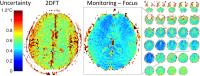 |
Efficient Volumetric Thermometry for MR-Guided FUS Brain
Treatment Monitoring, Using Multiple-Echo Spirals and Mixed
Update Rates
Michael Marx1, Pejman Ghanouni1, and
Kim Butts Pauly1
1Radiology, Stanford University, Stanford, CA,
United States
Multi-slice thermometry was developed that overcomes several
limitations of single-slice 2DFT thermometry in MR-guided
focused ultrasound brain treatment. Using multiple-echo
spiral imaging provides much greater imaging performance,
which was applied to improved focal spot localization and to
improved ablation monitoring. High-resolution
higher-precision multi-slice focal spot localization can
shorten treatment time and improve patient safety.
High-speed high-precision focal spot monitoring, combined
with full-brain monitoring and 3-dimensional focal spot
characterization during ablations can improve treatment
guidance and feedback while also improving patient safety.
The new sequences were validated both in
vivo and in
a phantom within a clinical transducer.
|
| |
10:54
|
0818.
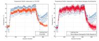 |
Motion Compensation using Principal Component Analysis and
Projection onto Dipole Fields for Abdominal Magnetic Resonance
Thermometry during High-Intensity Focused Ultrasound
Jeremy Tan1,2,3, Adam C. Waspe1,2,
Charles Mougenot4, Kullervo Hynynen1,5,
James M. Drake1,2, and Samuel Pichardo3,6
1University of Toronto, Toronto, ON, Canada, 2Hospital
for Sick Children, Toronto, ON, Canada, 3Thunder
Bay Regional Research Institute, Thunder Bay, ON, Canada, 4Philips
Healthcare, Toronto, ON, Canada,5Sunnybrook
Research Institute, Toronto, ON, Canada, 6Electrical
Engineering, Lakehead University, Thunder Bay, ON, Canada
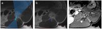
Accurate thermometry during abdominal high-intensity focused
ultrasound is severely compromised by motion and
susceptibility artifacts. A hybrid artifact correction
method has been developed using principal component analysis
as a multi-baseline method and projection onto dipole fields
as a near-referenceless approach. The hybrid algorithm was
tested using free-breathing porcine and human subjects and
achieved an average temperature stability and precision of
0.31 (±0.22) °C and 1.18 (±0.94) °C, respectively in the
kidney.
|
| |
11:06
|
0819.
 |
Non-invasive cardiac stimulation with MR guided HIFU: a rapid,
cardiac triggered, MR-ARFI method for direct visualization of
stimulation site and assessment of tissue stiffness.
Pierre Bour1,2, Fabrice Marquet2,
Fanny Vaillant2, Valery Ozenne2,
Solenn Toupin2,3, Matthieu Lepetit coiffe3,
Erik Dumont1, and Bruno Quesson2
1IGT, PESSAC, France, 2IHU-LIRYC,
PESSAC, France, 3Siemens
Healthcare, Saint-Denis, France
HIFU cardiac stimulation may enable diagnostic and
therapeutic applications such as noninvasive
electrophysiological exam, emergency care and temporary
stimulation. In-vivo proof
of concept of HIFU cardiac stimulation has already been done
on pig. We propose here a first proof of feasibility to
monitor the displacement induced by acoustic radiation force
impulse (MR-ARFI) during contactless stimulation ex-vivo,
on a beating pig heart model. ARFI displacement maps will be
used for precise localization of the depolarization source
and a quantification of displacement will be done during
refractory (contraction) and non-refractory (resting time)
period.
|
| |
11:18
|
0820.
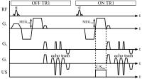 |
Simultaneous Acquisition of Acoustic Radiation Force Imaging and
Proton Resonance Frequency Shift Thermometry Using Interleaved
Acquisition with Temporally Constrained Reconstruction for
Increased Temporal Resolution
Joshua de Bever1,2, Henrik Odéen 1,2,
and Dennis L. Parker1,2
1Department of Radiology, University of Utah,
Salt Lake City, UT, United States, 2Utah
Center for Advanced Imaging Research, Salt Lake City, UT,
United States
Using focused ultrasound and MR acoustic radiation force
imaging (MR-ARFI), the mechanical properties of tissues can
be interrogated. Changes to tissue properties, for instance
before and after a MR guided focused ultrasound thermal
therapy, could help evaluate treatment success. This
abstract presents a novel method for measuring acoustic
radiation force simultaneously with proton resonance shift
thermometry. This would enhance the safety of MR-ARFI, and
provide additional temperature information that may indicate
when, and at what temperature, a tissue property change
occurred. Temporal resolution was enhanced by a factor of 5
by employing a temporally constrained reconstruction
algorithm.
|
| |
11:30
|
0821.
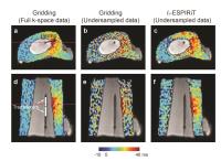 |
Acceleration of 3D UTE Imaging to Quantify Temperature-Dependent
T1 Changes in Cortical Bone
Misung Han1, Wenwen Jiang2, Roland
Krug1, Peder Larson1,2, and Viola
Rieke1
1Radiology and Biomedical Imaging, University of
California, San Francisco, San Francisco, CA, United States, 2Joint
Graduate Program in Bioengineering, University of
California, San Francisco/Berkeley, San Francisco, CA,
United States
High-intensity focused ultrasound (HIFU) is a promising,
noninvasive technique to ablate bone tumors and palliate
painful bone metastases. During HIFU treatment, temperature
mapping is desirable for proper heat deposition to targeted
bone regions. Even though conventional PRF-based thermometry
cannot be applied for cortical bone due to its short T2
relaxation time, it was demonstrated using 3D UTE imaging
can be used to measure T1 changes due to heating. In this
work, we accelerated 3D UTE imaging by combining parallel
imaging and compressed sensing and compared calculated T1
changes due to heating with those from fully sampled data.
|
| |
11:42
|
0822.
 |
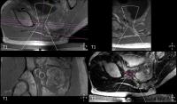
Preclinical Study of MRgFUS Ablation of the Lumbar Medial Branch
Nerve: Functional Outcomes and Histology
Elena Kaye1, Sebastien Monette2, Majid
Maybody3, Stephen B Solomon3, and
Amitabh Gulati4
1Medical Physics, Memorial Sloan Kettering Cancer
Center, New York, NY, United States, 2Comparative
Pathology, Sloan Kettering Institute, New York, NY, United
States, 3Radiology,
Memorial Sloan Kettering Cancer Center, New York, NY, United
States, 4Anesthesiology,
Memorial Sloan Kettering Cancer Center, New York, NY, United
States
The main goals of this preclinical study were to determine
whether direct MRgFUS ablation of the lumbar MB nerve leads
to functional changes and to study the extent of the thermal
damage to the targeted and adjacent tissues, including
neurologic structures. We found that direct FUS ablation of
the lumbar MBN achieves thermal necrosis of the targeted
nerve with minimal thermal damage of the adjacent bone and
muscle tissue. The extent of the cellular changes in bone is
limited to a few millimeters with no changes in the spinal
cord, confirming the protective effects of spine bone
rapidly attenuating the ultrasound. No functional changes
were observed.
|
| |
11:54
|
0823.
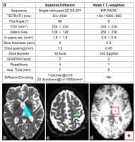 |
Diffusion MRI Tractography for Improved MRI-guided Focused
Ultrasound Thalamotomy Targeting for Essential Tremor
Qiyuan Tian1,2, Max Wintermark2, Kim
Butts Pauly2, Diane Huss3, W. Jeffrey
Elias4, and Jennifer A. McNab2
1Electrical Engineering, Stanford University,
Stanford, CA, United States, 2Radiology,
Stanford University, Stanford, CA, United States, 3Physical
Therapy, University of Virginia, Charlottesville, VA, United
States,4Neurosurgery, University of Virginia,
Charlottesville, VA, United States
We retrospectively studied 13 essential tremor patients
treated with MRI-guided focused ultrasound. The purpose was
to demonstrate the value of using diffusion MRI tractography
to help localize the ventral intermediate (Vim) nucleus of
the thalamus (the treatment target). Tractography between
the thalamus and hand-knob region of the motor cortex was
consistent from subject-to-subject and followed the expected
anatomy. The thalamic voxels with high tractography
streamline counts qualitatively matched the location of Vim
as depicted on the Schaltenbrand-Wahren Atlas. A trend was
found towards better treatment outcome scores with higher
pre-treatment probabilistic tractography streamline counts
within the visualized MRgFUS treatment-induced lesion.
|
| |
12:06
|
0824.
 |
MR-HIFU mild hyperthermia for sensitization of radiation and
chemotherapy for recurrent rectal cancer: First phase I clinical
trial results.
William Chu1, Robert Staruch2, Samuel
Pichardo3,4, Yuexi Huang5, Charles
Mougenot6, Matti Tillander7, Max O.
Köhler7, Mika Ylihautala7, Merrylee
McGuffin1, Gregory Czarnota1, and
Kullervo Hynynen5
1Radiation Oncology, Sunnybrook Health Science
Centre, Toronto, ON, Canada, 2Philips
Research, Cambridge, MA, United States, 3Thunder
Bay Regional Research Institute, Thunder Bay, ON, Canada, 4Electrical
Engineering, Lakehead University, Thunder Bay, ON, Canada, 5Physical
Sciences, Sunnybrook Research Institute, Toronto, ON,
Canada, 6Philips
Healthcare, Toronto, ON, Canada, 7Philips
Healthcare, Vantaa, Finland
We present the first results of a Phase I trial that
includes delivery of mild hyperthermia using magnetic
resonance-guided high intensity focused ultrasound (MR-HIFU)
combined with radiation and chemotherapy in the treatment of
locally recurrentrectal cancer. MR-HIFU mild hyperthermia
was delivered in three sessions (day 1, 8 and 15) during a
17-day treatment protocol (total dose 30.6 Gy combined with
fluropyrimidine-based chemotherapy). MR-HIFU mild
hyperthermia was successfully delivered and the procedure
was well tolerated by the patient. No adverse effects have
been reported 3 months after the treatment.
|
| |
12:18
|
0825.
 |
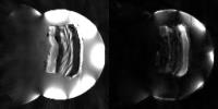
MRI-guided laser thermal ablation for T1a renal cell carcinoma
(RCC): A 4-year experience with longitudinal follow-up of
patients
Juan C. Camacho1,2, Nima Kokabi1,
Tracy E. Powell2, and Sherif G. Nour1,2
1Radiology and Imaging Sciences, Emory University
School of Medicine, Atlanta, GA, United States, 2Interventional
MRI Program, Emory University Hospital, Atlanta, GA, United
States
The objective of this study is to present outcomes of
MRI-guided laser ablation for early stage renal cell
carcinomas and to describe associated prognostic factors in
a consecutive cohort of patients with relative long-term
longitudinal follow-up. A prospective cohort of patients
presenting with pathology-confirmed RCC underwent MRI-guided
biopsy and subsequent laser ablation. Twenty-four
consecutive patients presenting with 35 RCC were recruited.
Follow-up MRI imaging was obtained in all cases with a
median follow-up period of 20 months. Of the different
analyzed prognostic factors, R.E.N.A.L nephrometry score was
the only one predicting the incidence of complications.
|
|