| |
10:30
 |
0846.
 |
Translating State-Of-The-Art Spinal Cord MRI Techniques To
Clinical Use: A Systematic Review Of Clinical Studies Utilizing
DTI, MT, MWF, MRS, and fMRI 
Allan R. Martin1, Izabela Aleksanderek1,
Julien Cohen-Adad2, Zenovia Tarmohamed3,
Lindsay Tetreault1, Nathaniel Smith4,
David W. Cadotte1, Adrian Crawley5,
Howard Ginsberg1, David J. Mikulis5,
and Michael G. Fehlings1
1Neurosurgery, University of Toronto, Toronto,
ON, Canada, 2Electrical
Engineering, Polytechnique Montreal, Montreal, QC, Canada, 3Royal
College of Surgeons Ireland, Dublin, Ireland, 4McMaster
University, Hamilton, ON, Canada, 5Medical
Imaging, University of Toronto, Toronto, ON, Canada
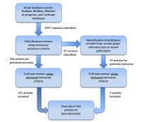
5 state-of-the-art spinal cord MRI techniques have been
identified with great clinical potential. This systematic
review finds trends in the technical methods employed and
measures the progress of these techniques toward clinical
translation. 104 studies were identified, with 69 DTI, 25
MT, 1 MWF, 11 MRS, and 8 fMRI studies. The DTI metric FA has
the strongest evidence of utility, correlating with
disability in numerous spinal conditions. Large,
well-designed studies with a priori hypotheses, standardized
acquisition methods, detailed clinical data collection, and
robust automated analysis techniques are needed to fully
demonstrate the potential of these rapidly evolving
techniques.
|
| |
10:42
|
0847.
 |
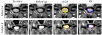
Combining biomechanical finite element analysis and
multi-parametric MRI to assess mechanical and structural damage
in cervical spondylotic myelopathy
Manuel Taso1,2,3,4, Pierre-Jean Arnoux2,4,
Léo Fradet4,5, Arnaud Le Troter1,3,
Jean-Philippe Ranjeva1,3,4, Kathia Chaumoître4,6,
Pierre-Hugues Roche4,7, and Virginie Callot1,3,4
1CRMBM UMR 7339, Aix-Marseille Université, CNRS,
Marseille, France, 2LBA
UMR T 24, Aix-Marseille Université, IFSTTAR, Marseille,
France, 3CEMEREM,
AP-HM, Pôle d'imagerie médicale, Marseille, France,4iLab-Spine
international associate laboratory, Marseille/Montréal,
France, 5Mechanical
Engineering, Ecole Polytechnique de Montréal, Montréal, QC,
Canada, 6Service
de Radiologie, Hôpital Nord, AP-HM, Pôle d'imagerie médicale,
Marseille, France, 7Service
de Neurochirurgie, Hôpital Nord, AP-HM, Trauma Center,
Marseille, France
While diagnosis of cervical spondylotic myelopathy is easily
done with MRI, patient outcome is still difficult to
predict. It is nonetheless associated to a strong mechanical
cause as spinal cord’s (SC) compression is the first event
leading to tissue alterations and neurological deficits.
This work proposes an original approach using biomechanical
numerical simulation, to apprehend the mechanisms of SC
compression by the disk, and multi-parametric MRI, to probe
the consequent microstructural alterations (axonal loss,
demyelination …). Thanks to spatial normalization, first
results on 3 patients are presented, allowing
co-localization of personalized simulation of mechanical
stress and structural MR alterations.
|
| |
10:54
 |
0848.
 |
Application of APT CEST in Cervical Spinal Cord Normal Appearing
White Matter of MS Patients at 3T
Samantha By1,2, Alex K. Smith1,2,
Adrienne N. Dula2,3, Bailey D. Lyttle2,
Siddharama Pawate4, and Seth A. Smith2,3
1Biomedical Engineering, Vanderbilt University,
Nashville, TN, United States, 2Vanderbilt
University Institute of Imaging Science, Vanderbilt
University, Nashville, TN, United States, 3Radiology
and Radiological Sciences, Vanderbilt University, Nashville,
TN, United States, 4Neurology,
Vanderbilt University, Nashville, TN, United States
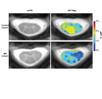
Amide proton transfer (APT) CEST was applied to healthy and
multiple sclerosis (MS) cohorts to determine its sensitivity
to changes in normal appearing white matter in MS. Using a
Lorentzian difference analysis, differences in the z-spectra
of the MS and healthy cohorts around the APT frequency
(Δω=+3.5 ppm) were observed. Significant differences in APT
effect between MS and healthy controls were seen in the
whole cord (p=0.0159), dorsal column (p=0.0159), and gray
matter (p=0.0317). Lastly, a group-wise analysis highlights
the ability to detect a decrease in mean APT effect in the
MS cohort, despite the difficulty in detecting lesions in
the anatomical.
|
| |
11:06
|
0849.
 |
Assessing Structure and Function of Myelin in Cervical
Spondylotic Myelopathy: Evidence of Focal Demyelination in the
Dorsal Column
Hanwen Liu1, Erin MacMillan 1,
Emil Ljungberg1, Burkhard Mädler2,
Shannon Kolind 1,
Marcel Dvorak1, David Li1, Alex MacKay1,
John Kramer1, Cornelia Laule1, and
Armin Curt3
1University of British Columbia, Vancouver, BC,
Canada, 2University
of Bonn, Bonn, Germany, 3Balgrist
University, Zürich, Switzerland
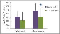
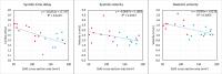
Cervical spondylotic myelopathy (CSM) is a major cause of
spinal cord dysfunction. To better understand the
pathophysiology underlying CSM, we used somatosensory evoked
potentials (SSEPs) and myelin water imaging to study
patients with CSM and healthy controls. Significant
differences were found in the myelin water fraction (MWF) of
the dorsal column between subjects classified as normal or
pathological based on SSEPs. A strong correlation between
tibial SSEP latency and MWF was found in CSM. Our findings
suggest that MWF can monitor cervical spinal cord
demyelination and may be a valuable tool to assess clinical
interventions in spinal cord injury.
|
| |
11:18
|
0850.
 |
Atrophy computation in the spinal cord using the Boundary Shift
Integral
Ferran Prados1,2, Marios C Yiannakas2,
Manuel Jorge Cardoso1, Francesco Grussu2,
Floriana De Angelis2, Domenico Plantone2,
David H Miller2, Olga Ciccarelli2,
Claudia Angela Michela Gandini Wheeler-Kingshott2,3,
and Sebastien Ourselin1
1Translational Imaging Group, Medical Physics and
Biomedical Engineering, University College London, London,
United Kingdom, 2NMR
Research Unit, Queen Square MS Centre, Department of
Neuroinflammation, UCL Institute of Neurology, University
College London, London, United Kingdom, 3Brain
Connectivity Center, C. Mondino National Neurological
Institute, Pavia, Italy
In this work, we introduce a new pipeline based on the
latest iteration of the BSI for computing atrophy in the SC
and compare its results with the most popular atrophy
measurements for this region, mean CSA. We demonstrated for
the first time the use of BSI in the SC, as a sensitive,
quantitative and objective measure of longitudinal tissue
volume change. The BSI pipeline presented in this work is
repeatable, reproducible and standardises a pipeline for
computing SC atrophy.
|
| |
11:30
|
0851.
 |
Comparison of cervical cerebrospinal fluid flow between healthy
controls and chronic spinal cord injury participants using cine
phase contrast MRI
Kwan-Jin Jung1, Andrea Willhite2, and
Susan Harkema2
1Radiology, University of Louisville, Louisville,
KY, United States, 2Neurological
Surgery, University of Louisville, Louisville, KY, United
States
The cerebrospinal fluid (CSF) flow in the cervical spine was
compared between healthy controls and persons with spinal
cord injury (SCI) using phase contrast MRI. The
subarachnoid cross-section of SCI participants was smaller
than that of healthy controls. The flow velocities in both
diastolic and systolic cardiac phases were faster in SCI
participants than that of healthy controls. Considering a
slower heart rate and a reduced ejection fraction and stroke
volume of the heart in SCI participants, the reduced
subarachnoid area may be a main contributing factor to the
increased velocity of CSF flow in SCI participants.
|
| |
11:42
|
0852.
 |
Quantitative measurements of the spinal cord blood flow of an
animal model of relapsing-remitting MS.
Mohamed Tachrount1, Andrew Davies2,
Roshni Desai2, Kenneth Smith2, David
Thomas1, and Xavier Golay1
1Dept. of Brain Repair and Rehabilitation, UCL
Institute of Neurology, London, United Kingdom, 2Dept.
of Neuroinflammation, UCL Institute of Neurology, London,
United Kingdom
Perfusion-weighted imaging studies have demonstrated that
there is a widespread cerebral hypoperfusion in patients
with MS, regardless of the clinical subtype. The mechanism
and the role of hypoxia are still unclear. The purpose of
this work was to longitudinally investigate the SC blood
flow (SCBF) during the different phases of disease
progression in EAE rats using an optimized ASL technique.
These measurements demonstrated for the first time on EAE
animal model that the neurological deficits are strongly
correlated with impaired blood flow.
|
| |
11:54
|
0853.
 |
A Prospective Longitudinal Study in Degenerative Cervical
Myelopathy Using Quantitative Microstructural MRI with
Tract-Specific Metrics
Allan R. Martin1, Benjamin De Leener2,
Izabela Aleksanderek1, Julien Cohen-Adad2,
David W. Cadotte1, Sukhvinder Kalsi-Ryan1,
Lindsay Tetreault1, Adrian Crawley3,
Howard Ginsberg1, David J. Mikulis3,
and Michael G. Fehlings1
1Neurosurgery, University of Toronto, Toronto,
ON, Canada, 2Electrical
Engineering, Polytechnique Montreal, Montreal, QC, Canada, 3Medical
Imaging, University of Toronto, Toronto, ON, Canada
This study investigates if DTI, MT, and T2*-weighted imaging
of the rostral cervical cord can 1) detect injury of WM
tracts, 2) correlate with global and focal disability, and
3) predict outcomes in degenerative cervical myelopathy
(DCM). Data includes detailed clinical assessments,
electrophysiology, and MRI, repeated at 1-year. Quantitative
MRI in 37 DCM patients and 29 healthy controls provided
reliable results and showed decreased CSA, FA, and MTR, and
increased T2* WM/GM ratio. FA of individual tracts
correlates well with clinical measures. Quantitative
multimodal assessment of WM injury with a clinically
feasible protocol is possible, with many potential clinical
applications.
|
| |
12:06
|
0854.
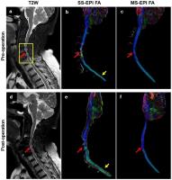 |
High Resolution Diffusion Tensor Imaging for Cervical
Spondylotic Myelopathy: A Preliminary Follow-up Study
Yuhui Xiong1, Xiaodong Ma1, Xiaolong
Chen2, Li Guan2, Yong Hai2,
Zhe Zhang1, Le He1, Chun Yuan1,3,
and Hua Guo1
1Center for Biomedical Imaging Research,
Department of Biomedical Engineering, School of Medicine,
Tsinghua University, Beijing, China, People's Republic of, 2Department
of Orthopedics, Beijing Chao-Yang Hospital, Capital Medical
University, Beijing, China, People's Republic of, 3Vascular
Imaging Laboratory, Department of Radiology, University of
Washington, Seattle, WA, United States
As a conventional method in spinal cord assessment in
cervical spondylotic myelopathy (CSM) patients,
intramedullary high signal intensity (HSI) in T2W images is
limited in diagnosis accuracy and predictive capacity for
postoperative recovery. Single-shot EPI DTI can detect
microstructural information, but it has low image resolution
and distortion. In this work, a multi-shot interleaved EPI
DTI using SYMPHONY reconstruction method is used to assess
the pathologic conditions and the function of spinal cords
of CSM patients quantitatively. The results show that the
high resolution MS-EPI DTI can performs better than HSI or
SS-EPI DTI in CSM diagnosis and recovery monitoring.
|
| |
12:18
|
0855.
 |
Diffusion Tensor Imaging Predicts Outcome ASIA Motor Scores in
Acute Traumatic C-Spinal Injury
Jiachen Zhuo1, Hegang Chen2, Bizhan
Aarabi3, Jay Menaker4, Rao Gullapalli1,
and Kathirkamanathan Shanmuganathan1
1Diagnostic Radiology and Nuclear Medicine,
University of Maryland School of Medicine, Baltimore, MD,
United States, 2Epidemiology
& Public Health, University of Maryland School of Medicine,
Baltimore, MD, United States, 3Neurosurgery,
University of Maryland School of Medicine, Baltimore, MD,
United States, 4Surgery,
University of Maryland School of Medicine, Baltimore, MD,
United States
Convention MRI is the imaging modality of choice to
demonstrate the anatomical location and extent in spinal
cord injury (SCI) following trauma. However, quantitative
and qualitative lesion parameters within the cord are of
limited use in predicting patient neurological outcomes. In
this study we demonstrated that acute DTI measurements
improve model prediction for 1 year AISA score following
blunt cervical SCI. Among all DTI measurements, axial
diffusivity, while not radial diffusivity, showed strong
effect in predicting outcome, indicating that axonal injury
in the cord may be the main factor affecting patient
recovery.
|
|