| |
08:00
|
1118.
 |
Reconstruction and validation of T2-weighted 4D Magnetic
Resonance Imaging for radiotherapy treatment planning 
Zdenko van Kesteren1, Daniël Tekelenburg1,2,
Oliver Gurney-Champion1,3, Aart Nederveen3,
Eelco Lens1, Astrid van der Horst1,
Aleksandra Biegun2, and Arjan Bel1
1radiotherapy, Academic Medical Centre,
Amsterdam, Netherlands, 2KVI-Center
for Advanced Radiation Technology, University of Groningen,
Groningen, Netherlands, 3radiology,
Academic Medical Centre, Amsterdam, Netherlands
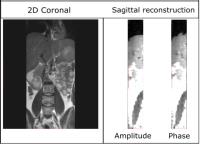
We developed a respiratory-correlated 4DMRI for abdominal
imaging by retrospective sorting 2D T2-weighted TSE images.
Each image is assigned to a respiratory state, which is
either binned in phase or the amplitude domain. The
diaphragm motion per image was determined by registering the
diaphragm to the begin-inhale image of a series. Per slice
and per bin multiple images were acquired and we defined the
intra-bin variation as the standard deviation of diaphragm
positions. Amplitude binning results in lower intra-bin
variation with respect to phase binning, 0.8 versus 2.4 mm
respectively.
|
| |
08:12
|
1119.
 |
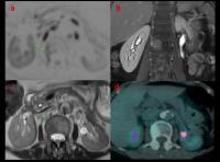
Clinical evaluation of ultra high field MRI for
three-dimensional visualization of tumour size in uveal melanoma
patients, with direct relevance to treatment planning.
Jan-Willem Beenakker1, Teresa Ferreira1,
Karina Soemarwoto1, Lorna Grech Fonk1,
Stijn Genders1, Wouter Teeuwisse1,
Andrew Webb1, and Gregorius Luyten1
1Leiden University Medical Centre, Leiden,
Netherlands
Recent advances in ocular MRI make it possible to acquire
high resolution three dimensional images of uveal melanoma
in eye tumour patients, allowing a much better assessment of
the maximal tumour prominence compared to conventional
clinical ultrasound measurements. Nine uveal melanoma
patients were examined on a 7 Tesla using a custom-built
eye-coil. Eye-motion artefacts were minimized by the use of
a cued-blinking protocol. For all patients the MR-images
showed a slightly lower tumour prominence. For two of these
patients this resulted in a substantial change in treatment
planning, saving an eye that would otherwise have been
removed.
|
| |
08:24
|
1120.
 |
Non-muscle-invasive and Muscle-invasive Bladder Cancer: Image
Quality and Clinical Value Compared between Reduced
Field-of-view DWI and Single-shot Echo-planar-imaging DWI
Yanchun Wang1, Zhen Li1, Daoyu Hu1,
and Xiaoyan Meng1
1radiology, Tongji hospital, Wuhan, China,
People's Republic of
Bladder cancer is the most common malignant tumor of the
urinary tract, and the incidence rate of bladder cancer is
6% for men and 2% for women. Clinical treatment of bladder
cancer depends on the level of muscle invasion (stage T2 or
higher) or non-invasion of the muscle of the bladder wall
(stage T1 or lower). Non-invasive tumors are mainly treated
with transurethral resection (TUR), whereas invasive tumors
are mainly treated with radical cystectomy . Therefore, it’s
important to precisely differentiate between
non-muscle-invasive and muscle-invasive bladder cancer
before treatment.
|
| |
08:36
|
1121.
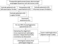 |
Precise staging of preoperative 3.0-T MR imaging for esophageal
carcinoma by using ex vivo MR imaging-matched pathologic
findings as the reference standard
Yi Wei1,2, Shao-Cheng Zhu1,2, Sen Wu1,2,
Da-Peng Shi1,2, and Dan-Dan Zheng3
1Radiology, Zhengzhou University People's
Hospital, Zhengzhou, China, People's Republic of, 2Henan
Provincial People's Hospital, Zhengzhou, China, People's
Republic of, 3GE
Healthcare,MR Research China, Beijing, China, People's
Republic of
Magnetic resonance imaging (MRI) was reported to evaluate
the esophageal layers invasion in vitro and demonstrated
that high-resolution T2-weighted imaging can clearly depict
8 layers of esophagus. However, former studies were mostly
carried on ultra-high-field scanner ex vivo, which can not
satisfy the need of preoperative staging and provide
essential information for clinic. In this study, an in vivo
experiment was conducted on 3.0T clinical scanner to
prospectively establish the MRI signal characteristics of
the normal esophageal wall and to assess the diagnostic
accuracy of high-resolution MR imaging for depicting the
depth of esophageal wall invasion by corresponding to ex
vivo MR imaging-matched certain histopathological slice.
|
| |
08:48
|
1122.
 |
Comparison of Whole-body MRI and PET-CT for staging adult
lymphoma: Preliminary result at 3.0T
Arash Latifoltojar1, Natacha Rosa1,
Maria Klusmann2, Mark Duncan2, Kirit
Ardeshna2, Jonathan Lambert2, Alan
Bainbridge2, Magdalena Sokolska2,
Sajir Mohamedbhai2, and Shonit Punwani1
1University College London, London, United
Kingdom, 2University
College London Hospital, London, United Kingdom
Whole body MRI (WB-MRI) offers a radiation-free imaging
technique for staging lymphoma. However, there are
conflicting reports concerning appropriate sequence(s) being
used in various WB-MRI protocols. In this work we
investigated diagnostic performance of different
morphological and functional MRI sequences as part of a
WB-MRI protocol.
|
| |
09:00
 |
1123.
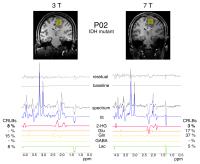 |
The benefits of in vivo 2-hydroxyglutarate detection using
semi-LASER at 7T and 3T: a comparative study
Adam Berrington1, Natalie Voets1,
Sarah J Larkin2, Nick de Pennington2,
James Mccullagh3, Khalid Al-Qahtani3,
Richard Stacey4, Peter Jezzard1,
Stuart Clare1, Christopher J Schofield3,
Olaf Ansorge2, Tom Cadoux-Hudson4,
Puneet Plaha4, and Uzay E Emir1
1FMRIB Centre, University of Oxford, Oxford,
United Kingdom, 2Nuffield
Department of Clinical Neurosciences, University of Oxford,
Oxford, United Kingdom, 3Department
of Chemistry, University of Oxford, Oxford, United Kingdom, 4Department
of Neurosurgery, University of Oxford, Oxford, United
Kingdom
We assess the ability of semi-LASER to detect
2-hydroxyglutarate (2-HG), a metabolic product of mutation
in the enzyme IDH, in gliomas at 3T and 7T. Robust detection
could lead to increased patient stratification yet is
hindered by signal overlap and compartmental artifacts. We
find semi-LASER (TE=110ms), with broadband adiabatic
refocussing, is able to correctly identify IDH-mutants at 3T
and 7T in a sample of six patients. Fitting errors are
greatly reduced at 7T and additional metabolites (GABA, Gly)
are detected in some IDH-mutated tumours. We conclude
semi-LASER provides a unique clinical opportunity for 2-HG
detection at both 3T and 7T.
|
| |
09:12
|
1124.
 |
ACRIN 6684: Multicenter, phase II assessment of tumor hypoxia in
newly diagnosed glioblastoma using magnetic resonance
spectroscopy - Permission Withheld
Eva-Maria Ratai1,2, Zheng Zhang3,
James Fink4, Mark Muzi4, Lucy Hanna3,
Erin Greco3, Todd Richards4, Akiva
Mintz5, Lale Kostakoglu6, Edward
Eikman7, Melissa Prah8, Benjamin
Ellingson9, Kathleen Schmainda8,
Gregory Sorensen1,2, Daniel Barboriak10,
David Mankoff11, and Elizabeth Gerstner12
1Radiology, Massachusetts General Hospital,
Boston, MA, United States, 2A.
A. Martinos Center for Biomedical Imaging, Charlestown, MA,
United States, 3Brown
University, Providence, RI, United States,4University
of Washington, Seattle, WA, United States, 5Wake
Forest University, Winston-Salem, NC, United States, 6Mt.
Sinai Medical Center, New York, NY, United States, 7Moffitt
Cancer Center, Tampa, FL, United States, 8Medical
College of Wisconsin, Milwaukee, WI, United States, 9UCLA
Medical Center, Los Angeles, CA, United States, 10Duke
University, Durham, NC, United States, 11University
of Pennsylvania, Philadelphia, PA, United States, 12MGH
Cancer Center, Massachusetts General Hospital, Boston, MA,
United States
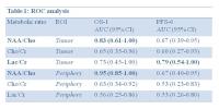
The Phase II multi-center trial ACRIN 6684 conducted by the
American College of Radiology Imaging Network was designed
to assess tumor hypoxia in newly diagnosed glioblastoma
(GBM) using [18F] Fluoromisonidazole (FMISO)-PET and MRI.
Data from magnetic resonance spectroscopic imaging (MRSI)
were available on 17 participants from four sites. The MRS
marker of tumor burden (NAA/Cho) was a significant predictor
of one-year survival (OS-1). Furthermore, the MRS marker of
tumor hypoxia (Lac/Cr) was a significant predictor of
six-month progression-free-survival (PFS-6) using receiver
operating characteristic (ROC) analysis.
|
| |
09:24
|
1125.
 |
Texture analysis of hepatocellular carcinomas in
Contrast-enhanced MR images for malignant differentiation
Wu Zhou1, Kaixin Wang1, Lijuan Zhang1,
Zaiyi Liu2, Guangyi Wang2, and
Changhong Liang2
1Key Laboratory for Health Informatics, Shenzhen
Institutes of Advanced Technology, Shenzhen, China, People's
Republic of, 2Department
of Radiology, Guangdong General Hospital, Guangdong Academy
of Medical Sciences, Shenzhen, China, People's Republic of
Lesion characterization based on imaging features is
essential to the successful treatment of hepatocellular
carcinomas (HCC). In this work, we investigate the malignant
of HCC from Contrast-enhanced MR images based on the
analysis of texture features. Our study demonstrated that
the texture feature (average intensity value and grey level
nonuniformity) of HCC in contrast-enhanced MR images was a
good predictor to characterize the malignant of HCC. By
quantitatively comparing the texture parameters in well
differentiated and moderately differentiated HCCs, the
values of average intensity remarkably decreased and GLN
significantly increased according to the increasing degree
of malignant for HCCs.
|
| |
09:36
|
1126.
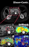 |
A feasibility study to perform combined MR Elastography, IVIM
and DCE-MRI in pancreatic cancer patients. - Permission Withheld
Jurgen H Runge1, Remy Klaassen2,
Oliver J Gurney-Champion1,3, Hanneke WM van
Laarhoven2, Ralph Sinkus4, Aart J
Nederveen1, and Jaap Stoker1
1Radiology, Academic Medical Center, Amsterdam,
Netherlands, 2Medical
Oncology, Academic Medical Center, Amsterdam, Netherlands, 3Radiation
Oncology, Academic Medical Center, Amsterdam, Netherlands, 4Biomedical
Engineering, King's College London, London, United Kingdom
Pancreatic cancer remains one the most deadly cancers. New
therapeutic agents cause confusion as prior morphological
criteria to determine the presence of a response appear to
be unreliable. MR Elastography (MRE) is uniquely able to
determine tissue stiffness, a property potentially useful
for therapy response monitoring. Here we present our first
preliminary results of combining MRE with IVIM and DCE MRI.
|
| |
09:48
|
1127.
 |
Estimate of liver functional reserve using T1 mapping on
Gd-EOB-DTPA-enhanced MRI in HCC patients
Chenyang Chen1, Jie Chen1, Chunchao
Xia1, Panli Zuo2, and Bin Song1
1Department of Radiology, West China Hospital,
Sichuan University, Chengdu, China, People's Republic of, 2Siemens
Healthcare, MR Collaboration NE Asia, Beijing, China,
People's Republic of
We found that liver tumors showed clearer borderline on T1
map during hepatobiliary phase of Gd-EOB-DTPA enhanced MRI.
A significant correlation between the reduction rate of
liver parenchyma with ICG-15 indicated that the T1 mapping
is useful to estimate the liver functional reserve in HCCs
patients.
|
|