|
|
13:30
|
0438.
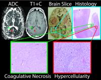 |
Progressing bevacizumab induced diffusion restriction is
associated with coagulative necrosis surrounded by viable tumor
and decreased overall survival in recurrent glioblastoma
patients 
Ha Son Nguyen1, Nelson Milbach2, Sarah
L Hurrell2, Elizabeth Cochran3,
Jennifer Connelly4, Mona Al-Gizawiy2,
Joseph Bovi5, Scott D Rand2, Kathleen
M Schmainda2, and Peter S. LaViolette2,6
1Neurosurgery, Medical College of Wisconsin,
Milwaukee, WI, United States, 2Radiology,
Medical College of Wisconsin, Milwaukee, WI, United States, 3Pathology,
Medical College of Wisconsin, Milwaukee, WI, United States, 4Neurology,
Medical College of Wisconsin, Milwaukee, WI, United States, 5Radiation
Oncology, Medical College of Wisconsin, Milwaukee, WI,
United States, 6Biophysics,
Milwaukee, WI, United States
It is the standard of care to initiate bevacizumab therapy
for patients with recurrent glioblastoma. Some patients
develop areas of diffusion restriction on diffusion imaging
following the onset of therapy. We recruited five patients
with this condition to donate their brains postmortem. A
histological analysis was performed and compared to MR
images to discover what caused the diffusion restriction.
It was found to be coagulative necrosis surrounded by viable
hypercellular tumor. A second population study shows that
patients with progressively expanding diffusion restriction
had a significantly lower survival compared to those
without.
|
|
|
13:42
|
0439.
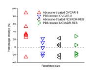 |
In vivo quantification of antimitotic-treatment-induced
microstructural changes using temporal diffusion
xiaoyu jiang1, hua li1, jingping Xie1,
ping zhao1, junzhong xu1, dineo
khabele2, and John Gore1
1vanderbilt university institute of imaging
science, nashville, TN, United States, 2vanderbilt
university, nashville, TN, United States
Reliable and sensitive methods for assessing the response of
tumors to treatment are critical in rapid selection of the
most appropriate therapy for individual patients, and
development of novel therapies. Temporal diffusion
spectroscopy, which measures the variation of apparent
diffusion coefficient (ADC) over a range of effective
diffusion times, is proposed to measure tumor
microstructural variations in response to chemotherapy. The
proposed method is shown to detect the increase in cell size
in response to the antimitotic-treatment in both
well-characterized cell culture and solid tumors in vivo.
The MR observations are supported by flow cytometric,
microscopic, and histological analysis.
|
|
|
13:54
|
0440.
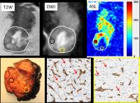 |
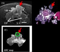
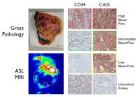
Quantitative Arterial Spin Labeled (ASL) Perfusion and Diffusion
Weighted Imaging (DWI) in Clear Cell Renal Cell Carcinoma:
Correlation with Heterogeneous Tumor Vascularity and Cellularity
at Histopathology
Qing Yuan1, Payal Kapur2,3, Yue Zhang1,
Yin Xi1, Sabina Signoretti4, Ananth
Madhuranthakam1,5, Ivan E Dimitrov5,6,
Jeffrey A Cadeddu1,3, Vitaly Margulis3,
and Ivan Pedrosa1,5
1Radiology, UT Southwestern Medical Center,
Dallas, TX, United States, 2Pathology,
UT Southwestern Medical Center, Dallas, TX, United States, 3Urology,
UT Southwestern Medical Center, Dallas, TX, United States, 4Pathology,
Brigham and Women's Hospital, Boston, MA, United States, 5Advanced
Imaging Research Center, UT Southwestern Medical Center,
Dallas, TX, United States, 6Philips
Medical Systems, Cleveland, OH, United States
We investigated intratumor heterogeneity of perfusion and
diffusion in
vivo using
ASL and DWI in clear cell renal cell carcinoma (ccRCC), and
correlated these measures with tumor vascularity and
cellularity at histopathology. Focused histopathologic
analysis of tumor areas corresponding to high perfusion
regions on ASL confirmed higher microvessel density (MVD)
and demonstrated higher cellularity compared to tumor areas
with low perfusion on ASL. A negative correlation between
MRI diffusion measures and tissue cellularity further
supports noninvasive MRI techniques as potential imaging
biomarker in ccRCC for assessment of heterogeneity in tumor
angiogenesis and microenvironment in
vivo.
|
|
|
14:06
|
0441.
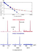 |
Apparent diffusion coefficient of hyperpolarized lactate reports
on lactate production and efflux in renal cell carcinomas
Renuka Sriram1, Bertram Koelsch1,
Jeremy W Gordon1, Mark Van Criekinge1,
Celine Baligand1, Robert A Bok1, Dan B
Vigneron1, Kayvan R Keshari2, Peder E
Larson1, Zhen Jane Wang1, and John
Kurhanewicz1
1University of California, San Francisco, San
Francisco, CA, United States, 2Memorial
Sloan-Kettering Cancer Center, New York, NY, United States
This study demonstrated that diffusion weighted HP 13C
MRI can provide an estimate of the amount of extra- versus
intracellular HP 13C
lactate based on its apparent diffusion coefficient (ADC).
In metastatic renal cell carcinoma, a large portion of the
HP 13C
lactate signal arises from an extracellular lactate pool,
based on reliable estimates of ADC in the same cell line in
a the MR compatible bioreactor. The juxtaposition of cells
in bioreactor and the in vivo animal model is a powerful
tool for interpretation of the hyperpolarized ADC
measurements. This unique combination can be further
extended to investigate the relationship between lactate
transport and tumor metastatic potential.
|
|
|
14:18
|
0442.
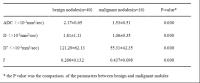 |
Diagnostic value of intravoxel incoherent motion (IVIM) for differentiating benign and malignant thyroid nodules
hui Tan1, jun CHEN1, YUN-fei ZHA1,
liang ZHANG1, jing LU1, Chang-sheng
LIU1, and hui LIN2
1Renmin Hospital of Wuhan University, wuhan,
China, People's Republic of, 2GE
healthcare, shanghai, China, People's Republic of
To preliminary explore the value of intravoxel incoherent motion
(IVIM) in the differention between benign and malignant thyroid
lesions, 45 patients with 56 thyroid nodules underwent
preoperative IVIM (b –1000 s/mm2). Data was
postprocessed by IVIM model for quantitation of apparent
diffusion coefficient (ADC), perfusion fraction f, diffusivity D
and pseudo diffusivity D*. Significant intergroup difference was
observed in ADC, D, D*, and f, the f value is the most valuable
parameter in identifying the malignant from benign nodules. The
IVIM sequence has potential to differentiate the benign from
malignant thyroid nodules.
|
|
|
14:30
 |
0443.
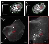 |
Combined MRI and optical CT imaging of tumour vasculature in a
preclinical model of neuroblastoma
Ciara M McErlean1, Yann Jamin1,
Jessica KR Boult1, Alexander Koers1,
Laura S Danielson1, David J Collins1,
Martin O Leach1, Simon P Robinson1,
and Simon J Doran1
1Institute of Cancer Research, London, United
Kingdom
This study compares MRI functional measurements of the
vasculature in a preclinical model of neuroblastoma with ex
vivo optical
CT high-resolution 3D imaging of the functional vasculature
using India ink staining. MRI showed a heterogeneously
perfused tumour with high fractional blood volume and vessel
size index, characteristic of hypervascular neuroblastoma.
The high resolution optical CT images allowed visualisation
of individual vessels and corroborated the MRI findings.
With improved registration, optical CT could help validate
MRI functional biomarkers of the vasculature and accelerate
both our understanding of vessel biology and the evaluation
of vascular-targeted treatment in cancer and other
vascular-related pathologies.
|
|
|
14:42
 |
0444.
 |
Exploring the Relationship between MR-derived Apparent Diffusion
Coefficient, Cellularity, and Extracellular Porosity: A
Preliminary Animal Study in Prostate Cancer
Deborah K. Hill1,2, Andreas Heindl3,
Daniel N. Rodrigues3, Øystein Størkersen2,
Yinyin Yuan3, Siver A. Moestue 1,2,
Martin O. Leach3, Tone F. Bathen1,
David J. Collins3, and Matthew D. Blackledge3
1Norwegian University of Science and Technology,
Trondheim, Norway, 2St.
Olavs University Hospital, Trondheim, Norway, 3The
Institute of Cancer Research and Royal Marsden NHS
Foundation Trust, London, United Kingdom
An increased ADC can imply reduced cellularity; DWI is
considered a useful tool for assessing tumour treatment
response, although there is little validation of this
relationship in cancer. We compared ADC, cellularity, and
extracellular porosity using a transgenic adenocarcinoma of
the mouse prostate model. ADC values were derived from DWI
data, and cellularity was assessed from histology using
novel visualisation and segmentation tools. We investigated
the relationship between extracellular porosity and ADC, and
validated our findings using cell segmentation analysis of
histology slides. This analysis is useful to inform on
tissue cellularity for cases where histology samples are not
available.
|
|
|
14:54
|
0445.
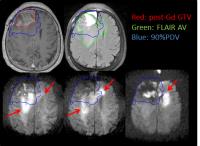 |
Non-enhanced Hypercellular Volume in Glioblastoma identified by
High b-value Diffusion Weighted Imaging
Yue Cao1,2, Daniel Wahl1, Priyanka
Pramanik1, Michelle Kim1, Theodore S
Lawrence1, and Hemant Parmar2
1Radiaiton Oncology, University of Michigan, Ann
Arbor, MI, United States, 2Radiology,
University of Michigan, Ann Arbor, MI, United States
It is a challenge to differentiate non-enhanced components
of glioblastoma (GBM) from edema and normal tissue using
conventional MRI. The ill-differentiation could lead to
inadequate treatment for GBM by surgery and radiation
therapy. This study evaluated the enhanced and non-enhanced
hypercellular volume (HCV) of GBM identified by high b-value
diffusion weighted (DW) imaging with gross tumor volume
defined on post-Gd T1 weighted images, abnormality volume on
T2 FLAIR images, high dose coverage planned according to
conventional MRI, and progression. This study found that
the HCV was an aggressive component of GBM and predicted
progression free survival.
|
|
|
15:06
|
0446.
 |
Differential tumor perfusion in vivo on Arterial Spin Labeled
MRI correlates with heterogeneity in the molecular phenotype of
clear cell Renal Cell Carcinoma
Manoj Bhasin1, Rupal Bhatt2, Phillip M
Robson3, Deepa Rajamani1, Sabina
Signoretti4, David C Alsop3, and Ivan
Pedrosa5
1Division of Interdisciplinary Medicine &
Biotechnology, and Genomics, Proteomics, Bioinformatics and
Systems Biology Center, Department of Medicine, Beth Israel
Deaconess Medical Center, Boston, MA, United States, 2Division
of Hematology and Oncology, Beth Israel Deaconess Medical
Center, Boston, MA, United States, 3Department
of Radiology, Beth Israel Deaconess Medical Center, Boston,
MA, United States,4Pathology, Brigam and Women's
Hospital, Boston, MA, United States, 5Radiology,
UT Southwestern Medical Center, Dallas, TX, United States
We used Arterial Spin Labeled (ASL) MRI to explore the
association between heterogeneous in vivo perfusion in clear
cell renal cell carcinoma (ccRCC) and the underlying genomic
profile to identify key genes linked to tumor angiogenesis.
Ephrin-A5 (EFNA5) expression correlated with ASL perfusion
(R2 = 0.504, P value= .002) and exhibited highest
significant differences between low and high perfusion (Fold
Change = 2.88, P value < 0.02). Higher expression of EFNA5
is associated with poor 3 and 5 years survival (P = 0.0009).
We propose MRI-based targeted tissue sampling to
characterize the heterogeneous genetic alterations driving
angiogenesis in ccRCC.
|
|
|
15:18
|
0447.
 |
DCE-MRI High-resolution Metabolic Prostate Imaging is
Insensitive to AIF Uncertainty
Xin Li1, Mark G. Garzotto2,3, Fergus
V. Coakley4, Brendan Moloney1, William
J. Woodward1, Yiyi Chen5, Wei Huang1,
William D. Rooney1, and Charles S. Springer, Jr.1
1Advanced Imaging Research Center, Oregon Health
& Science University, Portland, OR, United States, 2Portland
VA Medical Center, Portland, OR, United States, 3Urology,
Oregon Health & Science University, Portland, OR, United
States, 4Department
of Diagnostic Radiology, Oregon Health & Science University,
Portland, OR, United States, 5Division
of Biostatistics, Dept. of Public Health and Preventive
Medicine, Knight Cancer Institute, Oregon Health and Science
University, Portland, OR, United States
Accurate arterial input function (AIF) measurement in
Dynamic Contrast Enhanced MRI (DCE-MRI) remains challenging.
This hinders DCE-MRI’s wider adoption. Since the contrast
reagent (CR) is detected indirectly through water proton R1 relaxation
rate constant change, DCE-MRI intrinsically works as a
dual-probe (CR and water) method. In this study, we
demonstrate that while the common pharmacokinetic parameters
associated with CR extravasation are highly sensitive to AIF
accuracy, the transcytolemmal water exchange parameter is
not. With the recent correlation of water exchange kinetics
and cellular metabolic activity, this current work
demonstrates the practicability of high-resolution
metabolic imaging of the prostate.
|
|