| |
13:30
|
0398.
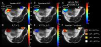 |
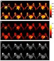
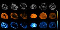
Assessment of tumor perfusion, oxygenation, and metabolism using
DCE, BOLD, and hyperpolarized 13C MRI in a mouse model of breast
cancer 
Erin B Adamson1, Roberta M Strigel1,2,3,
David J Niles1, Kai D Ludwig1, Ben L
Cox1,4,5, Amy R Moser2,6, and Sean B
Fain1,3,7
1Medical Physics, University of
Wisconsin-Madison, Madison, WI, United States, 2Carbone
Cancer Center, University of Wisconsin-Madison, Madison, WI,
United States, 3Radiology,
University of Wisconsin-Madison, Madison, WI, United States, 4Morgridge
Institute for Research, Madison, WI, United States, 5Laboratory
for Optical and Computational Instrumentation, University of
Wisconsin-Madison, Madison, WI, United States, 6Human
Oncology, University of Wisconsin-Madison, Madison, WI,
United States, 7Biomedical
Engineering, University of Wisconsin-Madison, Madison, WI,
United States
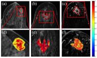
Hyperpolarized (HP) 13C
MRSI, dynamic contrast-enhanced (DCE) MRI, and
blood-oxygen-level dependent (BOLD) MRI have the potential
to non-invasively characterize tumor metabolism, perfusion,
and oxygenation, respectively, and aid in the development of
individualized treatment plans for cancer patients. However,
a regional comparison of these non-invasive techniques for
probing the tumor microenvironment has not been explored.
This work aims to test the feasibility of performing
quantitative, spatial analysis and comparison of HP 13C
MRSI and BOLD and DCE MRI in a murine breast cancer model.
|
| |
13:42
|
0399.
 |
3D Magnetic Resonance Fingerprinting for Quantitative Breast
Imaging
Yong Chen1, Shivani Pahwa1, Jesse
Hamilton2, Sara Dastmalchian1, Donna
Plecha3, Nicole Seiberlich2, Mark
Griswold1, and Vikas Gulani1
1Department of Radiology, Case Western Reserve
University, Cleveland, OH, United States, 2Department
of Biomedical Engineering, Case Western Reserve University,
Cleveland, OH, United States, 3Department
of Radiology, University Hospitals Case Medical Center,
Cleveland, OH, United States
In this study, a rapid relaxometry method was developed for
breast imaging using the MRF technique, which allows
simultaneous and volumetric quantification of T1 and
T2 relaxation
times for breast tissues.
|
| |
13:54
|
0400.
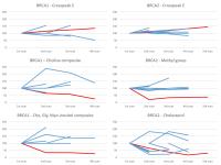 |
Breast tissue lipid and metabolite deregulation precedes
malignant transformation in women with BRCA gene mutations: a
longitudinal study
Gorane Santamaria1, Jessica Buck2,3,
Leah Best4, David Clark5, Judith
Silcock5, Peter Lau4, Saadallah
Ramadan6, Scott Quadrelli3,7, Peter
Malycha3, and Carolyn Mountford3
1Hospital Clinic de Barcelona, Barcelona, Spain, 2Oxford
University, Oxford, United Kingdom, 3Translational
Research Institute, Brisbane, Australia, 4Hunter
New England Area Health, Newcastle, Australia, 5The
Breast and Endocrine Centre, Gateshead, Gateshead,
Australia, 6University
of Newcastle, Australia, Newcastle, Australia, 7Queensland
University of Technology, Brisbane, Australia
Women carrying the BRCA1 and BRCA2 gene mutations exhibited
lipid and metabolite profiles consistent with very early
deregulation recorded earlier in cancer cell models. The
deregulation was different for BRCA1 and BRCA2. Here we
report a longitudinal study where these same women are
monitored every six month using the L-COSY MRS method and
every 12 month with contrast enhanced MRI. For most women
in the study the biomarkers remained relatively stable over
time. Of the 6 BRCA1 and 10 BRCA2 patients examined, one
BRCA1 patient and one BRCA2 patient showed further
deregulation.
|
| |
14:06
|
0401.
 |
Fat-Based Registration of Breast DCE Water Images
Subashini Srinivasan1, Brian A Hargreaves1,
and Bruce L Daniel1
1Radiology, Stanford University, Stanford, CA,
United States
Three-dimensional breast dynamic contrast-enhanced imaging
is susceptible to deformable motion and affects both
semi-quantitative and pharmacokinetic parameters. B-Spline
motion registration with a mutual information metric is
often used to register DCE images but is sometimes
susceptible to introduction of new motion. Here we have
introduced a fat-based motion registration, using a
mean-squared-difference signal metric, to register the water
images without introducing new motion. The acquired images
and both registration methods were qualitatively assessed in
16 breasts. Voxel-by-voxel pharmacokinetic mapping was also
performed in 21 tumors. Our results show that fat-based
registration can be used to register the water images with
improved image quality and reduced errors in quantification.
|
| |
14:18
|
0402.
 |
gagCEST imaging in patients with breast tumors at 7 Tesla -
preliminary results
Olgica Zaric1, Katja Pinker-Domenig2,3,
Esau Poblador 1,
Vadimir Mlynarik1, Thomas Helbich4,
Siegfrid Trattnig1,5, and Wolfgang Bogner1
1High Field Magnetic Resonance Centre, Medical
University of Vienna, Vienna, Austria, 2Department
of Biomedical Imaging and Image-guided Therapy, Medical
University Vienna, Vienna, Austria, 3Molecular
Imaging and Therapy Service, Memorial Sloan Kettering Cancer
Center, New York, NY, United States, 4Department
of Biomedical Imaging and Image-guided Therapy, Medical
University of Vienna, Vienna, Austria,5Christian
Doppler Lab for Clinical Molecular MRI, Christian Doppler
Forschungsgesellschaft, Vienna, Austria, Vienna, Austria
Proteoglycans content in malignant tumors may provide
information regarding the altered metabolism and neoplastic
cell behavior. The aim of this study was to investigate the
feasibility of gagCEST imaging in patients with breast
tumors at 7 Tesla. Eleven patients with 15 lesions were
examined. gagCEST imaging was performed with 1.7mm in-plane
resolution and nine minutes of measurement time. Results
based on MTRasym showed excellent differentation between
malignant and benign lesions (CI=95%, p=0.001) and
insignificant difference between benign and healthy tissue
(CI=95%, p=0.159). gagCEST has a great potential in breast
tumors evaluations providing substantially different
information obtained with standard MRI techniques.
|
| |
14:30
|
0403.
 |
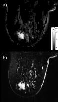
Directional-gradient based radiogenomic descriptors on DCE-MRI
appear to distinguish different PAM50-identified subtypes of
HER2+ Breast Cancer
Prateek Prasanna1, Nathaniel Braman1,
Salendra Singh1, Donna Plecha2, Hannah
Gilmore2, Lyndsay Harris2, Tao Wan3,
Vinay Varadan1, and Anant Madabhushi1
1Case Western Reserve University, Cleveland, OH,
United States, 2University
Hospitals, Cleveland, OH, United States, 3Beihang
University, Beijing, China, People's Republic of
We present the initial results of using a novel
radiogenomic descriptor, CoLlAGe, on breast DCE-MRI to
identify associations with HER2+ breast cancer subtypes.
Current method involves using a PAM50 assay to analyze
primary tumor tissues. CoLlAGe is a quantitative measurement
of the degree of order/disorder of localized image gradient
orientations. We extract CoLlAGe entropy from the regions of
interest. Unsupervised hierarchical clustering of the
entropy statistics show that we can segregate the cohort
into three distinct subtypes (enriched, basal and luminal),
as identified by PAM50 assay. CoLlAGe resulted in higher
clustering accuracy as compared to pharmacokinetic
parameters and signal intensities.
|
| |
14:42
|
0404.
 |
Rapid high-resolution sodium relaxometry in human breast
Glen Morrell1, Josh Kaggie2, Matthew
Stein1, Scott Parker1, and Neal
Bangerter3
1Radiology, University of Utah, Salt Lake City,
UT, United States, 2Radiology,
University of Cambridge, Cambridge, United Kingdom, 3Electrical
and Computer Engineering, Brigham Young University, Provo,
UT, United States
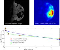
We have performed rapid high-resolution breast sodium MRI
relaxometry using a custom sodium breast phased array coil.
Clear delineation of short- and long-T2* components of the
sodium signal is possible with a spatial resolution of 3.75
x 3.75 x 4mm over the entire breast with a total imaging
time of under 10 min. This method will allow the
investigation of the potential of sodium relaxometry to
improve the specificity of breast MRI for the detection of
breast cancer.
|
| |
14:54
|
0405.
 |
The performance of MRI screening in the detection of breast
cancer in an intermediate and high risk screening program
Suzan Vreemann1, Albert Gubern-Merida1,
Susanne Lardenoije1, Nico Karssemeijer1,
and Ritse M. Mann1
1Radiology and Nuclear Medicine, Radboudumc,
Nijmegen, Netherlands
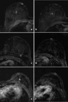
Women at increased risk for breast cancer require annual
mammography and MRI. The purpose of this study is to
evaluate cancers detected in MRI screening and assess the
visibility on prior MRI-examinations. MRI-scans of breast
cancers detected in our MRI screening program were
re-evaluated and lesions on the diagnostic MRI and prior MRI
were scored according to Breast Imaging Reporting and Data
(BI-RADS) MR-lexicon. The visibility of the lesions on the
prior MRI was rated as visible, minimal sign and invisible.
Our results show that almost one third of the breast cancers
should have been recalled based on consensus review.
|
| |
15:06
|
0406.
 |
Comparison of Conventional DCE-MRI and a Novel Golden-Angle
Radial Compressed-Sensing and Parallel Imaging Method for the
Evaluation of Breast Lesion Conspicuity and Morphology
Laura Heacock1, Yiming Gao1, Samantha
Heller1, Amy Melsaether1, Sungheon Kim1,2,
and Linda Moy1
1Bernard and Irene Schwartz Center for Biomedical
Imaging, Department of Radiology, New York University School
of Medicine, New York, NY, United States, 2Center
for Advanced Imaging Innovation and Research (CAI2R), New
York University School of Medicine, New York, NY, United
States
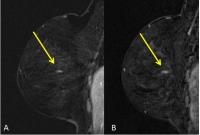
GRASP DCE-MRI (Golden-angle Radial Sparse Parallel) DCE-MRI
allows simultaneous high spatial and temporal resolution.
The purpose of this study was to evaluate breast lesion
conspicuity between GRASP and conventional Cartesian
sampling DCE-MRI. Readers assessed conspicuity of 48
biopsy-proven lesions on conventional DCE-MRI and subsequent
GRASP biopsy. No significant difference was found between
the two techniques for all lesions (p=0.21, p=0.19, p=0.46),
masses (p=1.0, p=0.48, p=0.7) or NME (p=0.18,p=0.08,
p=0.64). There was strong reader agreement in evaluating
conspicuity (ICC=0.735). GRASP DCE-MRI is comparable to
conventional DCE-MRI imaging for masses and NME with
diagnostic-quality high spatial resolution and flexibility
of temporal resolution.
|
| |
15:18
 |
0407.
 |
Different anti-angiogenic drugs have different effects on the
relationship between vascular structure and function in a
patient-derived breast cancer model
Eugene Kim1, Jana Cebulla1, Astrid
Jullumstrø Feuerherm2, Berit Johansen2,
Olav Engebråten3, Gunhild Mari Mælandsmo3,
Tone Frost Bathen1, and Siver Andreas Moestue1
1MR Cancer Group, Department of Circulation and
Medical Imaging, Norwegian University of Science and
Technology, Trondheim, Norway, 2Avexxin
AS, Department of Biology, Norwegian University of Science
and Technology, Trondheim, Norway, 3Department
of Tumor Biology, Institute for Cancer Research, Oslo
University Hospital, Oslo, Norway
This study investigated the relationship between tumor
vascular function (DCE-MRI) and structure (ex vivo
micro-CT). Control tumors did not exhibit any significant
correlations between micro-CT and DCE-MRI parameters. Tumors
treated with bevacizumab or a cPLA2 inhibitor (AVX235), both
anti-angiogenic drugs, displayed reduced perfusion and
vascularization. But interestingly, there was a significant
positive correlation between vascular surface area and
Ktrans in AVX235-treated tumors, whereas the corresponding
correlation was negative in bevacizumab-treated tumors. This
suggests that different therapies can differentially
modulate the vascular structure-function relationship, which
highlights the challenge in interpreting DCE-MRI
measurements and adopting them as clinical biomarkers of
therapeutic response.
|
|