| |
16:00
 |
0516.
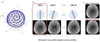 |
“Windowed” Composite Reconstruction Improves Rotating Short-Axis
High-Resolution DWI (RSA-DWI) in both Simulation and Human data 
Qiuting Wen1, Chandana Kodiweera2, and
Yu-Chien Wu1
1Radiology and Imaging Sciences, Indiana
University, Indianapolis, IN, United States, 2Darmonth
College, Hanover, NH, United States
High-resolution DWI often relies on multi-shot acquisitions,
which suffer from long acquisition time and motion-related
phase issues. However, highly correlated information exists
in DWIs as they are weighting the same structure. To take
advantages of this feature, rotating short-axis DWI was
proposed to accelerate DWI acquisition by acquiring only one
rotating blade per diffusion direction. In the previous
reconstruction, high-resolution DWI was achieved by
integrating the full set of DWIs. In this work, we propose a
“windowed” composite reconstruction where only a subset of
DWIs was selected to reconstruct each high-resolution DWI.
Improved image quality was appreciated in both simulation
and human data.
|
| |
16:12
|
0517.
 |
Multi-frequency reconstruction for frequency-modulated
stack-of-stars bSSFP
Anne Slawig1, Tobias Wech1, Valentin
Ratz1, Johannes Tran-Gia1,2, Henning
Neubauer1, Thorsten Bley1, and Herbert
Köstler1
1Departement for Diagnostic and Interventional
Radiology, University of Würzburg, Würzburg, Germany, 2Department
of Nuclear Medicine, Würzburg, Germany
Banding artefacts in images acquired by bSSFP are a big
challenge in fast MRI as they can considerably reduce image
quality and deteriorate the diagnostic value. As the steady
state tolerates small shifts in frequency, it is possible to
acquire a frequency-modulated bSSFP. Unfortunately a simple
gridding reconstruction of such a measurement suffers from
signal loss. Our study proposes a multi-frequency
reconstruction and demonstrates its capability of
reconstructing banding-free 3D images while retaining the
high signal levels of standard bSSFP.
|
| |
16:24
|
0518.
 |
Distortion correction of Golden Angle radial images with GIRF-predicted
k-space trajectories using the gradient waveform history - Permission Withheld
Adrienne E Campbell-Washburn1, Robert J Lederman1,
Anthony Z Faranesh1, and Michael S Hansen1
1Cardiovascular and Pulmonary Branch, Division of
Intramural Research, National Heart, Lung, and Blood
Institute, National Institutes of Health, Bethesda, MD,
United States
Balanced SSFP Golden Angle radial imaging uses a rapidly
varying gradient scheme and thus is susceptible to image
distortion caused by gradient delays and eddy currents. We
propose that storing a history of the gradient waveforms in
each axis can enable us to better predict our true k-space
coordinates during sampling. We use the gradient system
impulse response function to predict k-space coordinates and
demonstrate reduced image distortion (shading and streaking)
in a phantom and in vivo when utilizing the gradient
waveform history. This method will be useful for dynamic and
real-time imaging with Golden Angle balanced SSFP imaging
schemes.
|
| |
16:36
|
0519.
 |
Cosine-modulated acquisition cleans spectra for better
respiratory cine
Cihat Eldeniz1, Yasheng Chen1, and
Hongyu An1
1Washington University in St. Louis, St. Louis,
MO, United States
Breath-hold or navigator-based MR acquisition has been
widely used to remove the effect of motion from the images.
However, breath holding can be challenging for patients. On
the other hand, navigator-based methods suffer from
lengthened acquisition time and the disturbance of
magnetization history. In this respect, we will developed a
self-gated free-breathing MR imaging method to obtain 4D MRI
(3D spatial+1D respiratory phases) for deformable motion
derivation.
|
| |
16:48
|
0520.
 |
Self-calibrated off-resonance correction method for linogram MRI
Ali Ersoz1 and
L Tugan Muftuler2,3
1Department of Biophysics, Medical College of
Wisconsin, Milwaukee, WI, United States, 2Department
of Neurosurgery, Medical College of Wisconsin, Milwaukee,
WI, United States, 3Center
for Imaging Research, Medical College of Wisconsin,
Milwaukee, WI, United States
Although radial MRI has favorable properties, a major
disadvantage is the image blurring caused by off-resonance
effects. This is less tolerable than image distortions
typically seen in Cartesian scans. Linogram MRI, which
carries advantages of radial MRI, has an off-resonance
behavior similar to Cartesian sampling. Thus, linogram
combines the beneficial properties of two sampling
techniques and avoids the disadvantages. In this study, we
propose a self-calibrated off-resonance correction method
for linogram sampling, which doesn’t require a field map.
Both experimental phantom and human studies demonstrated
that the proposed method significantly improved the image
quality and provided sharper images.
|
| |
17:00
|
0521.
|
Fast, Iterative Subsampled Spiral Reconstruction via Circulant
Majorizers
Matthew J. Muckley1,2, Douglas C. Noll1,
and Jeffrey A. Fessler1,2
1Biomedical Engineering, University of Michigan,
Ann Arbor, MI, United States, 2Electrical
Engineering and Computer Science, University of Michigan,
Ann Arbor, MI, United States
Majorize-minimize algorithms are a powerful tool for solving
image reconstruction problems with sparsity-promoting
regularization; however, when non-Cartesian trajectories are
used it becomes challenging to design a suitable majorizer
for these methods due to the high density of samples near
the center of k-space. We derive a new circulant majorizer
that is related to the density compensation function of the
k-space trajectory. We then use the frequency localization
properties of wavelets and the circulant majorizer to design
an algorithm that converges faster than conventional FISTA
for reconstructing images from undersampled, non-Cartesian
k-space data.
|
| |
17:12
|
0522.
 |
T2*-Weighted Imaging with A Distributed Spiral In-Out Trajectory
Dinghui Wang1, Zhiqiang Li1, and James
G. Pipe1
1Imaging Research, Barrow Neurological Institute,
Phoenix, AZ, United States
T2*-weighted (T2*w) gradient-echo (GRE) sequences are
commonly used in neuroimaging to depict hemorrhage,
calcification and iron deposition. Compared to
three-dimensional (3D) GRE sequences, 2D GRE sequences are
more sensitive to the deleterious T2* effects at air-tissue
interfaces. However, 3D Cartesian high-resolution T2*w GRE
sequences usually require long scan times, because of the
preferred long TRs and TEs. In this study, we implement a
fast, scan efficient 3D T2*w imaging method with a
distributed spiral in-out trajectory.
|
| |
17:24
|
0523.
 |
Model-based Spiral Trajectory Correction using Scanner-specific
Gradient Calibration
Craig H. Meyer1, Samuel W Fielden1,
Josef Pfeuffer2, John P. Mugler III3,
Alto Stemmer2, and Berthold Kiefer2
1Department of Biomedical Engineering, University
of Virginia, Charlottesville, VA, United States, 2Application
Predevelopment, Siemens Healthcare GmbH, Erlangen, Germany, 3Department
of Radiology & Medical Imaging, University of Virginia,
Charlottesville, VA, United States
The purpose of this work was to apply a spiral k-space
characterization method to a variety of scanner models to
assess the consistency of characterization parameters and
the ability of the method to yield high-quality spiral
images on the different scanners. Characterization of
gradient-system performance on 11 MR scanners yielded only
minor variation in parameter values among scanners, and in
all cases model-based correction of spiral trajectories
yielded very similar image results to reconstruction based
on measured trajectories. These results suggest that
model-based reconstruction may represent a viable approach
for obtaining high-quality spiral images without the need
for characterization of specific spiral-trajectory
implementations.
|
| |
17:36
|
0524.
 |
3D MRI with non-linear gradient field, 3D O-space
Sangwon Oh1, Gigi Galiana1, Dana
Peters1, and R. Todd Constable1
1Department of Radiology and Biomedical Imaging,
Yale University, New Haven, CT, United States
MRI with non-linear spatial encoding magnetic (SEM) fields
was originally introduced to realize faster gradient
switching time without peripheral nerve stimulation (PNS) 1.
Since then various MRI encoding method such as O-Space,
4D-RIO, and FRONSAC have been introduced for more efficient
accelerated spatial encoding 2,
3, 4. However, these methods are mostly focused on
2-dimensional MRI and there is uncertainty in its
applicability to 3-dimensional MRI. We apply O-Space to 3D
MRI and find practical challenges and improvement over 3D
radial sequence.
|
| |
17:48
|
0525.
 |
PowerGrid: A open source library for accelerated iterative
magnetic resonance image reconstruction
Alex Cerjanic1,2, Joseph L Holtrop1,2,
Giang Chau Ngo1, Brent Leback3, Galen
Arnold4, Mark Van Moer4, Genevieve
LaBelle2,5, Jeffrey A Fessler6, and
Bradley P Sutton1,2
1Bioengineering, University of Illinois at
Urbana-Champaign, Urbana, IL, United States, 2Beckman
Institute, University of Illinois at Urbana-Champaign,
Urbana, IL, United States, 3PGI
Compilers & tools; an NVIDIA brand, Portland, OR, United
States, 4National
Center for Supercomputing Applications, University of
Illinois at Urbana-Champaign, Urbana, IL, United States, 5Electrical
and Computer Engineering, University of Illinois at
Urbana-Champaign, Urbana, IL, United States, 6Electrical
Engineering and Computer Science, University of Michigan,
Ann Arbor, MI, United States
PowerGrid is an accelerated, open source, freely available
toolkit for iterative reconstruction supporting
non-Cartesian trajectories. Using high level compiler
directives, GPU accelerated Fourier transform operators were
implemented in a high level syntax designed to correlate
with the popular Image Reconstruction Toolbox (IRT). A
speed-up of up to 8.96x over the unaccelerated IRT
reconstruction was obtained using an NVIDIA Tesla K40c
accelerator.
|
|