| |
16:00
|
0760.
 |
Detection and modeling of 0.75 Hz neural oscillations using
rapid fMRI at 7 Tesla 
Laura Lewis1,2, Kawin Setsompop2,3,
Bruce R Rosen2,3, and Jonathan R Polimeni2,3
1Society of Fellows, Harvard University,
Cambridge, MA, United States, 2Athinoula
A. Martinos Center for Biomedical Imaging, Massachusetts
General Hospital, Boston, MA, United States, 3Department
of Radiology, Harvard Medical School, Boston, MA, United
States
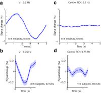
Recent work has suggested that fMRI can detect
neural activity on faster timescales than previously
thought. We tested the temporal limits of fMRI using
oscillating visual stimuli to generate an oscillatory neural
response in human visual cortex. Using rapid (TR=227 ms)
fMRI acquisition at 7 Tesla, we were able to detect 0.75 Hz
oscillations in visual cortex that were an order of
magnitude larger than predicted by canonical linear models.
Using the balloon/Windkessel model we show that continuous
and rapidly varying neural activity can generate larger fMRI
signals than expected. We conclude that fMRI can be used to
measure oscillations of up to at least 0.75 Hz, and suggest
alterations to hemodynamic response models for experiments
studying continuous and rapidly varying neural activity.
|
| |
16:12
 |
0761.
 |
Validation and Optimization of Calibrated fMRI from
oxygen-sensitive Two-Photon Microscopy of the mouse brain
Louis Gagnon1,2,3, Sava Sakadzic4,
Frederic Lesage2, Philippe Pouliot2,
Anders M Dale5, Anna Devor5, Richard B
Buxton5, and David A Boas3
1Department of Medicine, Laval University,
Quebec, QC, Canada, 2Department
of Electrical Engineering, Ecole Polytechnique Montreal,
Montreal, QC, Canada, 3Athinoula
A. Martinos Center for Biomedical Imaging, Department of
Radiology, Massachusetts General Hospital, Harvard Medical
School, Charlestown, MA, United States, 4Athinoula
A. Martinos Center for Biomedical Imaging, Department of
Radiology, Massachusetts General Hospital, Harvard Medical
School, Chalestown, MA, United States, 5Department
of Radiology and Neuroscience, UCSD, La Jolla, CA, United
States
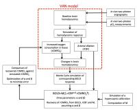
Calibrated fMRI allows to estimate relative changes in the
Cerebral Metabolic Rate of Oxygen Consumption (rCMRO2)
from combined BOLD and ASL measurements during a functional
task. Here, we improved the accuracy of the approach by
using Two-Photon microscopic measurements of the cortical
microvasculature together with first principle Monte Carlo
simulations of proton diffusion across the two-photon
volumes. Our method allowed (1) to validate Calibrated fMRI
from the microscopic point of view and (2) to optimize the
free parameters of the biophysical model assumed, therefore
increasing the accuracy of this method to estimate rCMRO2.
|
| |
16:24
|
0762.
 |
Graded hypercapnia-calibrated BOLD: Beyond the iso-metabolic
hypercapnia assumption
Ian D Driver1, Richard G Wise1, and
Kevin Murphy1
1CUBRIC, School of Psychology, Cardiff
University, Cardiff, United Kingdom
We propose a method for correcting for bias introduced by an
iso-metabolic assumption in hypercapnia calibrated BOLD
studies. A graded hypercapnia design and an assumption of
linear CMRO2 dependence
on hypercapnia level are used to separate the calibration
parameter M from CMRO2 changes
during hypercapnia. This method avoids intra-subject and
experimental variability introduced by making a prior
assumption of iso-metabolism or a CMRO2 decrease
with hypercapnia based on literature values. We implement
this method using two distinct levels of hypercapnia,
measuring lower M values than when making the iso-metabolic
assumption, with a significant dose-wise reduction in CMRO2 with
hypercapnia level.
|
| |
16:36
 |
0763.
 |
The acute effects of caffeine on brain oxygen metabolism: a dual
calibrated FMRI study
Alberto Merola1, Michael A Germuska1,
Esther AH Warnert1, Sharmila Khot1,2,
Daniel Helme2, Lewys Richmond2, Kevin
Murphy1, and Richard G Wise1
1CUBRIC, Cardiff University, Cardiff, United
Kingdom, 2Department
of Anesthesia and Intensive Care Medicine, Cardiff
University, Cardiff, United Kingdom
Caffeine acute effects on oxygen metabolism are not well
characterized across the brain with MRI. We aim at measuring
these in a double-blind, crossover, placebo-controlled study
on sixteen healthy, moderate caffeine consumers using a dual
calibrated fMRI approach and a novel forward estimation
model. Results show spatial variations in OEF0,
CBF, CVR, venous CBV and CMRO2 across
grey matter at different levels of resolution (grey matter,
ROI and voxel), in agreement with most of the literature
findings. Therefore we propose this approach as the first
viable method to assess the effects of drugs on brain
metabolism with a voxel-wise resolution.
|
| |
16:48
|
0764.
 |
Visual cortical responses to auditory stimulation during deep
isoflurane anesthesia: an fMRI study - Permission Withheld
Celia M. Dong1,2, Patrick P. Gao1,2,
Leon C. Ho1,2, Alex T.L. Leong1,2,
Russell W. Chan1,2, Xunda Wang1,2, and
Ed X. Wu1,2
1Laboratory of Biomedical Imaging and Signal
Processing, The University of Hong Kong, Hong Kong, China,
People's Republic of, 2Department
of Electrical and Electronic Engineering, The University of
Hong Kong, Hong Kong, China, People's Republic of
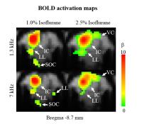
Anesthesia is needed in many neuroscience studies but its
effect on brain network response properties has not been
fully understood. In particular, how it modulates crossmodal
sensory responses remains largely unknown. This study
investigated the brain responses to auditory stimulation at
different isoflurane levels using large-view BOLD fMRI.
Robust responses to multiple pure tone sound stimuli were
detected in the bilateral visual cortex at 2.5% isoflurane
but not at 1.0% isoflurane level. These results revealed the
broad and profound modulation effects of anesthesia on brain
crossmodal response properties during external sensory
stimulation.
|
| |
17:00
|
0765.
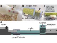 |
Resting state and stimulus evoked fMRI in awake, head-posted and
habituated rats.
Pei-Ching Chang 1,
Daniele Procissi2, Maria Virginia Centeno1,
and Vania Apkarian1
1Physiology, Northwestern University, Chicago,
IL, United States, 2Radiology,
Northwestern University, Chicago, IL, United States
fMRI in rodents is a major tool for basic neuroscience
research. It allows investigation of brain networks in
different animal models of disease and injury using
translational methods with clinical relevance. In many
instances it is essential to image animals in an awake
condition (i.e. without anesthesia). While several have
shown it is possible to image animals in the awake condition
they nearly all require initial anesthesia and forced
restraint. In this study we describe a strategy to image
rats trained to be "comfortably" restrained and head posted
and show how it is possible to enhance the performance of
the fMRI experiments.
|
| |
17:12
|
0766.
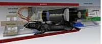 |
Concurrent fMRI and intrinsic optical imaging spectroscopy with
high resolution at ultra high field (14.1T)
Matthias F. Valverde Salzmann1, Klaus Scheffler1,
and Rolf Pohmann1
1High-field Magnetic Resonance, Max Planck
Institute for Biological Cybernetics, Tuebingen, Germany
A setup for concurrent functional MRI and intrinsic optical
imaging spectroscopy inside a 14.1 T animal scanner was
developed, based on a magnetic field proof camera and
optics. fMRI and optical imaging were simultaneously
performed on rats with electrical forepaw stimulation,
resulting in excellent signals for both BOLD and optical
reflectance in two wavelengths (red and green). Only minor
interactions between both modalities were observed. The
combination of these two techniques can be used to
investigate the origins of the BOLD effect and to open up
novel ways of exploring brain function.
|
| |
17:24
|
0767.
 |
Resting-state BOLD local synchrony as a strong proxy of glucose
uptake and as a biomarker of aging using functionally-driven
gray matter parcelization
Michaël Bernier1, Étienne Croteau2,
Christian-Alexandre Castellano2, Stephen C
Cunnane2, and Kevin Whittingstall3
1Nuclear medecine and radiobiology, Université de
Sherbrooke, Sherbrooke, QC, Canada, 2Research
center of aging, Université de Sherbrooke, Sherbrooke, QC,
Canada, 3Diagnostic
radiology, Université de Sherbrooke, Sherbrooke, QC, Canada
Currently, PET is the primary imaging modality used to infer
energy metabolism in the brain. It is also known to be a
reliable biomarker of aging and cognitive diseases.
However, the cost and invasive nature of PET limits its use
in basic research. There is therefore great interest in
developing alternative less invasive approaches for
estimating brain glucose metabolism. Using resting state
fMRI metrics such as regional local homogeneity (ReHo),
amplitude of low-frequencies fluctuations (ALFF) and
regional global connectivity (closeness) we found that both
regional- and subject-variations in ReHo strongly correlate
with brain glucose uptake in healthy young and aging
participants.
|
| |
17:36
|
0768.
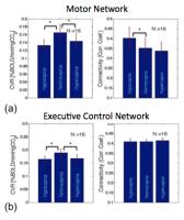 |
The association between cerebrovascular reactivity and
resting-state fMRI connectivity in healthy adults
Ali Golestani1, Jonathan Kwinta1,2,
Stephen Strother1,2, Yasha Khatamian1,
and Jean Chen1,2
1Rotman Research Institute at Baycrest, Toronto,
ON, Canada, 2Medical
Biophysics, University of Toronto, Toronto, ON, Canada
Changes in the cerebrovascular reactivity (CVR) in known to
alter the amplitude of the task-based blood oxygenation
level dependent (BOLD) fMRI signal. The effect of CVR on
resting-state functional connectivity however is still
unknown. In this study, we altered within-individual CVR by
manipulating the end-tidal CO2 (PETCO2)
level, and in each PETCO2 level
we calculated CVR and resting-state connectivity in the
motor and executive control networks. rs-fMRI connectivity
is significantly influenced with CVR, irrespective of neural
function. The strength of this association varies between
motor and executive control networks. This study stresses
the importance of vascular measurements to remove biases in
interpreting rs-fMRI connectivity.
|
| |
17:48
|
0769.
 |
Cortical Laminar Resting-State Fluctuations Scale with
Hypercapnic Response
Maria Guidi1, Laurentius Huber2,
Leonie Lampe1, and Harald E. Möller1
1Max Planck Institute for Human Cognitive and
Brain Sciences, Leipzig, Germany, 2NIMH,
Bethesda, MD, United States
Cortical layer-dependent fMRI can investigate effective
connectivity of the brain. However, in order to obtain
layer-dependent activity information, the unspecific fMRI
sensitivity to draining veins must be accounted for, e.g.,
with calibrated BOLD methods. Regional variations of
resting-state fMRI signal fluctuations have been suggested
to resemble features of baseline physiology, such as venous
blood volume and vascular reactivity. In this study, we
investigate the possibility to use resting-state signal
fluctuations to normalize/calibrate layer-dependent fMRI
task-responses. In calibration studies with induced
hypercapnia, we validate the new approach to obtain cortical
profiles of vascular reactivity by comparisons with the
established M-value.
|
|