| |
10:45
 |
0062.
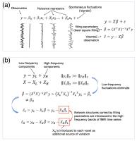 |
Nuisance Regression of High-frequency FMRI Data: De-noising Can
Be Noisy 
Jingyuan E. Chen1,2, Hesamoddin Jahanian2,
and Gary H. Glover1,2
1Electrical Engineering, Stanford University,
Stanford, CA, United States, 2Radiology,
Stanford University, Stanford, CA, United States
A growing number of studies using fast sampling have
demonstrated the persistence of functional connectivity (FC)
in resting state (RS) networks beyond the conventional 0.1
Hz. However, some RS studies have reported frequencies
(e.g., up to 5 Hz) not easily supported by canonical
hemodynamic response functions. Here, we investigated the
influence of a common preprocessing step – whole-band (the
entire frequency band resolved by a short TR) linear
nuisance regression (LNR) – on RSFC. We demonstrated via
both simulation and real data that LNR can introduce network
structures in HF bands, which may largely account for the
observations of HF-RSFC.
|
| |
10:57
|
0063.
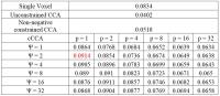 |
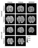
A family-constrained local canonical correlation model to
improve activation detection in fMRI
Xiaowei Zhuang1, Zhengshi Yang1, Tim
Curran2, and Dietmar Cordes1,2
1Cleveland Clinic Lou Ruvo Center for Brain
Health, Las Vegas, NV, United States, 2Department
of Psychology and Neuroscience, University of Colorado
Boulder, Boulder, CO, United States
A family constrained CCA (cCCA) method was introduced to
improve the accuracy of activation detection in noisy fMRI
data. The cCCA was converted into a constrained multivariate
multiple regression problem and solved efficiently with a
numerical optimization algorithm. Results from both
simulated data and real episodic memory data indicated that
a higher detection sensitivity for a fixed specificity can
be achieved with the proposed cCCA method as compared to the
widely used mass-univariate or other conventional
multivariate (CCA) approaches.
|
| |
11:09
|
0064.
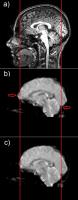 |
Use of T2-weighted 3D acquisition for correction of EPI-induced
distortion in fMRI
Andrea Nordio1,2,3, Denis Peruzzo2,
Filippo Arrigoni2, Fabio Triulzi2,4,
and Alessandra Bertoldo1,2
1Department of Information Engineering (DEI),
University of Padova, Padova, Italy, 2IRCCS
E.Medea, Bosisio Parini, Lecco, Italy, 3IRCCS
Casa Sollievo della Sofferenza, San Giovanni Rotondo, Foggia,
Italy, 4IRCCS
Cà Granda Ospedale Maggiore, Policlinico, Milano, Italy
Echo Planar Imaging (EPI) sequences used for acquiring fMRI
time series data have a high temporal resolution but are
also highly sensitive to the magnetic field inhomogeneity
resulting in geometric distortions. In this work we propose
an approach for correction of EPI distortion in fMRI
sequences. Our method takes advantage of a non-distorted
T2-weighted (T2W) 3D sequence as intermediate step between
the acquired fMRI data and the anatomical image. This
strategy allows to use non-linear registration functions. We
validated our method on a group of healty subjects during
finger-tapping task, proving that the proposed method
significantly improves the group analysis results of
functional data.
|
| |
11:21
|
0065.
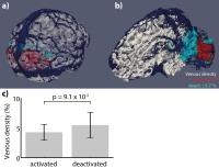 |
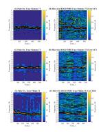
Investigating the effects of venous vasculature on the BOLD
response: A combined SWI and multi-band fMRI approach
David Provencher1, Alexandre Bizeau1,
Yves Bérubé-Lauzière2, and Kevin Whittingstall1,3
1Radiation Sciences and Biomedical Imaging,
Université de Sherbrooke, Sherbrooke, QC, Canada, 2Electrical
and Computer Engineering, Université de Sherbrooke,
Sherbrooke, QC, Canada, 3Diagnostic
Radiology, Université de Sherbrooke, Sherbrooke, QC, Canada
We previously showed that venous density correlates with
BOLD signal amplitude1. Since the BOLD contrast
inherently originates in veins, we hypothesized that its
temporal dynamics would also be affected by venous density.
Here, we use fast multi-band fMRI imaging (TR=0.45s), SWIp
vein reconstruction and different visual stimuli yielding
co-localized activation, yet different BOLD dynamics. From
this, we assess the effects of venous density on BOLD
timing. Results show a robust association between higher
vein density and shorter hemodynamic delay when comparing
activated and deactivated regions. BOLD response timing
differences may thus not entirely reflect neural activity,
but also structural differences.
|
| |
11:33
 |
0066.
 |
The hidden heart rate in the slice-wise BOLD-fMRI global signal.
Michael Hütel1,2, Andrew Melbourne1,
David L Thomas1,2, Jonathan Rohrer2,
and Sebastien Ourselin1,2
1Translational Imaging Group, University College
London, London, United Kingdom, 2Dementia
Research Centre, University College London, London, United
Kingdom
Previous studies have shown that slow variations in the
cardiac cycle are coupled with signal changes in the
blood-oxygen level dependent (BOLD) contrast. The detection
of neurophysiological hemodynamic changes, driven by
neuronal activity, is hampered by such physiological noise.
It is therefore of great importance to model and remove
these physiological artefacts. The cardiac cycle causes
pulsatile arterial blood flow. This pulsation is translated
into brain tissue and fluids bounded by the cranial cavity.
We exploit this pulsality effect and provide evidence that
the heart rate is inherent in BOLD fMRI images.
|
| |
11:45
|
0067.
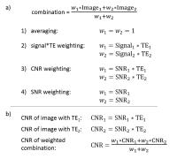 |
Advanced combinations of dual-echo fMRI data provide no
advantages over the simple average at group-level analyses
Ádám Kettinger1,2, Christian Windischberger3,
Christopher Hill4, and Zoltán Nagy4
1Department of Nuclear Techniques, Budapest
University of Technology and Economics, Budapest, Hungary, 2Brain
Imaging Centre, Research Centre for Natural Sciences,
Hungarian Academy of Sciences, Budapest, Hungary, 3Center
for Medical Physics and Biomedical Engineering, Medical
University of Vienna, Vienna, Austria, 4Laboratory
for Social and Neural Systems Research, University of
Zurich, Zurich, Switzerland
Multi-echo EPI acquisitions are used in fMRI research due to
their superior BOLD sensitivity. Several advanced methods of
echo combinations have been proposed. We confirmed, using
dual-echo data, that CNR weighting is the optimal
combination on a single subject level. However, we have
shown that these advantages do not carry over to a group
analysis where a simple averaging of the echos provides
equally good statistical results. This is likely due to the
increase of inter-subject variance of contrast-to-noise
ratio. Future work aims to quantitatively compare
inter-subject and intra-subject variance of dual-echo data
in group studies.
|
| |
11:57
|
0068.
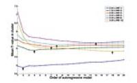 |
Effect of temporal resolution and serial autocorrelations in
fast fMRI
Ashish Kaul Sahib1, Klaus Mathiak2,
Michael Erb1, Adham Elshahabi3, Silke
Klamer3, Klaus Scheffler4, Niels Focke3,
and Thomas Ethofer1
1Biomedical magnetic resonance, University of
tuebingen, Tuebingen, Germany, 25Department
of Psychiatry, Psychotherapy and Psychosomatics, University
Hospital Aachen, Aachen, Germany, 3Department
of Neurology/Epileptology, University of tuebingen,
Tuebingen, Germany, 4Max-Planck-Institute
for Biological Cybernetics, Tuebingen, Germany
To assess the impact of colored noise on statistics and
determine optimal imaging parameters in event-related fMRI
(visual stimulation using checkerboards) acquired by
simultaneous multi-slice imaging enabling repetition times
(TR) between 2.64 to 0.26s. Optimal statistical power was
obtained for a TR of 0.33s, but short TRs required
higher-order autoregressive (AR) models to achieve stable
statistics. Colored noise in event-related fMRI obtained at
short TRs calls for more sophisticated correction of serial
autocorrelations.
|
| |
12:09
|
0069.
 |
Individual Subject Functional Connectivity Parcellation with
Group-Level Spatial and Connectivity Priors
Ru Kong1, Alexander Schaefer1, Avram
J. Holmes2, Simon B. Eickhoff3,4,
Xi-Nian Zuo5, and B.T. Thomas Yeo1
1Department of Electrical and Computer
Engineering, ASTAR-NUS Clinical Imaging Research Centre,
Singapore Institute for Neurotechnology and Memory Networks
Program, National University of Singapore, Singapore,
Singapore, 2Department
of Psychology, Yale University, New Haven, CT, United
States, 3Institute
for Clinical Neuroscience and Medical Psychology,
Heinrich-Heine University Düsseldorf, Düsseldorf, Germany, 4Institute
for Neuroscience and Medicine (INM-1), Research Center
Jülich, Jülich, Germany, 5Lab
for Functional Connectome and Development Division of
Cognitive and Developmental Psychology, CAS, Beijing, China,
People's Republic of
We propose a hidden Markov Random Field (MRF) model to
parcellate the cerebral cortex of individual subjects using
resting-state fMRI (rs-fMRI). Our MRF model imposes a
smoothness prior on the individual-specific parcellation,
while imposing group-level population priors that capture
inter-subject variability in both functional connectivity
profiles and spatial distribution of functional brain
networks. Experiments on a test-retest dataset suggest that
the resulting parcellation estimates are better than
alternative approaches at capturing stable properties of
individual subjects’ intrinsic brain organization, instead
of transient noise or session-dependent variations.
|
| |
12:21
|
0070.
 |
High-resolution T1-mapping using inversion-recovery EPI and
application to cortical depth-dependent fMRI at 7 Tesla
Sriranga Kashyap1, Dimo Ivanov1,
Martin Havlícek1, Benedikt A Poser1,
and Kâmil Uludag1
1Department of Cognitive Neuroscience, Maastricht
University, Maastricht, Netherlands
Cortical-depth dependent fMRI usually relies on the
definition of depths on an anatomical image (eg. MPRAGE).
The geometric dissimilarities of the functional compared to
the anatomical data require further spatial processing of
the functional data to ensure good co-registration. We
propose an alternative approach that uses an optimised
inversion-recovery EPI derived T1 image,
whose resolution and readout, hence distortions, are
identical to that of the functional data, in order to
delineate cortical depths. As a result, the cortical-depth
specific fMRI data can be analysed in the native space
without any spatial confounds stemming from distortion
correction and inaccurate registration.
|
| |
12:33
|
0071.
 |
Distortion-matched T1-maps and bias-corrected T1w-images as
anatomical reference for submillimeter-resolution fMRI
Wietske van der Zwaag1, Pieter Buur1,
Maarten Versluis2, and José P. Marques3
1Spinoza Centre for Neuroimaging, Amsterdam,
Netherlands, 2Philips
Healthcare, Best, Netherlands, 3Donders
Institute for Brain, Cognition and Behaviour, Nijmegen,
Netherlands
Achieving sufficiently good quality co-registration between
the anatomical and functional images is currently a large
stumbling block for laminar fMRI. Here, we present a
distortion-matched T1weighted/T1-estimation
mapping approach using two 3D-EPI readouts per inversion,
following the MP2RAGE signal combination. 0.7mm isotropic T1 data
with matching distortions to a 0.7mm isotropic fMRI protocol
can be acquired in less than two minutes.
|
|