| |
16:00
|
0750.
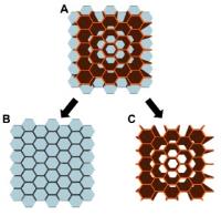 |
The relationships between microstructure and the diffusion
tensor in simulated skeletal muscle 
David B Berry1, Benjamin M Regner2,
Vitaly L Galinsky3, Samuel R Ward1,4,5,
and Lawrence R Frank3
1Bioengineering, UCSD, La Jolla, CA, United
States, 2Institute
of Engineering in Medicine, UCSD, La Jolla, CA, United
States, 3Center
for Scientific Computation in Imaging, UCSD, La Jolla, CA,
United States,4Orthopaedic Surgery, UCSD, La
Jolla, CA, United States, 5Radiology,
UCSD, La Jolla, CA, United States
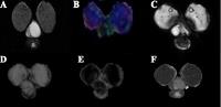
Diffusion tensor imaging (DTI) has been used to measure
changes in restricted diffusion in skeletal muscle after
injury, which are thought to track microstructural, and
therefore functional changes. However, there are few direct
comparisons between muscle microstructure and DTI
measurements because it is difficult to precisely control in
vivo experiments. Here, we use a computational (in silico)
modeling approach to explore changes in DTI measurements as
muscle microstructure is systemically changed. Muscle fiber
diameter and edema have the largest effects on the DT.
Additionally, we have shown multi-echo DTI is required to
resolve changes in microstructure when edema is present.
|
| |
16:12
|
0751.
 |
Towards robust Diffusion Tensor Imaging of skeletal muscles via
an automatic artifact removal tool.
Chiara Giraudo1, Stano Motyka1,
Siegfried Trattnig1, and Wolfgang Bogner1
1Department of Biomedical Imaging and
Image-guided Therapy- MR Centre of Excellence, Medical
University Vienna, Vienna, Austria
STEAM-DTI sequence recently provided excellent results for
DTI analysis of muscle fibers (e.g., high signal-to-noise
ratio, low apparent diffusion coefficient, high fractional
anisotropy values) but demonstrated also to be affected by
strong artifacts, which can be assumed to be due to
involuntary muscle contractions. The hereby proposed
automatic post-processing method, based on weighted mean of
the averages for each DTI-direction and b-value,
demonstrated to successfully detect and correct these
artifacts, improving fiber tracking of the calf muscles.
|
| |
16:24
|
0752.
 |
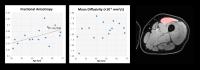
Super-Resolution Magnetic Resonance Elastography (SR-MRE) of
Exercise Induced Muscle Damage (EIMD)
M. Perrins1, E. Barnhill2, P. Kennedy1,
J. Braun2, I. Sack2, A. Hunter3,
C. Brown4, E. van Beek1, and Neil
Roberts1
1University of Edinburgh, Edinburgh, United
Kingdom, 2Department
of Radiology, Charité - Universitätsmedizin Berlin, Berlin,
Germany, 3School
of Sport, University of Stirling, Stirling, United Kingdom, 4The
Mentholatum Company Ltd., East Kilbride, United Kingdom
Super-Resolution (SR) Magnetic Resonance Elastography (MRE)
was applied to measure thigh muscle viscoelastic properties
in 20 subjects in whom Exercise Induced Muscle Damage (EIMD)
was produced using a well-established muscle damage
protocol. SR-MRE is made possible by analysing
Multi-frequency MRE (MMRE) in a manner such that multiple
low-resolution images of the same scene are interpolated and
fused to create a single, high-resolution image. Muscle
tissue is well suited to study using SR-MRE and the sites of
muscle damage could be clearly identified suggesting
potential useful clinical applications for the technique.
SR-MRE also has potential to provide insight regarding the
mechanisms underlying tissue damage in EIMD.
|
| |
16:36
|
0753.
 |
A Quantitative Investigation of the Fatty Degeneration of the
Supraspinatus Muscle after Rotator Cuff Tear: SPLASH-MRI,
Model-Based T$$$_1$$$ Mapping and Shear Wave Ultrasound
Andreas Max Weng1, Fabian Gilbert2,
Johannes Tran-Gia1,3, Tobias Wech1,
Detlef Klein1, Thorsten Alexander Bley1,
and Herbert Köstler1
1Department of Diagnostic and Interventional
Radiology, University of Würzburg, Würzburg, Germany, 2Department
of Trauma, Hand, Plastic and Reconstructive Surgery,
University of Würzburg, Würzburg, Germany, 3Department
of Nuclear Medicine, University of Würzburg, Würzburg,
Germany
Fatty degeneration of the rotator cuff is often investigated
by a visual inspection of T$$$_1$$$-weighted MR images.
Since this approach is in debate the aim of this study was
to investigate fatty degeneration of the supraspinatus
muscle by quantitative techniques: SPLASH, model-based
acceleration for parameter mapping (MAP) T$$$_1$$$
measurement and shear wave ultrasound. The obtained values
from SPLASH and T$$$_1$$$ mapping are in good accordance
(Pearson’s r=0.82). However, shear wave ultrasound does
neither correlate well with SPLASH (Spearman’s rho= 0.30)
nor with MAP (rho=0.19). Since data acquisition time of the
T$$$_1$$$ mapping technique used in our study is very short
(4s), this might be the technique of choice for
investigation of the fatty degeneration of the supraspinatus
after rotator cuff tear.
|
| |
16:48
|
0754.
 |
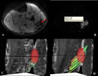
Assessment of passive muscle elongation using DTI: Correlation
between fiber length and diffusion coefficients
Valentina Mazzoli1,2,3, Jos Oudeman1,
Marco A Marra3, Klaas Nicolay2, Nico
Verdonschot3, Andre M Sprengers3,
Martijn Froeling4, Aart J Nederveen1,
and Gustav J Strijkers5
1Department of Radiology, Academic Medical
Center, Amsterdam, Netherlands, 2Biomedical
NMR, Department of Biomedical Engineering, Eindhoven
University of Technology, Eindhoven, Netherlands,3Orthopaedic
Research Lab, Radboud University Medical Center, Nijmegen,
Netherlands, 4Department
of Radiology, University Medical Center, Utrecht,
Netherlands, 5Biomedical
Engineering and Physics, Academic Medical Center, Amsterdam,
Netherlands
The aim of this study is to explore Diffusion Tensor Imaging
in the assessment of passive muscle elongation. We
investigated two dorsiflexor and two plantarflexor muscles
of the lower leg with the foot in dorsiflexion, neutral and
plantarflexion position. Significant negative correlation
was found between changes in fiberlength caused by passive
muscle lengthening and radial diffusivity for all muscles.
Furthermore the rate of change in radial diffusivity was
compatible with a cylindrical model with constant volume.
These findings give more insight into diffusion mechanisms
in skeletal muscles and are highly relevant for
biomechanical models.
|
| |
17:00
|
0755.
 |
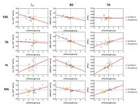
Age-Related Changes in Diffusion Tensor Imaging Measures in
Human Skeletal Muscle
Donnie Cameron1, David A. Reiter1,
Kenneth W. Fishbein1, Christopher M. Bergeron1,
Richard G. Spencer1, and Luigi Ferrucci1
1National Institute on Aging, National Institutes
of Health, Baltimore, MD, United States
This work investigates how ageing influences diffusion
tensor imaging (DTI) measures through application of a
robust protocol to the human thigh. Fifteen participants,
from 27-73 years old, were recruited, and mean diffusivity
(MD) and fractional anisotropy (FA) were calculated in their
quadriceps and plotted against age. Fibre tractography was
also calculated. Rectus femoris FA showed a significant
correlation with age (R2=0.27, p=0.04),
while FA approached significant correlations in other muscle
heads. MD had a more complicated relationship with age, if
any, in contrast to previous work where lipid influence was
neglected. This highlights the need for high-quality
fat-suppression in DTI.
|
| |
17:12
 |
0756.
 |
A multimodal MR approach to evaluate complex muscle degeneration
processes in Duchenne Muscular Dystrophy
Melissa Tamara Hooijmans1, Melissa Tamara
Hooijmans1, Nathalie Doorenweerd1,
Jedrek Burakiewicz1, Andrew Webb1, Jan
Vershuuren2, Erik Niks2, and Hermien
Kan1
1Radiology, Leiden University Medical Center,
Leiden, Netherlands, 2Neurology,
Leiden University Medical Center, Leiden, Netherlands
Quantitative MRI and MRS are increasingly important as
non-invasive and objective outcome measures in therapy
development for DMD. Several MR indices, have been shown to
correlate individually with age and functional measures.
However, much less attention has been given to how these
indices relate to each other. Our work combined quantitative
MRI and spatially resolved 31P MRS in the lower leg muscles
of DMD patients and showed that combining multimodal MR
measures is very important to objectively assess muscle
degeneration processes and potentially the effect of
therapeutic interventions in DMD.
|
| |
17:24
|
0757.
 |
Multi parametric MRI evaluation of skeletal muscle growth and
myopathies in mice
Kerryanne V. Winters1,2, Olivier Reynaud1,2,
Dmitry S. Novikov1,2, Els Fieremans1,2,
and Sungheon G. Kim1,2
1Center of Biomedical Imaging, Department of
Radiology, NYU School of Medicine, New York, NY, United
States, 2Center
for Advanced Imaging Innovation and Research, NYU Langone
Medical Center, New York, NY, United States
The random permeable barrier membrane (RPBM) model for
diffusion tensor imaging (DTI) provides a non-invasive
modality potentially useful for early and accurate diagnosis
for the wide range of myopathies. We have utilized the
DTI-RPBM method to assess myofiber changes in the
Surface-to-Volume ratio S/V and
sarcolemma permeability κ as markers in growing and wasting
skeletal muscle. Preliminary results show that S/V and
κ decrease in both wild-type and mdx mice,
with a more pronounced change between weeks 3 and 4 in mdx mice.
The conventional IDEAL-Dixon and T2 mapping measures were
not sensitive enough to observe the same change.
|
| |
17:36
|
0758.
 |
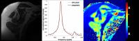
31P-MRI using A Spectrally Selective 3D non-Cartesian FLORET
Sequence at 7 T
Prodromos Parasoglou1, Ryan Brown1,2,
and Guillaume Madelin1
1Department of Radiology, New York University
School of Medicine, New York, NY, United States, 2NYU
WIRELESS, Polytechnic Institute of New York University,
Brooklyn, NY, United States
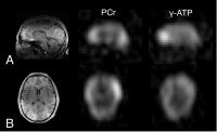
We developed a spectrally selective 3D non-Cartesian FLORET
pulse sequence to map phosphorus-containing metabolites in
the human tissue. In particular, through this highly
efficient pulse sequence we mapped phosphocreatine and
γ-adenosine triphosphate at 1.4 cm isotropic nominal voxel
size in the human brain. In addition, we were able to map
phosphocreatine in the skeletal muscle during exercise and
recovery with 6 s temporal resolution. We showed that
spectrally selective 3D-FLORET is an efficient pulse
sequence that can be used to image 31P-containing
metabolites in the human tissue when high spatiotemporal
resolution is needed.
|
| |
17:48
|
0759.
 |
Association of quadriceps muscle fat with isometric strength
measurements in healthy males using chemical shift
encoding-based water-fat MRI
Thomas Baum1, Stephanie Inhuber2,
Michael Dieckmeyer1, Christian Cordes1,
Stefan Ruschke1, Elisabeth Klupp3,
Holger Eggers4, Hendrik Kooijman5,
Ernst J Rummeny1, Ansgar Schwirtz2,
Jan S Kirschke3, and Dimitrios C Karampinos1
1Department of Radiology, TU Munich, Munich,
Germany, 2Department
of Sports and Health Sciences, TU Munich, Munich, Germany, 3Section
of Neuroradiology, TU Munich, Munich, Germany, 4Philips
Research Laboratory, Hamburg, Germany, 5Philips
Healthcare, Hamburg, Germany
MR-based assessment of quadriceps muscle fat has been
proposed as surrogate marker in sarcopenia, osteoarthritis,
and neuromuscular disorders. The present study demonstrated
strong associations between chemical shift encoding-based
water-fat MRI quadriceps inter- and intramuscular fat
parameters and corresponding physical strength measurements
in healthy males. Thus, chemical shift encoding-based
water-fat MRI can provide clinically important information
beyond quadriceps muscle morphology and T1-weighted muscle
fat quantifications and may potentially track early changes
in muscles that are not severely atrophied or fatty
infiltrated in the beginning of a disease process.
|
|