| |
13:30
|
0675.
 |
Whole Body Skeletal Imaging Using Zero TE 
Florian Wiesinger1, Sandeep Kaushik2,
Anne Menini1, Sangtae Ahn3, Lishui
Cheng3, Cristina Cozzini1, Thomas Hope4,
Jaewon Yang4, Peder Larson4, and
Dattesh Shanbhag2
1GE Global Research, Munich, Germany, 2GE
Global Research, Bangalore, India, 3GE
Global Research, Schenectady, NY, United States, 4University
of California San Francisco, San Francisco, CA, United
States
Recently we presented a method for zero TE MR bone imaging
in the head. In this abstract, we describe the extension of
this work towards whole body skeletal imaging as required
for applications like PET/MR Attenuation Correction, or MR-based
Radiation Therapy Planning.
|
| |
13:42
|
0676.
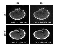 |
30-Second Bound- and Pore-Water Maps of Cortical Bone
Mary Kate Manhard1, Kevin D Harkins2,
Daniel F Gochberg2, Jeffry S Nyman3,
and Mark D Does1
1Biomedical Engineering, Vanderbilt University,
Nashville, TN, United States, 2Vanderbilt
University Institute of Imaging Science, Vanderbilt
University, Nashville, TN, United States, 3Department
of Orthopaedics & Rehabilitation, Vanderbilt University,
Nashville, TN, United States
Imaging bound and pore water concentrations in cortical bone
using UTE MRI has shown potential for evaluating fracture
risk, but 3D methods require a relatively long scan time
(~30 minutes total). 2D UTE with optimized half-pulses was
implemented to acquire both bound and pore water maps in ~30
seconds and results were compared to 3D UTE, both ex vivo
and in vivo. Mean differences in bound/pore water
concentration were less than 10%. Applying these fast
sequences in 2D has the potential to greatly increase the
utility of these methods in clinical settings for evaluating
fracture risk in patient populations.
|
| |
13:54
 |
0677.
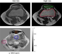 |
On the Feasibility of Quantitative Susceptibility Mapping For
Trabecular Bone Volume Density Mapping at 3 T
Maximilian Nikolaus Diefenbach1, Anh T. Van2,
Jakob Meineke3, Hendrik Kooijman4,
Axel Haase2, Ernst J. Rummeny5, Jan S.
Kirschke6, Thomas Baum1, and Dimitrios
C. Karampinos5
1Department of Diagnostic and Interventional
Radiology, Technische Univeristät München, Munich, Germany, 2Zentralinstitut
fu¨r Medizintechnik, Technische Universita¨t Mu¨nchen,
Munich, Germany, 3Philips
Research Laboratory, Hamburg, Germany, 4Philips
Healthcare, Hamburg, Germany, 5Department
of Diagnostic and Interventional Radiology, Technische
Universität München, Munich, Germany, 6Section
of Neuroradiology, Technische Universität München, Munich,
Germany
Trabecular bone imaging has a high clinical significance for
predicting fracture risk in patients with osteoporosis.
Quantitative susceptibility mapping (QSM) has been recently
emerging for mapping diamagnetic and paramagnetic
substances. Recent reports attempted to use QSM combined
with ultra-short echo time imaging for mapping the
susceptibility of cortical bone. However, it remains unknown
whether QSM is feasible for measuring bone volume density in
trabecular bone, where the bone density is much lower than
cortical bone. The purpose of the present work is to study
the feasibility of QSM for trabecular bone density mapping,
using numerical simulations, specimen measurements and
preliminary in vivo measurements.
|
| |
14:06
|
0678.
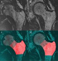 |
High-Resolution MRI to Assess Trabecular Bone Microstructure in
the Proximal Femur of HIV-Infected Men
Roland Krug1, Andrew Lai1, Lorenzo
Nardo1, Luca Facchetti1, Misung Han1,
Galateia Kazakia1, and Julio Carballido-Gamio1
1University of California, UCSF, San Francisco,
CA, United States
MRI is currently the only modality to assess trabecular bone
structure with high-resolution in the proximal femur
in-vivo. We have optimized image acquisition and image
analysis techniques to assess microstructural bone
parameters in HIV-infected individuals compared to healthy
controls. We have found significant differences in the
femoral head, neck and trochanteric regions between patients
and controls. We conclude that MRI can be an important tool
to assess bone structure in the central skeleton at
important fracture sites such as the proximal femur with
very high resolution.
|
| |
14:18
|
0679.
 |
Age-related loss of bound water in human trabecular bone
Mathilde Granke1, Kuniko Hunter2,
Sasidhar Uppuganti1, Jeffry S Nyman1,3,
and Mark D Does1
1Vanderbilt University, Nashville, TN, United
States, 2Rensselaer
Polytechnic Institute, Troy, NY, United States, 3VA
Tennessee Valley Healthcare System, Nashville, TN, United
States
1H NMR- derived bound water measurements in
cadaveric human trabecular bone are sensitive to age-related
changes in the quality of bone tissue, and therefore could
be predictive of fracture risk in trabecular sites prone to
fracture.
|
| |
14:30
|
0680.
 |
Fast volumetric mapping of bound and pore water content in
cortical bone in vivo using 3D Cones sequences
Jun Chen1, Michael Carl2, Hongda Shao1,
Eric Chang1, Graeme Bydder1, and Jiang
Du1
1Radiology, University of California, San Diego,
San Diego, CA, United States, 2GE
Healthcare, San Diego, CA, United States
Bone water exists in the form of free water in the Haversian
canals or lacunar-canalicular system, as well as bound water
either loosely bound to collagen or more tightly bound to
mineral. Ultrashort echo time (UTE) sequences with TEs as
short as 8 µs can potentially detect signal from pore water
and loosely bound water. In this study, we introduce an
approach for fast volumetric mapping of bound and pore water
content in vivo using a clinical 3T MR scanner.
|
| |
14:42
|
0681.
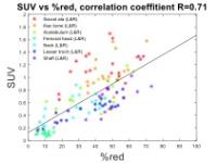 |
Quantitative and Functional Assessment of Red and Yellow Bone
Marrow Using PET-MR Imaging
Chuan Huang1,2, Anuradha Janardhanan1,3,
and Mark Schweitzer1
1Radiology, Stony Brook Medicine, Stony Brook,
NY, United States, 2Psychiatry,
Stony Brook Medicine, Stony Brook, NY, United States, 3Diagnostic
Imaging, Kuala Lumpur Hospital, Kuala Lumpur, Malaysia
Understanding the distribution of red marrow is important
for various hematopoetic diseases and especially osseous
metastases as areas of red marrow are the primary sites for
hematogenous seeding of tumor cells, accounting for
approximately 90% of skeletal metastases. Using a
simultaneous PET-MR we sought to evaluate voxel of red
marrow in the femora and pelvis using fat/water sequences
correlated with FDG PET uptake. This quantitative assessment
of red and yellow marrow was done in specific anatomic
subregions. The bone marrow composition and metabolism were
found to be symmetric in each individual. Good correlation
between SUV and %red were found for each ROI among all
subjects. The metabolism (FDG uptake) was found to be
different for the ROIs with the same amount of red marrow.
Further research will study whether this leads to higher
chance of tumor seeding.
|
| |
14:54
|
0682.
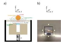 |
Novel Approach in Detection of Bone Marrow Changes Related to
Osteoporosis, Using a Stray Field NMR Scanner
Inbar Hillel1, Yifat Sarda1, Elad
Bergman1, Itzhak Binderman2, and Uri
Nevo1
1Department of Biomedical Engineering, Tel-Aviv
University, Tel Aviv, Israel, 2School
of Dental Medicine, Tel-Aviv University, Tel Aviv, Israel
Osteoporosis is a disease characterized by loss of bone
mineral density, caused by loss of the equilibrium between
osteogenesis and adipogenesis. In this work T2, T1 and ADC
were measured using a low-field NMR scanner, for the
detection of bone marrow changes related to osteoporosis.
Results showed that this method is capable of significantly
classifying between bones of rats that were ovariectomized,
ovariectomized and treated with parathyroid hormone, and
sham-operated rats.
|
| |
15:06
|
0683.
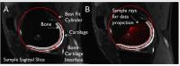 |
A Method to Quantitatively Compare Bone and Cartilage Changes
Post Knee Injury: Initial Results
Uchechukwuka Monu1, Feliks Kogan2,
Emily McWalter2, Brian Hargreaves2,
and Garry Gold2
1Electrical Engineering, Stanford University,
Stanford, CA, United States, 2Radiology,
Stanford University, Stanford, CA, United States
New PET/MR systems have made the simultaneous acquisition
and quantitative assessment of bone and cartilage possible.
Using projection maps and cluster analysis, the
comprehensive visualization and quantification of PET 18F-NaF
uptake within an injured and contralateral knee are
determined and compared with corresponding T2 and T1rho
relaxation times within the cartilage. Significant increase
in PET uptake is observed in the injured knee compared to
the contralateral knee and some areas of high PET uptake
correspond with elevated T2 and T1rho relaxation times. This
developed tool shows promise in assessing bone metabolic
activity and its relationship with quantitative MR
parameters.
|
| |
15:18
|
0684.
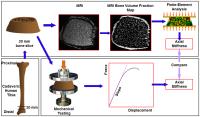 |
Validation of MRI-based Assessment of Mechanical Competence of
Distal Tibia using Cadaveric Human Bone.
Chamith S. Rajapakse1, Benjamin T. Newman1,
Wenli Sun1, Michael Ispiryan1,
Michelle Slinger2, Elizabeth A. Kobe2,
Kelly Borges1, Karyll Davis2, Keren De
Jesus2, Jeremy Magland1, and Felix W.
Wehrli1
1Laboratory for Structural NMR Imaging,
Department of Radiology, University of Pennsylvania,
Philadelphia, PA, United States, 2University
of Pennsylvania School of Engineering and Applied Science,
University of Pennsylvania, Philadelphia, PA, United States
High-resolution MRI-derived finite element analysis allows
for the in vivo estimation of bone strength. This
information is useful for planning treatments and
interventions in individuals suffering from conditions that
affect bone mineral homeostasis. However these methods have
not been previously validated. This study subjected distal
tibia specimens to both MRI-based finite element analysis
and mechanical testing ex vivo. Estimated bone
stiffness was strongly correlated to the experimental values
(R2=0.84) supporting usefulness of MRI-based bone strength
assessment in human subjects.
|
|