| |
16:30
|
0257.
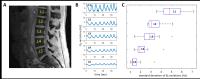 |
Measurement and Compensation of Respiration-Induced B0
Variations for Bone Marrow Fat Quantification in Lumbar Spine - Permission Withheld
Yoonho Nam1, Joon-Yong Jung1, Hyun
Seok Choi1, Eojin Hwang1, Hongpyo Lee2,
and Dong-Hyun Kim2
1Department of Radiology, Seoul St. Mary's
Hospital, College of Medicine, The Catholic University of
Korea, Seoul, Korea, Republic of, 2Department
of Electrical and Electronic Engineering, Yonsei University,
Seoul, Korea, Republic of
Fat fraction of the bone marrow has been suggested as an
important quantitative parameter in the assessment of
treatment response and determination of the benignity in
oncologic imaging. Therefore, accurate fat quantification is
a prerequisite for the fat fraction to be established as a
reliable imaging biomarker. For this purpose, spoiled
gradient echo sequences have been commonly used. However,
gradient echo imaging is susceptible to B0 variations
from various sources such as respiration, cardiac pulsation.
In this study, we investigate and compensate the effects of
respiration-induced B0 variations
on fat quantification of the bone marrow in the lumbar
spine.
|
| |
16:42
|
0258.
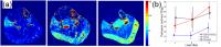 |
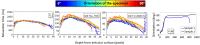
Quantitative Muscle Perfusion with DCE-MRI Shows Distinct
Load-Dependent Exercise-Stimulated Muscle Perfusion Patterns 
Jeff L. Zhang1, Christopher Hanrahan1,
Christopher C. Conlin1, Corey Hart2,
Gwenael Layec2, Kristi Carlston1,
Daniel Kim1, Michelle Mueller3, and
Vivian S. Lee1
1Radiology, University of Utah, Salt Lake City,
UT, United States, 2Internal
Medicine, University of Utah, Salt Lake City, UT, United
States, 3Vascular
surgery, University of Utah, Salt Lake City, UT, United
States
Noninvasive mapping of calf muscle perfusion with high
spatial resolution has potential for assessing the severity
of peripheral artery disease (PAD) and studying associated
capillary density abnormality. We tested our novel DCE-MRI
method to measure calf muscle hyperemia stimulated by
plantar flexion at three different workloads. Increases in
exercise load caused increased total perfusion in
gastrocnemius, with a heterogeneous pattern at medium load
and homogeneous at higher load. Perfusion in soleus did not
increase until very heavy load of 16 lbs. DCE-MRI provides
high spatial resolution measurement of post-exercise muscle
perfusion.
|
| |
16:54
|
0259.
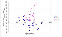 |
Gender Differences in Sodium Deposition in Muscle and Skin
Ping Wang1,2, Muge Serpil Deger3,
Hakmook Kang4, T. Alp Ikizler3, Jens
M. Titze5, and John C. Gore1,2
1Institute of Imaging Science, Vanderbilt
University Medical Center, Nashville, TN, United States, 2Department
of Radiology and Radiological Sciences, Vanderbilt
University Medical Center, Nashville, TN, United States, 3Division
of Nephrology and Hypertension, Department of Medicine,
Vanderbilt University Medical Center, Nashville, TN, United
States, 4Department
of Biostatistics, Vanderbilt University Medical Center,
Nashville, TN, United States, 5Division
of Clinical Pharmacology, Department of Medicine, Vanderbilt
University Medical Center, Nashville, TN, United States
Sodium ions play a vital role in cellular homeostasis and
electrochemical activity throughout the human body. Previous
studies have measured muscle and skin sodium contents in
vivo in
humans using MRI and have shown characteristic changes with
age and as a result of pathological changes. In
this study, we found significant gender differences in
sodium deposition between muscle and skin, with male has
higher sodium content in skin than in muscle, while female
has higher muscle sodium than skin sodium. This
observation seems to be more reliable with the increase of
age.
|
| |
17:06
|
0260.
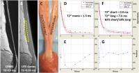 |
Correlation of Mono-exponential and Bi-exponential UTE-T2*
Analyses and Biomechanics in Human Achilles Tendons
Eric Y Chang1,2, Robert M Healey3,
Reni Biswas2, Sheronda Statum2, Betty
Tran2, Kenyu Iwasaki4, Jiang Du2,
Won C Bae2, and Christine B Chung1,2
1Radiology Service, VA San Diego Healthcare
System, San Diego, CA, United States, 2Department
of Radiology, University of California, San Diego Medical
Center, San Diego, CA, United States, 3Department
of Orthopaedic Surgery, University of California, San Diego
Medical Center, San Diego, CA, United States, 4Department
of Orthopaedic Surgery, Kyushu University, Fukuoka, Japan
In this pilot study, we sought to determine if
mono-exponential T2, mono-exponential UTE-T2*, or
bi-exponential UTE-T2* correlated with biomechanical
properties in human Achilles tendons. We found very high and
significant correlation coefficients between
mono-exponential T2* (rho = 0.90, p = 0.002) and
bi-exponential T2* fractions (rho = -0.97, p < 0.001)
obtained using the UTE-Cones sequence and ultimate tensile
strain. Ultimate tensile strain represents the percentage
change in tendon length prior to failure and high strains
have been previously associated with tendon degeneration.
Our results suggest that non-invasive MRI of the Achilles
tendon may serve as a surrogate measure.
|
| |
17:18
|
0261.
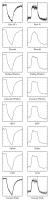 |
A comparison of denoising methods in dynamic MRS
Benjamin C Rowland1 and
Alexander P Lin1
1Centre for Clinical Spectroscopy, Brigham and
Women's Hospital, Boston, MA, United States
MR spectroscopy is often used to study dynamic systems,
such as muscle energetics using 31P.
The need to perform temporal averaging to improve signal to
noise ratios can compromise the temporal resolution of the
measurements. Indirect time domain denoising can help to
resolve this issue. In this study we evaluate six potential
denoising approaches for dynamic MRS.
|
| |
17:30
|
0262.
 |
Extracting Quantitative Information From MRI Bound- and
Pore-Water Maps of Cortical Bone
Mary Kate Manhard1, Sasidhar Uppuganti2,
Mathilde C Granke2, Daniel F Gochberg3,
Jeffry S Nyman2, and Mark D Does1
1Biomedical Engineering, Vanderbilt University,
Nashville, TN, United States, 2Department
of Orthopaedics & Rehabilitation, Vanderbilt University,
Nashville, TN, United States, 3Vanderbilt
University Institute of Imaging Science, Vanderbilt
University, Nashville, TN, United States
Bound and pore water concentration measures of cortical bone
found from MRI have been shown to correlate with material
properties of bone, but the ideal way to analyze and draw
information from 3D quantitative maps remains unclear.
Material properties of cadaver radii found from a 3-point
bend test were correlated with characteristics of the
distribution of bound and pore water concentrations (e.g.
mean, skewness) in ROIs found from different segmentations.
Results highlighted the importance of segmentation method as
well as quantitative measures drawn from the maps.
|
| |
17:42
|
0263.
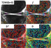 |
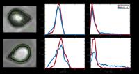
Detection of the meniscal blood supply changes in meniscal
problems with Intravoxel incoherent motion MR imaging
Tan Guo1, Dandan Zheng2, Bing Wu2,
and Min Chen1
1Radiology, Beijing Hospital, Beijing, China,
People's Republic of, 2GE
Healthcare, MR Research China, Beijing, Beijing, China,
People's Republic of
The blood supply of meniscus is an essential indicator for
the prognosis of meniscal problems. With a favorable blood
supply of the teared meniscus, it’s tend to preserve the
meniscus as much as possible at partial meniscetomy and
meniscal repair. Intravoxel incoherent motion (IVIM) theory
provide information about microcirculation of blood in
addition to the pure molecular diffusion. The perfusion
information detected with IVIM is emphasized on
microvascular bed, which is the typical blood supply pattern
of meniscus. In this study, IVIM model were used to estimate
the change of vasculature in normal, degenerated and teared
meniscus.
|
| |
17:54
|
0264.
 |
Orientation anisotropy of quantitative rotating and laboratory
frame relaxation parameters in articular cartilage
Jari Rautiainen1, Lassi Rieppo2,3,
Simo Saarakkala2,3,4, and Mikko Johannes Nissi1,5
1Department of Applied Physics, University of
Eastern Finland, Kuopio, Finland, 2Research
Unit of Medical Imaging, Physics and Technology, University
of Oulu, Oulu, Finland, 3Medical
Research Center Oulu, Oulu University Hospital and
University of Oulu, Oulu, Finland, 4Department
of Diagnostic Radiology, Oulu University Hospital, Oulu,
Finland, 5Diagnostic
Imaging Center, Kuopio University Hospital, Kuopio, Finland
Classical ($$$T_1$$$, $$$T_2$$$) and several rotating frame
quantitative MR parameters have been used for evaluation of
composition and structure of articular cartilage, and
demonstrated to have variable sensitivity to tissue
orientation. The orientation dependence of $$$T_1$$$,
$$$T_2$$$, $$$T_2^*$$$, CW-$$$T_{1\rho}$$$ with four
spin-lock amplitudes, adiabatic $$$T_{1\rho}$$$ with three
different pulse modulations, adiabatic $$$T_{2\rho}$$$ and
$$$T_{\rm RAFF}$$$ relaxation times were further
investigated at 9.4T at different orientations of articular
cartilage relative to B0 and compared with polarized light
microscopy of the same tissue. $$$T_1$$$, adiabatic
$$$T_{1\rho}$$$ with HS1-pulse and CW-$$$T_{1\rho}$$$ at 2
kHz spin-lock demonstrated the least orientation dependence.
|
| |
18:06
|
0265.
|
The value of DWI with ADC mapping for assessing synovitis and
bone erosion in early stage of RA
Xinwei Lei1, Jin QU1, Ying ZHAN1,
Huixia Li1, and Yu Zhang2
1Tianjin First Center Hospital, Tianjin, China,
People's Republic of, 2Philips
Healthcare, Beijin, China, People's Republic of
The aim of study was to explore whether synovitis and bone
erosion judged by ADC values correspond exactly or not to
those judged by CE-MRI. 25 patients were examined by 3.0T MR
including DWI and CE-MRI. ADC value of synovitis and bone
erosion was signi?cantly lower than that of joint effusion
and cysts. ADC values of 2.0 was found distinguishing joint
effusion from synovitis, and bone erosion from cysts.
Therefore, MR diffusion provides additional information to
the routine MRI sequences rendering it an effective
non-invasive tool in differentiating between synovitis and
joint effusion, as well as bone erosion and cysts.
|
| |
18:18
|
0266.
 |
Measurement of proteoglycan concentration in intervertebral
discs assessed by 1HMRS at 1.5T
Lisa Maria Harris1,2, Ella Hodder2,3,
Mara Cercignani2, Jan Bush2, Derek
Convill3, Paul Colley1, and Nicholas
Dowell2
1Radiological Sciences, Brighton and Sussex
University Hospitals NHS Trust, Brighton, United Kingdom, 2Clinical
Imaging Sciences Centre, Brighton and Sussex Medical School,
Brighton, United Kingdom,3Computing, Engineering
and Mathematics, University of Brighton, Brighton, United
Kingdom
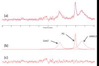
An assessment was made to determine whether proteoglycan
concentration could accurately be quantified at 1.5T using
1HMRS in a group of 13 healthy volunteers. A peak from the
N-acetyl resonance associated with proteoglycan was seen in
all thirteen spectra, and reliably measured (308.8±59.9).
This compares favourably with studies performed at higher
field strengths, thus showing that is it possible even at
1.5T to measure proteoglycans in intervertebral discs.
|
|