| |
16:00
 |
0479.
 |
Deciphering the Functional Role of Locus Coeruleus-derived
Norepinephrine using Chemogenetic fMRI and 18FDG-PET - Permission Withheld
Esteban Adrian Oyarzabal1,2, Manasmita Das3,
Sung-Ho Adrian Lee4, Natale Sciolino2,
Irina Evsyukova2, Patricia Jensen2,
and Yen-Yu (Ian) Shih3
1Neurology, UNC-Chapel Hill, Carrboro, NC, United
States, 2Laboratory
of Neurobiology, NIEHS/NIH, Research Triangle Park, NC,
United States, 3Neurology,
UNC-Chapel Hill, Chapel Hill, NC, United States, 4UNC-Chapel
Hill, Chapel Hill, NC, United States
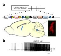
This study examines how selective chemogenetic stimulation
of noradrenergic neurons of the Locus Coeruleus (LC) in mice
modulates cerebral metabolism and vascular tone. This was
achieved by using a transgenic mouse line selectively
expresses Designer Receptors Exclusively Activated by
Designer Drugs (DREADDs) in the LC noradrenergic neurons. A
multimodal imaging approach was used, with MRI being used to
evaluate hemodynamic changes and PET being used to assess
glucose metabolism.
|
| |
16:12
|
0480.
 |
Light-driven single-vessel fMRI on the rat hippocampus 
Xuming Chen1,2, Hellmut Merkle1, and
Xin Yu1
1High-Field Magnetic Resonance, Max Planck
Institute for Biological Cybernetics, Tübingen, Germany, 2Neurology,
Renmin Hospital of Wuhan University, Wuhan, China, People's
Republic of
Previously, we have developed a single-vessel fMRI method to
visualize the hemodynamic signal propagation from individual
venules and arterioles in the deep layer cortex. Here, we
combined single-vessel fMRI with optogenetic
photo-activation to map vessel-specific fMRI signal from the
rat hippocampus. A MGE sequence was used to distinguish the
individual arterioles and venules penetrating the main
structure of the hippocampus. The BOLD-fMRI signal was
mapped to overlap with the individual venules. This result
makes it possible to study the coupled neuronal and vascular
interaction in the focal hippocampal stroke rat model, which
may mimic the pathophysiological basis of transient global
amnesia in human.
|
| |
16:24
|
0481.
 |
Combined auditory and optogenetic fMRI for investigation of
visual cortical descending modulation of auditory midbrain
processing
Patrick P. Gao1,2, Russell W. Chan1,2,
Alex T.L. Leong1,2, Celia M. Dong1,2,
and Ed X. Wu1,2
1Laboratory of Biomedical Imaging and Signal
Processing, The University of Hong Kong, Hong Kong, China,
People's Republic of, 2Department
of Electrical and Electronic Engineering, The University of
Hong Kong, Hong Kong, China, People's Republic of
In the auditory system, the midbrain inferior colliculus
(IC) receives massive corticofugal projections, yet their
functional implications remain unclear. Previous studies
utilizing single neuron recordings and electrical activation
or cryogenical inactivation of the cortex could not provide
a cell-type specific understanding of the large-scale
corticofugal modulation effects. This study combines
auditory and optogenetic fMRI to investigate the
corticofugal influences on auditory midbrain processing.
Large-view fMRI was used to monitor the IC noise response
during cell-type specific optogenetic stimulation of the VC.
The results demonstrate the feasibility of this novel
approach and show that VC normally facilitates auditory
midbrain responses.
|
| |
16:36
|
0482.
 |
Optogenetic fMRI reveals differences between paralemniscal and
lemniscal somatosensory thalamocortical circuit
Alex T. L. Leong1,2, Russell W. Chan1,2,
Patrick P. Gao1, Yilong Liu1,2, Xunda
Wang1,2, Kevin K. Tsia2, and Ed X. Wu1,2
1Laboratory of Biomedical Imaging and Signal
Processing, The University of Hong Kong, Hong Kong, China,
People's Republic of, 2Department
of Electrical and Electronic Engineering, The University of
Hong Kong, Hong Kong, China, People's Republic of
Identifying key differences between the paralemniscal and
lemniscal pathway in the somatosensory system remains a
challenge for electrophysiological studies due to
limitations in spatial coverage. The use of optogenetic fMRI
(ofMRI) however, provides an opportunity to map the large
scale differences between the two pathways. Our key findings
include, (1) differences in multisensory and motor system
interaction when stimulating paralemniscal compared to
lemniscal pathway and (2) differences in activity patterns
when stimulating paralemniscal pathway within the whisking
frequency range. In all, ofMRI provides an added dimension
to existing electrophysiological studies to advance our
understanding of information processing in thalamocortical
circuits.
|
| |
16:48
|
0483.
 |
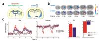
Pharmacological MRI combined with DREADD-technology enables
detection of induced brain activity in projections relevant for
feeding behavior
Tessa J.M. Roelofs1,2, Geralda A.F. van Tilborg1,
Mieneke C.M. Luijendijk2, Roger A.H. Adan2,
and Rick M. Dijkhuizen1
1Biomedical MR Imaging and Spectroscopy Group,
Center for Image Sciences, University Medical Center
Utrecht, Utrecht, Netherlands, 2Translational
Neurosciences, Brain Center Rudolf Magnus, University
Medical Center Utrecht, Utrecht, Netherlands
With the aim to develop a novel MRI-based approach for
detection of activation in neuronal networks associated with
feeding behavior in a rat model, we evaluated the potential
of pharmacological MRI (phMRI) to detect DREADD (Designer
Receptor Exclusively Activated by Designer Drug)-evoked
neuronal activity. BOLD phMRI was conducted under 1.5%
isoflurane anesthesia at 9.4T. Pharmacological activation
induced a significant BOLD response in DREADD-targeted
areas, which was confirmed by cFos-based
immunohistochemistry of neuronal activation. Our study shows
that phMRI allows detection of specific DREADD-evoked
neuronal activity, providing exciting opportunities to
assess network activity in association with feeding-related
behavioral phenotypes.
|
| |
17:00
|
0484.
 |
Deciphering the Functional Connectome of the External Globus
Pallidus with Electrical and Optogenetic Deep Brain
Stimulation-fMRI
Daniel Albaugh1, Nathalie Van Den Berge2,
Andrew Salzwedel3, Wei Gao3, Garret
Stuber4, and Yen-Yu Ian Shih5
1Curriculum in Neurobiology, UNC-Chapel Hill,
Chapel Hill, NC, United States, 2University
of Ghent, Ghent, Belgium, 3Cedars-Sinai
Medical Center, Los Angeles, CA, United States, 4Psychiatry,
UNC-Chapel Hill, Chapel Hill, NC, United States, 5Biomedical
Research Imaging Center, UNC-Chapel Hill, Chapel Hill, NC,
United States
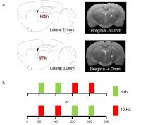
In this study, we unraveled the circuit and network
connectivity of the rodent external globus pallidus (GPe),
both in the healthy animals and a parkinson's disease model.
We also employed multiple stimulation types (electrical and
optogenetic), as well as fMRI modalities (evoked-fMRI and
functional connectivity analyses) to provide an exhaustive
analysis of this dynamic brain nucleus.
|
| |
17:12
|
0485.
 |
Study of the Transfer Functions of Hippocampal Subfields during
a Spatial Memory Task using High-Resolution fMRI
Xiaowei Zhuang1, Zhengshi Yang1, Tim
Curran2, and Dietmar Cordes1,2
1Cleveland Clinic Lou Ruvo Center for Brain
Health, Las Vegas, NV, United States, 2Department
of Psychology and Neuroscience, University of Colorado
Boulder, Boulder, CO, United States
In this abstract, the input/output transfer relationship in
human hippocampal subfields (mainly CA1, CA3, and DG) was
studied using fMRI during a spatial memory task with
increments in the change of FOV in the stimuli. Whole brain
activation was obtained for all lure v/s control contrasts.
Mean activation t value for each hippocampal subregions
(CA1, CA2&3 and CA4&DG) was extracted, averaged over all the
subjects and plotted against FOV changes to compare with the
existing models. K-means clustering was then applied. Data
from one of the k-means clusters showed a pattern
separation/completion curve similar to the animal model.
|
| |
17:24
|
0486.
 |
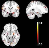
Visualizing adaptation of the central serotonin circuit in the
living brain
Bechara J. Saab1, Joanes Grandjean2,
Alberto Corcoba3, Martin C. Kahn4,
Louise A. Upton4, Erich Seifritz1,
Fritjof Helmchen5, Isabelle Mansuy1,
Edward O. Mann4, and Markus Rudin2
1University of Zurich, Zurich, Switzerland, 2University
and ETH Zurich, Zurich, Switzerland, 3EPFL,
Lausanne, Switzerland, 4University
of Oxford, Oxford, United Kingdom, 5University
and ETH Zurich, Zuerich, Switzerland
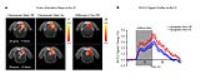
Mouse functional MRI was used to investigate the effect of
selective stimulation of serotonergic neurons of the dorsal
raphe via channelorodopsin-mediated optical control.
Electrophysiological recordings in the nucleus and in
projection areas confirmed neuronal activity changes upon
illumination with blue light. Acute pharmacological
modulation with fluoxetine, a serotonin reuptake inhibitor,
lead to increased CBV response upon 5HT release, while
animal restraint prior to measurements lead to a reduction
of the elicited response. This study demonstrates the
feasibility to assess a neurotransmitter function
non-invasively at a whole brain level and investigate
alterations in mood-controlling systems.
|
| |
17:36
|
0487.
 |
Direct mapping of functional connectivity with a novel
MR-compatible high resolution brain stimulation array
Sung-Ho Lee1, Hsin-Yu Lai1, Yu-Chieh
Jill Kai1, You-Yin Chen2, and Yen-Yu
Ian Shih1
1University of North Carolina at Chapel Hill,
Chapel Hill, NC, United States, 2Department
of Biomedical Engineering, University of National Yang Ming,
Taipei, Taiwan
In this study, we aim to provide in vivo evidence of using
this novel electrode array for selective deep brain
stimulation (DBS) in rats with simultaneous fMRI readouts.
This novel development opens up a new avenue to explore and
validate functional connectivity in the brain with a
resolution and specificity that cannot be achieved by
traditional fMRI or fcMRI approach.
|
| |
17:48
|
0488.
 |
Line scanning BOLD fMRI upon optogenetic stimulation - Permission Withheld
Franziska Albers1, Florian Schmid1,
Lydia Wachsmuth1, and Cornelius Faber1
1Department of Clinical Radiology, University of
Münster, Münster, Germany
With the line scanning technique BOLD responses can be
recorded with 50 ms temporal resolution. Here line scanning
fMRI was combined with optogenetic stimulation of excitatory
neurons in rat somatosensory cortex. The light for
optogenetic stimulation was delivered by an optical fiber
implanted in the brain. It was possible to compare BOLD
responses upon optogenetic and sensory stimulation with high
temporal resolution and shorter times to reach half maximum
were found for optogenetic stimulation. Furthermore the
spatial resolution offered the possibility to observe
cortical layer-specific BOLD signals.
|
|