| |
16:00
|
0489.
 |
DC plasma coils for MRI 
Vincent Oltman Boer1 and
Esben Thade Petersen1
1Danish Research Centre for Magnetic Resonance,
Centre for Functional and Diagnostic Imaging and Research,
Copenhagen University Hospital Hvidovre, Hvidovre, Denmark
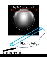
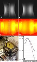
Coupling between conducting structures is one of the major
design limitations for in-bore MRI equipment. Here we show
how switchable plasma conductors can be used in a direct
current (DC) MR coil for B0field manipulation.
This can be applied in for example B0 shimming,
signal de-phasing or ultimately even gradient coil design.
|
| |
16:12
|
0490.
 |
Integration of Miniaturized Ultrasound and Single-Sided,
Low-Field MRI
Cheng Chen1, Mason Greer1, Michael
Twieg1, Mark A. Griswold1,2, and
Soumyajit Mandal1
1Department of Electrical Engineering and
Computer Science, Case Western Reserve University,
Cleveland, OH, United States, 2Department
of Radiology, Case Western Reserve University and University
Hospitals of Cleveland, Cleveland, OH, United States
Ultrasound (US) and magnetic resonance (MR) are two
well-established imaging modalities with largely
complementary contrast mechanisms. We propose and
experimentally evaluate the feasibility of a fundamentally
new tool; miniaturized two-dimensional (2-D) US collocated
with a one-dimensional (1-D) single-sided MR system for
bimodal imaging in portable or wearable form factors. The
proposed system will be capable of scheduling both
measurements in real-time, thus enabling closed-loop
operation in which the output of one sensor is used to
optimize the operation of the other. We study the
feasibility of such a system and show preliminary
experimental results obtained by combining a commercial US
imaging system with a custom single-sided planar MR sensor.
|
| |
16:24
|
0491.
 |
Traveling Internal Plane-wave Synthesis for Uniform B1(+) in
High Field MRI
Adam W Anderson1
1Biomedical Engineering, Vanderbilt University,
Nashville, TN, United States
Image quality in high field MRI is limited by B1 inhomogeneity.
This work describes a new approach to improving B1 homogeneity
using parallel transmission. Rather than transmitting a
conventional traveling wave, which is diffracted and
reflected by the human body, thereby creating a non-uniform
internal field, the new method seeks a solution to the
inverse problem—what external field produces a traveling
plane wave within the body? Simulations suggest dramatic
improvements in B1 homogeneity can be obtained given a
sufficient number of transmitted field modes.
|
| |
16:36
|
0492.
 |
Magnetic Pebbles – Materials with Controllable Magnetism for
Compact, Low-Power Shim Units
David Otto Brunner1, Simon Gross1,
Jonas Reber1, and Klaas Paul Pruessmann1
1Institute for Biomedical Engineering, University
and ETH Zurich, Zurich, Switzerland
B0 shimming
with very high channel count encounters many implementation
problems due to the size and current handling requirements
of the shim units. Here we present an approach using
distributions of ferromagnetic materials with controllable
magnetic moments to generate shim fields. These units are
small, require only low currents and can hence be
implemented in large numbers into RF receive arrays.
|
| |
16:48
|
0493.
 |
Size-adaptable “Trellis” receive array concept for knee imaging
Graham C Wiggins1, Bei Zhang1, and
Barbara Dornberger2
1Center for Advanced Imaging Innovation and
Research (CAI2R) and Center for Biomedical Imaging, New York
University School of Medicine, New York, NY, United States, 2Siemens
Healthcare, Erlangen, Germany
For optimal performance an array should conform closely to
the anatomy being imaged. Knee coils typically have rigid
formers which must be large enough to accommodate most
subjects, but which necessarily are not optimal for small
ones. We present here a cylindrical surface coil array which
can adapt in size while maintaining good tuning, match and
decoupling. It is built on a trellis-like structure which
controls the configuration and morphs the surface coils.
|
| |
17:00
 |
0494.
 |
Doppler Ultrasound Triggering for Cardiac Magnetic Resonance
Imaging at 7 Tesla
Fabian Kording1, Christian Ruprecht1,
Bjoern Schoennagel1, Mathias Kladeck Kladeck1,
Jin Yamamura1, Gerhard Adam1, Juliane
Goebel2,3, Kai Nassenstein2, Stefan
Maderwald3, Harald Quick3,4, and
Oliver Kraff3
1Department of Diagnostic and Interventional
Radiology, University Medical Center Hamburg, Hamburg,
Germany, 2Department
of Diagnostic and Interventional Radiology and
Neuroradiology, University Hospital, University
Duisburg-Essen, Essen, Germany, 3Erwin
L. Hahn Institute for Magnetic Resonance Imaging, University
Duisburg-Essen, Essen, Germany, 4High
Field and Hybrid MR Imaging, University Hospital, University
Duisburg-Essen, Essen, Germany, Essen, Germany
Cardiac synchronization for magnetic resonance imaging at
ultra-high-field MRI remains a challenge as disturbances in
the inherent electrical measurement of the ECG increase with
field strength. An ultrasound transducer and transmission
line was developed and the feasibility of Doppler Ultrasound
as an alternative method for cardiac synchronization was
evaluated in terms of safety concerns, signal and image
quality. The transmission line and transducer did not
disturb the transmit RF field or image homogeneity and were
approved for RF safety. Doppler Ultrasound was successfully
applied for cardiac synchronization without signal
disturbances and represents a promising alternative for
ultra-high field CMR.
|
| |
17:12
|
0495.
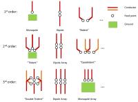 |
The Multi-Pole Antenna Array
Qi Duan1, Natalia Gudino1, and Hellmut
Merkle1
1Laboratory of Functional and Molecular Imaging,
National Institute of Neurological Disorders and Stroke,
National Institutes of Health, Bethesda, MD, United States
In this work, we propose concepts of transmit arrays based
on combination of monopole and dipole antennas and their
variations for high field imaging. Based on these concepts,
transmit arrays for a variety of applications can be derived
based on parameters such as desired and possible transmit
field-of-view, number of available transmit ports, etc. For
illustration purpose, a special case of the second order
array, a.k.a. the Trident antenna, was built for spine or
posterior cortex imaging and tested on phantom at a 7T
scanner.
|
| |
17:24
|
0496.
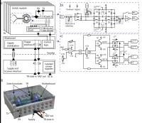 |
A High-Speed, High Power T/R Switching Frontend
David Otto Brunner1, Lukas Furrer2,
Markus Weiger1, Werner Baumberger2,
Thomas Schmid1, Jonas Reber1, Benjamin
Emanuel Dietrich1, Bertram Jakob Wilm1,3,
Romain Froidevaux1, and Klaas Paul Pruessmann1
1Institute for Biomedical Engineering, University
of Zurich and ETH Zurich, Zurich, Switzerland, 2ZSN
Center for Signal Processing and Communications, University
of Applied Sciences Winterthur, Winterthur, Switzerland, 3Skope
Magnetic Resonance Technologies, Zurich, Switzerland
Dead-times after the excitation pulse of the order of 1 µs
are required for imaging approaches for short T2 compounds
such as UTE, ZTE or SWIFT. Here we present a multi-channel
T/R interface box employing symmetrically biased T/R
switches which, in conjunction with a novel diode driver,
provide signal rise times of 350 ns. The unit further
comprises fiber-optic triggering, biasing, and malfunction
detection. Its performance is demonstrated by low artefact
ZTE scans with 500 kHz at 7T.
|
| |
17:36
|
0497.
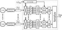 |
N-path frequency mixers for ultra-high density receive arrays
Michael Twieg1, Soumyajit Mandal1, and
Mark A Griswold1,2
1Electrical Engineering and Computer Science,
Case Western Reserve University, Cleveland, OH, United
States, 2Radiology,
Case Western Reserve University, Cleveland, OH, United
States
Dense MRI receiver arrays face challenges associated with RF
cabling, power consumption, and space required by on-coil RF
LNAs. On-coil frequency mixers and ADCs have been proposed
as solutions to these challenges. Here we propose the use of
passive N-path mixers implemented in CMOS for on-coil
frequency conversion. We demonstrate a prototype fabricated
in a 0.5µm CMOS process, and compare its measured and
simulated performance. We also show simulations of a similar
design in 65nm CMOS with greatly improved performance. The
improved version may handle multiple RF channels on a single
chip, and eliminates the need for RF LNAs entirely.
|
| |
17:48
|
0498.
 |
Progress Toward a Portable MRI System for Human Brain Imaging
J. Thomas Vaughan1, Bert Wang2,
Djaudat Idiyatullin1, Sung-min Sohn1,
Albert Jang1, Lance DelaBarre1, and
Michael Garwood1
1Center for Magnetic Resonance Research -
University of Minnesota, Minneapolis, MN, United States, 2Wang
NMR, Inc, Livermore, CA, United States
Critical magnet, imaging physics, RF and gradient technology
were built and tested to demonstrate the feasibility of a
portable 1.5T MRI system for imaging the brain in real world
environments. Feasibility is demonstrated.
|
|