| |
14:15
|
0147.
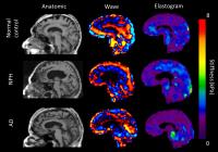 |
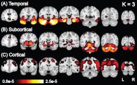
MR Elastography Demonstrates Unique Regional Brain Stiffness
Patterns in Dementias 
Mona ElSheikh1, Arvin Arani1, Avital
Perry2, Nealey Cray2, Fredric Meyer2,
David Lake1, Armando Manduca3, Kevin
Glaser1, Richard L Ehman1, and John
Huston1
1Radiology, Mayo Clinic, Rochester, MN, United
States, 2Neurosurgery,
Mayo Clinic, Rochester, MN, United States, 3Physiology
and Biomedical Engineering, Mayo Clinic, Rochester, MN,
United States
The development of
advanced MRI techniques has enabled noninvasive evaluation
of subtle changes of brain architecture in dementia. We
report a specific pattern of regional brain stiffness
changes using Magnetic Resonance Elastography (MRE) in three
different dementia groups: Alzheimer’s disease,
frontotemporal dementia, and normal pressure hydrocephalus.
MRE offers a potential biomarker to characterize the
viscoelastic properties of the brain in dementia patients,
and may have a role in the diagnosis and differentiation
between common subtypes of dementia.
|
| |
14:27
 |
0148.
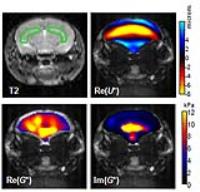
 |
High resolution MR elastography of the hippocampus reveals
differential tissue elasticity in Alzheimer's disease – a pilot
study
Andreas Fehlner1, Lea M Gerischer2,
Agnes Flöel2,3, Jürgen Braun4, and
Ingolf Sack1
1Department of Radiology, Charité -
Universitätsmedizin Berlin, Berlin, Germany, 2Department
of Neurology, Charité - Universitätsmedizin Berlin, Berlin,
Germany, 3NeuroCure
Clinical Research Center, Charité - Universitätsmedizin
Berlin, Berlin, Germany, 4Institute
of Medical Informatics, Charité - Universitätsmedizin
Berlin, Berlin, Germany
Multifrequency MR elastography (MMRE) was applied to 14
patients with Alzheimer's disease (AD) and compared to 14
age matched asymptomatic controls. We observed a marked
decrease of the white-matter complex shear modulus |G*|
in patients with AD. This reduction in |G*| was
even more pronounced in the hippocampal region. In this
region a diagnostic performance of 78% sensitivity and 92%
specificity (AUROC-value 0.918) was obtained based on a
viscoelasticity cutoff value of 0.9 kPa. In the future MMRE-measured
|G*| could serve as a quantitative imaging marker
for early diagnosis and progression monitoring of AD.
|
| |
14:39
|
0149.
 |
1H-[13C]-NMR Investigation of
Neuroprotective Action of Ayurvedic Formulation in AßPP-PS1
Mouse Model of Alzheimer’s Disease
Kamal Saba1, Niharika Rajnala1, and
Anant Bahadur Patel1
1NMR Microimaging and Spectroscopy, CSIR-Centre
for Cellular and Molecular Biology, Hyderabad, India
Alzheimer's disease (AD) is a progressive neurodegenerative
disorder. Currently no definite treatment available for AD.
We have examined the efficacy of Rasa Sindoor, an Ayurvedic
formulation, for the improvement of memory and neuronal
activity in AβPP-PS1 mouse model of AD. Neuronal metabolism
was followed by 1H-[13C]-NMR
spectroscopy together with an infusion of [1,6-13C2]glucose.
Our results indicate that the Rasa-Sindoor improved memory,
and excitatory and inhibitory neuronal metabolic activity in
AD mice.
|
| |
14:51
 |
0150.
 |
Brain phospholipid and energy metabolism in mild Alzheimer’s
disease and healthy aging: a 31P Magnetic Resonance Spectroscopy
study - Permission Withheld
Anne Rijpma1,2, Marinette van der Graaf3,4,
Olga Meulenbroek1,2, Marcel Olde Rikkert1,2,
and Arend Heerschap3
1Geriatric Medicine, Radboud university medical
center, Nijmegen, Netherlands, 2Radboud
Alzheimer Centre, Donders Institute for Brain, Cognition and
Behaviour, Radboud university medical center, Nijmegen,
Netherlands, 3Radiology
and Nuclear Medicine, Radboud university medical center,
Nijmegen, Netherlands, 4Paediatrics,
Radboud university medical center, Nijmegen, Netherlands
In this study we assessed phospholipid and energy metabolism
in patients with mild Alzheimer’s disease and healthy
age-matched control subjects by 3D 31P
MRS imaging. Four brain regions were investigated: left and
right hippocampus, anterior cingulate cortex, and
retrosplenial cortex. Disease specific differences as well
as differences between brain regions were found.
|
| |
15:03
|
0151.
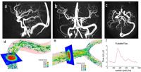 |
4D Flow MRI for assessing flow pulsatility along the carotid
siphon in Alzheimer’s disease
Leonardo A Rivera-Rivera1, Tilman Schubert2,
Kevin M Johnson1, Sterling C Johnson3,
Oliver Wieben1,2, and Patrick Turski2
1Medical Physics, University of Wisconsin
Madison, Madison, WI, United States, 2Radiology,
University of Wisconsin Madison, Madison, WI, United States, 3Medicine,
University of Wisconsin Madison, Madison, WI, United States
Cerebral arteries are often morphologically altered and
dysfunctional in Alzheimer’s disease (AD). In this study, 4D
flow MRI was used to assess flow pulsatility along the
carotid siphon in patients with AD, mild cognitive
impairment (MCI) and in healthy age matched controls. We
found the physiologic dampening of pulsatility along the
distal ICA is significantly diminished in patients with AD.
With the large volume coverage and high temporal and spatial
resolution, 4D flow MRI can provide additional biomarkers of
vascular health that can contribute to the identifying
patients who could benefit from interventions to improve
circulatory system functions.
|
| |
15:15
|
0152.
 |
In Vivo Visualization of Iron-Rich Amyloid Plaques In
Cholesterol-Fed Rabbits using Clinical Field-Strength Magnetic
Resonance Imaging
Yuanxin Chen1, Yong Wang1,2, Kem A
Rogers1, John A Ronald1, and Brian K
Rutt3
1Western University, London, ON, Canada, 2Lawson
Health Research Institute, London, ON, Canada, 3Stanford
University, Stanford, CA, United States
Hypercholesterolemia is a risk factor for AD and promotes
increased production of beta-amyloid protein. Our lab has
developed a rabbit model of AD by enriching the diets of
rabbits with low amounts of cholesterol. In this study, we
combined this cholesterol-fed rabbit model of AD with
iron-sensitive, high-resolution MRI and demonstrated
non-invasive in vivo visualization of AD plaques throughout
the brains of these animals. The imaging techniques have
been developed and optimized using a clinical field strength
scanner (3T), which is an important step towards clinical
application in human AD patients.
|
| |
15:27
|
0153.
 |
Latent Atrophy Factors in Alzheimer's Disease
Xiuming Zhang1, Elizabeth C. Mormino2,
Reisa A. Sperling2, Mert R. Sabuncu3,4,
and B.T. Thomas Yeo1,3,5
1ASTAR-NUS Clinical Imaging Research Centre,
Department of Electrical and Computer Engineering, Singapore
Institute for Neurotechnology and Memory Networks Program,
National University of Singapore, Singapore, Singapore, 2Department
of Neurology, Massachusetts General Hospital/Harvard Medical
School, Charlestown, MA, United States, 3Martinos
Center for Biomedical Imaging, Massachusetts General
Hospital/Harvard Medical School, Charlestown, MA, United
States, 4Computer
Science and Artificial Intelligence Laboratory,
Massachusetts Institute of Technology, Cambridge, MA, United
States, 5Centre
for Cognitive Neuroscience, Duke-NUS Graduate Medical
School, Singapore, Singapore
Alzheimer's disease (AD) is the most common form of dementia
and greatly heterogeneous. Here we develop a model of the
heterogeneity of AD-related atrophy, demonstrating that most
AD dementia patients and at-risk nondemented participants
express multiple latent atrophy factors to varying degrees.
Our study also demonstrates that these atrophy factors are
associated with distinct cognitive decline trajectories
across the preclinical and clinical stages. Our results
provide a framework by which biomarker readouts could
potentially predict disease progression at the individual
level. Our analytic strategy is general and might be
utilized to discover subtypes within and across other
heterogeneous brain disorders.
|
| |
15:39
|
0154.
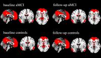 |
Association of Alzheimer’s disease GWAS loci with default mode
network
fan su1
1southeast university, nanjing, China, People's
Republic of
To investigate the altered pattern of DMN in amnestic mild
cognitive impairment (aMCI) subjects and the genetic factors
that lead to the DMN dysfunctions, 87 individuals with aMCI
and 131 matched healthy controls were recruited and an
average 3-year follow-up study was performed. We studied the
differences of DMN between aMCI subjects and healthy
controls at baseline and how the DMN changed over time.
Regression analyses were performed to explore whether the
GRS influence the DMN dysfunctions. We observed that DMN
disengage in the early stage of AD and the combined effect
of AD-related loci influence the DMN pattern.
|
| |
15:51
|
0155.
 |
The effect of Alzheimer's disease on the viscoelasticity of the
mouse brain under the influence of enriched environment. - Permission Withheld
Jing Guo1, Tonia Munder2, Charlotte
Klein2, Anna Pfeffer2, Jürgen Braun3,
Barbara Steiner2, and Ingolf Sack1
1Department of Radiology, Charité - University
Medicine Berlin, Berlin, Germany, 2Department
of Neurology, Charité - University Medicine Berlin, Berlin,
Germany, 3Institute
of Medical Informatics, Charité - University Medicine
Berlin, Berlin, Germany
MRE was used to study environmental influences on
viscoelasticity of the murine hippocampus in Alzheimer's
disease (AD). In wild type control mice, hippocampal
viscosity was significantly increased within 6 months while
elasticity remained unchanged. This suggests that
environment-stimulated neuronal proliferation adds mobile
elements to the mechanical matrix of the brain which
increases mechanical attenuation properties. Within 6
months, AD caused a decline of hippocampal viscosity only in
the enriched environment while standard mouse remained
unaffected suggesting that AD in an early phase primarily
affects new neurons in the murine hippocampus.
|
| |
16:03
|
0156.
 |
A preliminary study on MR amide proton imaging in patients with
Alzheimer’s disease and mild cognitive impairment - Video Not Available
Rui Wang1, Chunmei Li1, Yongming Dai2,
Dantao Peng3, Xuna Zhao4, and Min Chen1
1Radiology, Beijing Hospital, Beijing, China,
People's Republic of, 2Philips
Heathcare, Shanghai, China, People's Republic of, 3China-Japan
Friendship Hospital, Beijing, China, People's Republic of, 4Philips
Heathcare, Beijing, China, People's Republic of
The aim of this study is to evaluate the feasibility of MR
amide proton transfer (APT) imaging for the detection of
cerebral abnormalities in patients with Alzheimer’s disease
(AD) and amnestic mild cognitive impairment (aMCI), and to
explore its clinical utility. Twenty-one AD patients, 11
aMCI patients and 19 normal controls (NC) underwent APT MR
imaging. The magnetic resonance ratio asymmetry (MTRasym)
values at 3.5ppm of bilateral hippocampi, temporal white
matter regions, occipital white matter regions and cerebral
peduncles were measured on the oblique APT images. We found
that MTRasym(3.5ppm)asym in bilateral hippocampi showed a
consistently increasing trend from NC to MCI, to AD.
MTRasym(3.5ppm) values of bilateral hippocampi were
significantly negatively correlated with MMSE. Our results
suggested that APT imaging is a useful tool to diagnose
early AD and monitor the disease.
|
|