| |
16:00
|
0770.
 |
Perivascular Space Analysis in Non-lesional Epilepsy: Exploring
a Biomarker for Epilepsy 
Rebecca Emily Feldman1, Jack Rutland2,
Bradley Neil Delman3, Jiyeoun Yoo4,
Madeline Cara Fields4, Lara Vanessa Marcuse4,
and Priti Balchandani1
1Translational and Molecular Imaging Institute,
Icahn School of Medicine at Mount Sinai, New York, NY,
United States, 2Wake
Forest University, Winston-Salem, NC, United States, 3Radiology,
Icahn School of Medicine at Mount Sinai, New York, NY,
United States, 4Neurology,
Mount Sinai Hospital, New York, NY, United States
Epilepsy is a chronic condition, affecting approximately
150,000 people in the United States. 7T MRI facilitates the
visualization of the brain with unprecedented resolution and
contrast. Perivascular spaces (PVS) have been reported in
previous work but with uncertain significance. However, due
to the increased resolution enabled at 7T, PVSs are detected
with increasing frequency, both in healthy volunteers and in
epilepsy patients. We investigated the symmetry in the
distribution of PVSs in the brains of non-lesional epilepsy
patients.
|
| |
16:12
|
0771.
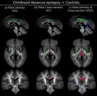 |
Structural Connectivity Changes in Refractory Childhood Absence
Epilepsy
Graeme Jackson1,2,3,4, Farnoosh Sadeghian1,
Patrick Carney1, David Raffelt1,
Fernando Calamante1,2, and Alan Connelly1,2
1The Florey Institute of Neuroscience and Mental
Health, Melbourne, Australia, 2The
Florey Department of Neuroscience and Mental Health, The
University of Melbourne, Melbourne, Australia, 3Department
of Medicine, The University of Melbourne, Melbourne,
Australia, 4Department
of Neurology, Austin Health, Melbourne, Australia
Childhood absence epilepsy (CAE) is a common neurological
condition. Here we assessed white matter connectivity using
fixel-based analysis (FBA) and grey matter structure using
voxel-based morphometry in adult patients with refractory
CAE. We identified increased grey matter volume in frontal
lobe as well as decreased fibre connectivity in superior
longitudinal fasciculi, right cingulum, motor area of corpus
callosum and cerebellar peduncles. Our results reinforce the
concept that the midline frontal areas are critically
involved in the phenotype of generalised spike and wave
discharges. These structural connectivity changes in CAE
could be either developmental or as a consequence of
seizures.
|
| |
16:24
|
0772.
 |
7 tesla MRI in the pre-surgical evaluation of 26 patients with
focal epilepsy
Tim J Veersema1, Cyrille H Ferrier1,
Pieter van Eijsden1, Peter H Gosselaar1,
Fredy Visser2,3, Jaco JM Zwanenburg2,4,
Hans Hoogduin2, Gerárd AP de Kort2,
Jeroen Hendrike2, and Kees PJ Braun1
1Department of Neurology and Neurosurgery, Brain
Center Rudolf Magnus, University Medical Center Utrecht,
Utrecht, Netherlands, 2Department
of Radiology, University Medical Center Utrecht, Utrecht,
Netherlands, 3Philips
Healthcare, Best, Netherlands, 4Image
Sciences Institute, University Medical Center Utrecht,
Utrecht, Netherlands
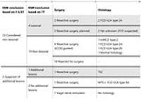
For this series we assessed all 26 epilepsy patients who
underwent 7T MRI for pre-surgical evaluation in our center,
and whose scans (both 7T and lower field) were discussed
during epilepsy surgery meetings (ESM). We compared the
conclusions of the visual assessments of 1.5T or 3T, and 7T
MRI as agreed upon by the ESM team. 7T MRI holds a promise
to improve identification of epileptogenic structural
abnormalities in patients with intractable epilepsy. In our
series of 26 patients with refractory focal epilepsy,
multidisciplinary evaluation of 7T MRI identified additional
lesions not seen on lower-field MRI in five patients
(19.2%).
|
| |
16:36
|
0773.
 |
In Vivo Whole-Brain T1-rho Mapping in evaluation of Mesial
Temporal Lobe epilepsy
Xixi Zhao1, Junling Wang1, Xiangliang
Tan1, Xiang Xiao1, Jiajun Zhang1,
Yingjie Mei2, Queenie Chan3, and Yikai
Xu1
1Department of Medical Imaging Center, Nanfang
Hospital, Southern Medical University, Guangzhou, China,
People's Republic of, 2Philips
Healthcare, Guangzhou, China, People's Republic of, 3Philips
Healthcare, HongKong, China, People's Republic of
In human brain, T1ρ has been proven to be relevant with the
macromolecular composition of tissues, and supposed to be
sensitive to neuronal degeneration. We used T1rho MR imaging
to investigate the variations in T1rho values of subcortical
gray matter structures automatic-drawn using FIRST
segmentation among temporal lobe epilepsy patients and the
underlying relation between the significantly altered T1rho
values or volumes of subcortical structures and duration of
epilepsy or age at epilepsy onset. Our results demonstrate
the feasibility of ROI-wise analysis by atlas-based
segmentation of T1rho imaging among mTLE patients
|
| |
16:48
|
0774.
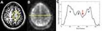 |
Sodium MRI for Evaluation of Sodium Ion Homeostasis in Epilepsy:
Clinical Implementation and Initial Impressions
Timothy Michael Shepherd1, Yongxian Qian1,
Karthik Lakshmanan1, Ruben Kuzniecky2,
Graham Wiggins1, and Fernando Boada1
1Radiology, New York University, New York, NY,
United States, 2Neurology,
New York University, New York, NY, United States
The detection and localization of sodium tissue
abnormalities in patients with epilepsy may have potential
to improve seizure localization, identify effective
pharmacotherapy and/or provide prognostic information for
individual patients. Here, we report initial results
evaluating a newly developed coil for performing 23Na MRI at
3-T in three patients with epilepsy.
|
| |
17:00
|
0775.
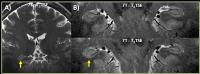 |
7T MRI detection of epileptogenic foci in previously
non-lesional patients with focal epilepsy - Permission Withheld
Rebecca Emily Feldman1, Bradley Neil Delman2,
Hadrien A Dyvorne1, Jiyeoun Yoo3,
Madeline Cara Fields3, Lara Vanessa Marcuse3,
and Priti Balchandani1
1Translational and Molecular Imaging Institute,
Icahn School of Medicine at Mount Sinai, New York, NY,
United States, 2Radiology,
Icahn School of Medicine at Mount Sinai, New York, NY,
United States,3Neurology, Mount Sinai Hospital,
New York, NY, United States
Epilepsy affects over 150,000 people in the United States.
Thirty percent of epilepsy is refractory to pharmacotherapy,
and in these cases surgery may be curative. There are focal
epileptogenic lesions, amenable to surgery, which are not
visualized by current imaging protocols. 7T MRI scanners may
increase the conspicuity of epileptogenic lesions and
provide more accurate delineation of lesion boundaries.
Reported are the results for a patient study, with
comparison to healthy controls, to assess the value of 7T
imaging to reveal subtle abnormalities acting as
epileptogenic foci in patients with focal epilepsy who have
non-lesional diagnostic MRI scans.
|
| |
17:12
 |
0776.
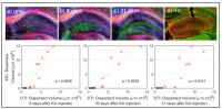 |
MRI monitoring of epileptogenesis with direct histological
validation
Niels Leonard Schwaderlapp1, Philipp Janz2,
Ute Häussler2, Jan Korvink3, Dominik
Elverfeldt1, Jürgen Hennig1, Carola
Haas2, and Pierre LeVan1
1Medical Physics, University Medical Center
Freiburg, Freiburg, Germany, 2Experimental
Epilepsy Research, University Medical Center Freiburg,
Freiburg, Germany, 3Institut
für Mikrostrukturtechnik, Karlsruher Institut für
Technologie, Karlsruhe, Germany
Cellular-level pathological changes in the kainate mouse
model of temporal lobe epilepsy (TLE) have been
well-characterized immunohistochemically (IHC) and include
neuronal injury followed by granule cell dispersion. In this
work, we demonstrate the possibility to non-invasively track
granule cell dispersion and neuronal injury using diffusion
imaging and 1H-spectroscopy. The volume of the
dispersed granule cell layer quantified by DTI and the
initial injury reflected by a reduction of NAA and glutamate
are quantitatively validated with IHC and can be used as
early markers of epileptogenicity in this mouse model of
TLE.
|
| |
17:24
|
0777.
 |
Decreased Fibre Density in Frontal Lobe Epilepsies related to
DEPDC5 mutations
David Raffelt1, Farnoosh Sadeghian1,
Brigid Regan2, Sarah Garry2, Samuel
Berkovic2, Ingrid Scheffer2, and Alan
Connelly1,2
1Florey Institute of Neuroscience, Melbourne,
Australia, 2Department
of Medicine, University of Melbourne, Melbourne, Australia
Mutations in the gene DEPDC5 cause up to 12% of Familial
Focal Epilepsy with Variable Foci. In this work we performed
a fixel-based analysis of diffusion MRI data to understand
how white matter might be altered in patients with DEPDC5
mediated frontal lobe epilepsy (FLE). We identified
significant reductions in fibre density in several pathways,
including the superior longitudinal fasciculi, corpus
callosum, inferior longitudinal fasciculus and cingulum. We
also investigated FLE mediated by KCNT1 mutation, and found
similar pathways affected. In KCNT1+ve subjects, pathways
had reduced cross-section, suggesting the observed effects
may be related to development and not seizure effects.
|
| |
17:36
|
0778.
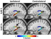 |
Automated fibre quantification of the fornix predicts outcome
after surgery for intractable temporal lobe epilepsy - Permission Withheld
Russell Glenn1, Leonardo Bonilha1,
Barbara Kreilkamp2, Mark P Richardson3,
Bernd Weber4, and Simon S Keller2
1Medical University of South Carolina,
Charleston, SC, United States, 2University
of Liverpool, Liverpool, United Kingdom, 3King's
College London, London, United Kingdom, 4University
Hospital Bonn, Bonn, Germany
Imaging markers of postoperative seizure control in
refractory temporal lobe epilepsy (TLE) would provide a
useful clinical tool for surgical decision making. In the
present diffusion tensor imaging study, we report that
regional tissue characteristics of the fornix ipsilateral to
the side of intended resection are related to postoperative
seizure control in patients with TLE. Interestingly, areas
found to be abnormal only in patients with a suboptimal
outcome were located outside the margins of resection. The
identification of fornical abnormalities outside the area of
intended resection may be an important prognostic marker of
suboptimal seizure control after temporal lobe surgery.
|
| |
17:48
|
0779.
 |
Individual measures of network efficiency in patients with
epilepsy based on cortical thickness
Gerhard Drenthen1,2, Marielle Vlooswijk2,3,
Marian Majoie2, Paul Hofman1,2, Albert
Aldenkamp2,3, Walter Backes1,2, and
Jacobus Jansen1,2
1Department of Radiology and Nuclear Medicine,
Maastricht University Medical Center, Maastricht,
Netherlands, 2School
for Mental Health and Neuroscience, Maastricht University,
Maastricht, Netherlands,3Department of Neurology,
Maastricht University Medical Center, Maastricht,
Netherlands
Brain network analysis that infers on interregional
correlations of anatomical features usually makes use of
intersubject correlation matrices that characterize
variations over subjects. Here, a novel method is introduced
that provides measures of network efficiency on an
individual basis in patients with epilepsy. To this end, for
each participant a measure of deviation from a group of
healthy controls is calculated, and compared to the
small-world parameters (clustering coefficient and minimum
path length) of a reference graph obtained for the native
control group. Results show that patients with epilepsy
exhibit a less efficient network compared to controls.
|
|