| |
10:00
 |
0337.
 |
Advances in Prospective Motion Correction with Gradient Tones 
Maximilian Haeberlin1, Alexander Aranovitch1,
Bertram Wilm1, David Otto Brunner1,
Benjamin Dietrich1, Barmet Christoph2,
and Klaas Paul Pruessmann1
1Institute for Biomedical Engineering, University
of Zurich and ETH Zurich, Zurich, Switzerland, 2Skope
Magnetic Resonance Technologies, Zurich, Switzerland
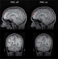
A system for prospective motion correction using field
probes and gradient tones is presented that is independent
of sequence parameters and thus compatible with clinically
relevant scans. An examples of a successfully corrected
MPRAGE sequence is shown and the bandwidth and the amount of
unintentional head motion is measured during a 32 min. scan.
|
| |
10:12
|
0338.
 |
High frequency orientation estimates for fast real-time motion
correction using vector observations of gravity and the static
magnetic field (B0).
Adam M.J. van Niekerk1, Paul Wighton2,3,
Ali Alhamud1, Matthew D. Tisdall2,3,
Andre J.W. van der Kouwe2,3, and Ernesta M.
Meintjes1
1Human Biology, MRC/UCT Medical Imaging Research
Unit, University of Cape Town, Cape Town, South Africa, 2Athinoula
A. Martinos Center, Massachusetts General Hospital, Boston,
MA, United States,3Radiology, Harvard Medical
School, Boston, MA, United States
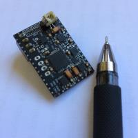
In this study we propose a novel approach to motion
correction in MRI that separates the challenges of tracking
orientation and translation. We developed an external
hardware device capable of high frequency orientation
estimates independent of the pulse sequence. The device
takes vector observations of gravity and the MRI scanner’s
static magnetic field (B0) and is therefore free from many
constraints of some existing external motion tracking
techniques. Most notably, no scanner specific calibration is
required and the device can be miniaturised. Translation
estimates are achieved through the use of 3 high-speed
orthogonal navigators. Line by line rigid body motion
correction is implemented in a spoiled gradient echo pulse
sequence.
|
| |
10:24
|
0339.
 |
Prospective Motion Correction Using External Tracking and
Intrinsic Motion Information
Michael Herbst1,2, Aditya Singh1,
Benjamin Knowles2, Maxim Zaitsev2, and
Thomas Ernst1
1JABSOM, University of Hawaii, Honolulu, HI,
United States, 2Medical
Physics, University Medical Center Freiburg, Freiburg,
Germany
Prospective motion correction with external tracking was
applied to high resolution diffusion weighted imaging, using
a phase-segmented EPI readout strategy. To detect and
correct for residual errors during prospective motion
correction, real-time volumetric registration provides
continuous feedback to the acquisition.
|
| |
10:36
|
0340.
 |
A Comparison of 19F NMR Field Probes and an Optical Camera
System for Motion Tracking
Martin Eschelbach1, Alexander Loktyushin1,
Paul Chang1,2, Jonas Handwerker3, Jens
Anders3, Anke Henning1,4, Axel
Thielscher1,5,6, and Klaus Scheffler1,7
1High Field MR Center, Max Planck Institute for
biol. Cybernetics, Tuebingen, Germany, 2IMPRS
for Cognitive and Systems Neuroscience University of
Tuebingen, Tuebingen, Germany, 3Institute
of Microelectronics, University of Ulm, Ulm, Germany, 4Institute
for Biomedical Engineering, ETH Zürich, Zurich, Switzerland, 5Univ
Copenhagen, Hvidovre Hosp, Danish Res Ctr Magnet Resonance,
Hvidovre, Denmark, 6Tech
Univ Denmark, Biomed Engn Sect, Lyngby, Denmark, 7Department
of Biomedical Magnetic Resonance, University Tuebingen,
Tuebingen, Germany
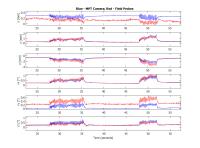
The goal of this study is to evaluate and compare motion
tracking with two different modalities: NMR field probes and
an optical MPT (Moiré Phase Tracking) camera system. This
was done by simultaneously measuring the manually induced
motion of a spherical phantom with both systems. Our
experimental results indicate that the motion patterns
measured with both methods are in good agreement. However,
the accuracy of the motion estimates from the field probe
measurements are of an order of magnitude worse than the
camera's tracking results.
|
| |
10:48
|
0341.
 |
Fast calculation of phase accumulation due to pulsed gradients
for arbitrary rigid body motion
Patrick Hucker1, Michael Dacko1,
Michael Herbst2, Ben Knowles1, and
Maxim Zaitsev1
1Dept. of Radiology · Medical Physics, University
Medical Center Freiburg, Freiburg, Germany, 2John
A. Burns School of Medicine, University of Hawaii, Honolulu,
HI, United States
A compact solution for phase calculation due to arbitrary
rigid body motion based on screw theory is presented. The
proposed approach allows for rapid and quantitatively
accurate calculations of the phase induced by the switching
magnetic field gradients using motion tracking information
e.g. from a motion tracking camera. The ability of
predicting phase accumulation due to motion in presence of
gradients is instrumental for achieving better correction of
the motion-induced data inconsistencies for MR pulse
sequences with extended signal preparation or readout
periods.
|
| |
11:00
|
0342.
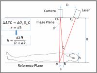 |
Motion Detection and Correction for Carotid Artery Wall Imaging
using Structured Light
Jin Liu1, Huijun Chen2, Jinnan Wang1,
Niranjan Balu1, Haining Liu1, and Chun
Yuan1
1University of Washington, Seattle, WA, United
States, 2Tsinghua
University, Beijing, China, People's Republic of
Carotid artery wall MRI is often affected by complex neck
motion. We aimed to separate different motion components and
correct them for better carotid artery wall delineation
using structured light system. A healthy volunteer was
scanned for 2D carotid MRI. It was demonstrated that
voluntary abrupt motion, unconscious bulk motion and
involuntary respiration can all be detected effectively.
Both abrupt motion and bulk neck shift can be corrected for
better vessel wall delineation, but the duration of abrupt
motion can affect motion correction effectiveness. Bulk neck
shift distance optimization by maximizing sharpness can
future reduce motion artifact.
|
| |
11:12
|
0343.
 |
Motion-corrected K-space Reconstruction for High Resolution
Multi-shot Diffusion Imaging
Fuyixue Wang1, Zijing Dong1, Xiaodong
Ma1, Erpeng Dai1, Zhe Zhang1,
and Hua Guo1
1Center for Biomedical Imaging Research,
Department of Biomedical Engineering, School of Medicine,
Tsinghua University, Beijing, China, People's Republic of
Recently, several techniques have been developed to be
capable of correcting shot-to-shot phase variations of
multi-shot acquisition in order to obtain diffusion images
with high spatial resolution. However, longer acquisition
time of multi-shot EPI makes these methods more sensitive to
bulk motion. In this work, we developed a novel k-space
based motion corrected reconstruction method for 2D
navigated multi-shot DWI. Motion simulations and in-vivo
head motion experiments validated the effectiveness of the
proposed method, which can remove the ghosting artifacts
from minuscule motion and the blurring from bulk motion.
|
| |
11:24
|
0344.
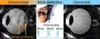 |
Robust MR eye scanning: blink detection and correction using
field probes
Joep Wezel1, Anders Garpebring2,
Andrew G. Webb1, Matthias J. van Osch1,
and Jan-Willem M. Beenakker3
1Radiology, Leiden University Medical Center,
Leiden, Netherlands, 2Radiation
Sciences, Umeå University, Umeå, Sweden, 3Ophthalmology,
Leiden University Medical Center, Leiden, Netherlands
Eye-blinks result in significant artifacts in ocular MRI
scans, often masking important clinical pathologies, such as
small ocular tumors. The aim of this study is to detect and
correct for these eye-blinks. We use a fluorine-based field
probe to detect these eye-blinks via changes in the local
magnetic field. The field probe measurements are linked to
the MR-scanner which subsequently automatically reacquires
the motion-corrupted part of k-space. This method
effectively corrects for the main origin of image artifacts
in ocular MRI, and thereby significantly improves the image
quality in a clinical setting.
|
| |
11:36
|
0345.
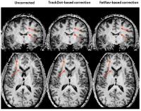 |
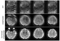
Head motion tracking and correction using discrete off-resonance
markers (trackDOTS) for high-resolution anatomical imaging at 7T
João Jorge1, Daniel Gallichan2, and
José P Marques3
1Laboratory for Functional and Metabolic Imaging,
École Polytechnique Fédérale de Lausanne, Lausanne,
Switzerland, 2Biomedical
Imaging Research Center, École Polytechnique Fédérale de
Lausanne, Lausanne, Switzerland, 3Donders
Institute, Radboud University, Nijmegen, Netherlands
High-resolution imaging can be significantly affected by
subject head motion. Here, we demonstrate the use of
discrete off-resonance MR markers (“trackDOTS”) in head
motion tracking and correction, for high-resolution
anatomical imaging. This approach relies on fast
1D-projection acquisitions (under 50ms per measurement)
which do not disturb the water signal. These measurements
were incorporated in an MP2RAGE sequence, and a 0.6mm
isotropic resolution image was acquired from a healthy
subject. Motion timecourses estimated from the trackDOTS
positions matched concomitant estimations performed with
FatNavs (with deviations of 0.09±0.08mm for translations and
0.20°±0.19° for rotations); MP2RAGE image quality was
visibly improved upon correction.
|
| |
11:48
|
0346.
 |
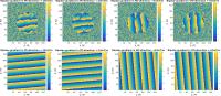
Amplified Magnetic Resonance Imaging (aMRI)
Samantha J Holdsworth1, Wendy W Ni1,
Greg Zaharchuk1, Michael E Moseley1,
and Mahdi S Rahimi1
1Lucas Center for Imaging, Department of
Radiology, Stanford University, Palo Alto, CA, United States
This work introduces a new visualization method called
amplified Magnetic Resonance Imaging (aMRI), which uses
Eulerian Video Magnification to amplify subtle spatial
variations in cardiac-gated brain MRI scans and magnify
brain motion. This approach reveals deformations of brain
structures and displacements of arteries due to cardiac
pulsatility, especially in the brainstem, cerebellum, and
spinal cord. aMRI has the potential for widespread neuro-
and non-neuro clinical application, because it can amplify
and characterize barely perceptible motion, and allows
visualization of biomechanical responses of tissues using
the heartbeat as an endogenous mechanical driver.
|
|