| |
16:00
 |
1020.
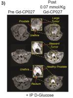 |
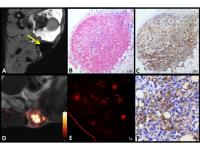
A zinc-sensitive MRI contrast agent differentiates healthy from
cancerous prostate in a transgenic prostate cancer model 
Veronica Clavijo Jordan 1,
Su-Tang Lo1, Christian Preihs1, Sara
Chirayil1, Wen-Hong Li1, Neil M Rofsky1,
and Dean Sherry1,2
1UT Southwestern Medical Center, Dallas, TX,
United States, 2UT
Dallas, Richardson, TX, United States
The prostate has the highest levels of Zn(II) in the
organism and there are marked differences in content between
the healthy, malignant, and benign hyperplastic prostate.
Given that accurate differential diagnosis between these
conditions is difficult non-invasively, we introduce
prostate Zn(II) as a MRI imaging biomarker. In this work we
use a Gd-based zinc sensor that can sensitively detect
glucose-stimulated intracellular release of Zn(II) in the
healthy, and malignant mouse prostate using a transgenic
adenocarcinoma model.
|
| |
16:12
|
1021.
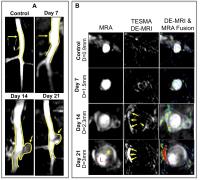 |
Assessment of abdominal aortic aneurysm progression using a
novel tropoelastin-specific MR contrast agent
Alkystis Phinikaridou1, Sara Lacerda1,
Begoña L Plaza1, Marcelo Andia2,
Silvia G Lorrio1, and René M Botnar1
1Biomedical Engineering, King's College London,
London, United Kingdom, 2Radiology,
Pontificia Universidad Católica de Chile, Santiago, Chile
The extracellular matrix proteins, elastin and collagen, are
the most important structural components of the vessel wall
that provide tensile strength and stability. During
abdominal aortic aneurysm (AAA) formation there is both,
progressive degradation and synthesis of new elastin fibers
that disrupts the structural integrity of the vessel wall
until it becomes unable to accommodate the high intraluminal
hemodynamic forces [1-4]. AAA formation is characterized by
dilation of the lumen area and thinning of the vessel wall.
Possible rupture of the AAA may have fatal consequences.
Rupture of aortic aneurysms is the third most common cause
of sudden death after myocardial infarction and stroke. We
have developed a tropoelastin-binding MR contrast agent (TESMA)
and sought to investigate if it can be used as a novel
biomarker to assess AAA development and the risk of rupture,
beyond aneurysmal diameter.
|
| |
16:24
 |
1022.
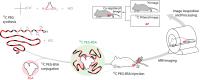 |
Direct Quantitative 13C-Filtered 1H Magnetic Resonance Imaging
of Pegylated Biomacromolecules In Vivo
Rohan Alvares1, Justin Lau2,3, Peter
Macdonald1, Charles Cunningham2,3, and
R. Scott Prosser1
1Department of Chemistry, University of Toronto,
Toronto, ON, Canada, 2Department
of Medical Biophysics, University of Toronto, Toronto, ON,
Canada, 3Physical
Sciences, Sunnybrook Research Institute, Toronto, ON, Canada
We demonstrate a new platform technology in which
macromolecular constituents, such as proteins and drug
delivery systems, are observed directly and quantitatively
in vivo using 1H
MRI of 13C-labeled
polyethylene glycol (13C-PEG) tags. The 28 kDa 13C-PEG
tags are non-immunogenic, and each bears approximately 2500
spectroscopically equivalent 1H
nuclei appearing at a single resonance position. By
filtering the 1H
PEG signal through the directly coupled 13C
nuclei, background water and fat signals are largely
eliminated. We demonstrate the approach by monitoring in
real-time the distribution of 13C-PEG
and 13C-pegylated
albumin injected into the hind leg of a mouse.
|
| |
16:36
|
1023.
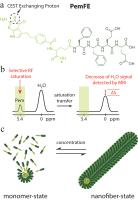 |
Label-free CEST MRI detection of self-assembly anticancer
drug-peptide nanofibers
Yuguo Li1,2, Lye Lin Lock3, Renyuan
Bai4, Xinpei Mao3, Verena Staedtke5,
Peter C.M Van Zijl1,2, Honggang Cui3,6,
and Guanshu Liu1,2
1The Russell H. Morgan Department of Radiology
and Radiological Science, Division of MR Research, Johns
Hopkins University School of Medicine, Baltimore, MD, United
States, 2F.M.
Kirby Research Center for Functional Brain Imaging, Kennedy
Krieger Institute, Baltimore, MD, United States, 3Department
of Chemical and Biomolecular Engineering, Johns Hopkins
University, Baltimore, MD, United States, 4Department
of Neurosurgery, Johns Hopkins School of Medicine,
Baltimore, MD, United States, 5Department
of Neurology, Johns Hopkins School of Medicine, Baltimore,
MD, United States, 6Institute
for NanoBioTechnology, Johns Hopkins University, Baltimore,
MD, United States
A new injectable and CEST MRI-detectable nanofiber hydrogel
has been developed for image-guided drug delivery of
anticancer drug Pemetrexed (Pem). Such a drug delivery
system is composed of only drug (Pem) and peptide (FFEE) and
the MRI detectability stems on the inherent CEST signal of
Pem. In the present study, PemFE nanofiber hydrogel was
first constructed and characterized. Then, the CEST MRI
detection of the constructed hydrogel in vivo was
demonstrated in an orthotopic brain tumor mouse model. Our
study clearly demonstrated the ability of using CEST MRI to
monitor drug delivery of PemFE hydrogel.
|
| |
16:48
|
1024.
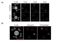 |
Assessment of Thrombus Stage by ‘Multicolor’ 19F MRI
Sebastian Temme1, Christoph Jacoby2,
Christoph Owenier1, Christoph Grapentin3,
Xiaowei Wang4, Rolf Schubert3,
Karlheinz Peter4, Jürgen Schrader1,
and Ulrich Flögel1,2
1Molecular Cardiology, University of Düsseldorf,
Düsseldorf, Germany, 2Department
of Cardiology, Pneumology and Angiology, University Hospital
Düsseldorf, Düsseldorf, Germany, 3Pharmaceutical
Technology and Biopharmacy, University of Freiburg, Freiburg
i. Br., Germany, 4Atherothrombosis
and Vascular Biology, Baker IDI Heart and Diabetes
Institute, Victoria, Australia
The present study was aimed at developing a non-invasive
approach for direct assessment of thrombus stage by
‘multicolor’ 19F
MRI. To this end, we used ligands binding specifically
during different phases of thrombosis and coupled them to
perfluorocarbons (PFCs) with indvidual spectral signatures.
Discrimination of the targeted agents was achieved by a
novel multi chemical shift selective imaging technique for
simultaneous, artifact-free detection of different PFCs. The
results show that this technique holds the potential to
differentiate thrombi in the acute, subacute and chronic
phase and may also be used for in situ labeling of a variety
of other targets.
|
| |
17:00
|
1025.
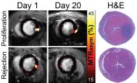 |
In vivo imaging of cell fate decisions in cardiac cell therapy
using cardioCEST MRI.
Ashley Pumphrey1, Zhengshi Yang2,
Shaojing Ye2, Ahmed Abdel-Latif2, and
Moriel Vandsburger3
1CVRC, University of Kentucky, Lexington, KY,
United States, 2University
of Kentucky, Lexington, KY, United States, 3Physiology,
University of Kentucky, Lexington, KY, United States
We developed a cardiac specific chemical exchange saturation
transfer pulse sequence and applied it to the tracking of
cell survival/proliferation or rejection in murine models of
cardiac cell therapy.
|
| |
17:12
|
1026.
 |
Quantitative Evaluation of Tumour Associated Macrophages in
Breast Cancer: Fluorine-19 versus Iron Oxide Nanoparticles
Ashley V Makela1,2, Jeffrey M Gaudet1,2,
and Paula J Foster1,2
1Medical Biophysics, Western University, London,
ON, Canada, 2Robarts
Research Institute, London, ON, Canada
Tumour associated macrophages (TAMs) are correlated with an
aggressive tumour type and poor outcomes. This study is the
first time iron and fluorine-19 (19F) based MRI
cell tracking methods have been compared for the detection
and quantification of TAMs in an orthotopic model of breast
cancer. Imaging was performed at 4 days and 3 weeks post
cell implantation. Both cell tracking methods showed a much
higher TAM density at 4 days; no other imaging study has
examined this at such an early time point. 19F
MRI provided quantitative information about TAM density and
tumoural distribution that was not possible with iron.
|
| |
17:24
|
1027.
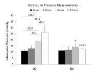 |
Age-related changes in anterograde transport, axonal integrity
and visuomotor function in the DBA/2J mouse model of chronic
glaucoma
Xiao-Ling Yang1,2, Yolandi van der Merwe1,3,
Leon C. Ho1,4, Ian P. Conner2,3,
Seong-Gi Kim1,5, Kira L. Lathrop2,
Gadi Wollstein2,3, Joel S. Schuman2,3,
and Kevin C. Chan1,2
1NeuroImaging Laboratory, University of
Pittsburgh, Pittsburgh, PA, United States, 2UPMC
Eye Center, Eye and Ear Institute, Ophthalmology and Visual
Science Research Center, Department of Ophthalmology,
University of Pittsburgh, Pittsburgh, PA, United States, 3Department
of Bioengineering, Swanson School of Engineering, University
of Pittsburgh, Pittsburgh, PA, United States, 4Department
of Electrical and Electronic Engineering, University of Hong
Kong, Pokfulam, Hong Kong, 5Center
for Neuroscience Imaging Research, Institute for Basic
Science, Sungkyunkwan University, Suwon, Korea, Republic of
Glaucoma is the leading cause of irreversible blindness
worldwide and is a slowly progressing neurodegenerative
disease of the visual system. While elevated intraocular
pressure (IOP) and age are major risk factors, their effects
on glaucoma pathogenesis remain incompletely understood. In
this study, we determined the onset of glaucomatous changes
and their progression in a chronic inherited glaucoma model
using DBA/2J mice. Our results indicate that elevation of
IOP may accelerate the deterioration of structure,
physiology and function of the visual system in the DBA/2J
mice across age. Comparatively, the visual system in
C57BL/6J mice appeared intact across the same ages.
|
| |
17:36
|
1028.
 |
Characterizing Iron Oxide NanoParticles using 4D Spectroscopic
SWIFT
Jinjin Zhang1, Hattie L. Ring1,
Michael Garwood1, and Djaudat Idiyatullin1
1Center for Magnetic Resonance Research,
Department of Radiology, University of Minnesota,
Minneapolis, MN, United States
The ability to accurately and sensitively quantify the
bio-distribution of iron oxide nanoparticles is essential
for their use as both diagnostic and therapeutic agents in
theranostics. In this study, a 4D spectroscopic SWIFT
technique was applied and optimized to characterize the
distribution of IONPs in mouse invivo up
to high concentration (>1.0 mg Fe/g of tissue). The
frequency shift due to susceptibility variation and T2*
shortening (down to 20 μs) caused by IONPs were detected in
mice organs depositing IONPs. The acquired T2*
map which provide quantitative information about IONP
bio-distribution makes the 4D spectroscopic SWIFT a
promising tool in nanoparticle-based theranostics.
|
| |
17:48
|
1029.
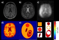 |
The framework and AnalytiCally Represented Oxygen-17 BrAin Tumor
(ACROBAT) phantom for optimization of CMRO2 quantification
protocols in dynamic 17O-MRI.
Dmitry Kurzhunov1, Robert Borowiak1,2,
Axel Krafft1,2, and Michael Bock1
1University Medical Center Freiburg, Dept. of
Radiology - Medical Physics, Freiburg, Germany, 2German
Cancer Research Center (DKFZ), German Cancer Consortium
(DKTK), Heidelberg, Germany
Direct dynamic 17O-MRI
allows quantification of the cerebral metabolic rate of
oxygen consumption (CMRO2). The influence of
acquisition parameters on the precision of CMRO2 quantification
needs to be investigated for routine application, but the
costly and rare 17O
gas prohibits extensive imaging studies. Thus, in this work
a flexible, Fourier domain-based simulation framework is
presented and analytical tumor and numerical 17O
MRI brain phantoms are utilized based on experimental 17O
relaxation times and signal-to-noise ratios. Precision of
CMRO2 quantification
is evaluated and optimal acquisition parameters are given.
|
|