| |
16:00
 |
0780.
 |
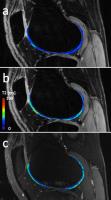
Highly Efficient Nonrigid Motion Corrected 3D Whole-Heart
Coronary Vessel Lumen and Wall Imaging 
Gastao Cruz1, David Atkinson2, Markus
Henningsson1, René Botnar1, and
Claudia Prieto1
1Division of Imaging Sciences & Biomedical
Engineering, King's College London, London, United Kingdom, 2University
College London, London, United Kingdom
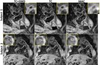
Non-invasive visualization of both coronary lumen and vessel
wall is desired for assessment of coronary atherosclerosis.
An interleaved acquisition was recently proposed to obtain
both 3D images with MRI. However, this approach is
susceptible to motion artifacts and dual respiratory gating
results in long and unpredictable scan times. Here, we
propose a ~100% scan efficiency, two-step motion correction
method using translational and nonrigid correction to
produce co-registered coronary lumen and vessel wall images.
The proposed method shows significant improvements over
translational correction and similar lumen quality to a
reference navigator-gated (6mm) scan, despite a scan time
reduction of ~1.8x.
|
| |
16:12
|
0781.
 |
Discontinuity Preserving Registration using Truncated L1
Regularization and Minimum Spanning Tree based Motion Clustering
Dongxiao Li1,2, Juerong Wu1, Kofi M.
Deh2, Thanh D. Nguyen2, Martin R.
Prince2, Yi Wang2,3, and Pascal
Spincemaille2
1College of Information Science and Electronic
Engineering, Zhejiang University, Hangzhou, China, People's
Republic of, 2Department
of Radiology, Weill Cornell Medical College, New York, NY,
United States,3Department of Biomedical
Engineering, Cornell University, Ithaca, NY, United States
Free breathing liver perfusion analysis requires non-rigid
motion registration of the unavoidable respiratory motion in
the dynamic data. Traditional non-rigid methods rely on
spatially smooth motion parameters, which is problematic for
the sliding motion of the liver against the abdominal wall.
In this work, truncated L1 regularized Minimum Spanning Tree
based motion clustering combined with a Markov Random Field
optimization is proposed to perform liver registration
without the need for manual segmentation. Results on
breath-hold liver images acquired at various positions of
the respiratory cycle demonstrated this method allows
superior liver motion estimation when compared to
traditional methods.
|
| |
16:24
 |
0782.
 |
Simultaneous in-vivo respiratory and cardiac motion correction
system for PET/MR
Thomas Küstner1,2, Christian Würslin1,3,
Martin Schwartz1,2, Petros Martirosian1,
Sergios Gatidis1, Konstantin Nikolaou1,
Fritz Schick1, Bin Yang2, Nina F.
Schwenzer1, and Holger Schmidt1
1University Hospital Tübingen, Tübingen, Germany, 2Institute
of Signal Processing and System Theory, University of
Stuttgart, Stuttgart, Germany, 3University
of Stanford, Palo Alto, CA, United States
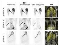
In oncologic imaging, simultaneous
Positron-Emission-Tomography/Magnetic Resonance (PET/MR)
scanners offer a great potential for improving diagnostic
accuracy. An accurate diagnosis requires a high PET image
quality reflecting in long PET examination times under free
movement conditions (respiration and heartbeat). Hence, to
ensure this high image quality one has to overcome the
motion-induced artifacts. The simultaneous acquisition
allows performing a MR-based non-rigid motion correction of
the PET image. We propose a clinical feasible respiratory
and cardiac motion correction system with a reduced scan
time of only 60s, freeing time for additional diagnostic MR
sequences. In-vivo patient
data substantiates the diagnostic improvements.
|
| |
16:36
|
0783.
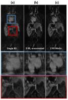 |
Image-Based Non-Rigid Motion Correction for Free-breathing 4D MR
Angiography
Fei Han1, Ziwu Zhou1, Paul J Finn1,
and Peng Hu1
1Radiology, University of California, Los
Angeles, Los Angeles, CA, United States
Cardiac-phase-resolved 4D MR angiography (MRA) is a
promising technique for evaluating patients with
cardiovascular disorder. However, current approaches usually
has low scan efficiency (20-40%) due to the gating based
respiratory motion compensation and therefore suffered from
prolonged yet unpredictable scan time. In this work, we
proposed a motion correction strategy in which complex
non-rigid respiratory motion is modeled using voxel-based
linear translations, which are estimated using 3D image
registration. Our preliminary result shows that the proposed
technique could compensate for complex motion across the
large field-of-view of 4D MRA and potentially improve the
scan efficiency by including more k-space data in the
reconstruction.
|
| |
16:48
|
0784.
 |
Motion-free Abdominal MRI using Manifold Alignment
Xin Chen1, Muhammad Usman1, Christian
Baumgartner2, Claudia Prieto1, and
Andrew King1
1Division of Imaging Sciences and Biomedical
Engineering, King's College London, London, United Kingdom, 2Biomedical
Image Analysis Group, Imperial College, London, United
Kingdom
We present a novel method based on manifold alignment, which
enables reconstruction of motion-free abdominal images
throughout the respiratory cycle to better capture
respiratory intra- and inter-cycle variations. The proposed
method was evaluated on both simulated and in-vivo 2D
acquisitions. Based on virtual navigator measurement, the
reconstructed dynamic sequence achieved Pearson correlation
coefficient of 0.9504 with the ground truth of the simulated
dataset. The proposed method enables much richer profile
data to be used for self-gating, resulting in less blurring
when compared to conventional central k-space self-gating
method for the in-vivo acquisition.
|
| |
17:00
|
0785.
 |
Free-Breathing Dynamic MRI with Sliding Slice Distorted
Simultaneous Multi-Slice
Kevin M Johnson1, James H Holmes2, and
Scott B Reeder1,3,4
1Medical Physics, University of Wisconsin -
Madison, Madison, WI, United States, 2Global
MR Applications and Workflow, GE Healthcare, Madison, WI,
United States, 3Radiology,
University of Wisconsin - Madison, Madison, WI, United
States, 4Biomedical
Engineering, University of Wisconsin - Madison, Madison, WI,
United States
Sliding slice MRI is a technique which uses a magnetization
prepared sliding 2D slice to cast respiratory motion
artifacts as geometric distortions rather than
diagnostically obscuring ghosting routinely associated with
3D phase-encoding. In this work, we present the combination
of simultaneous-multi-slice with pseudo-random Cartesian
based sliding slice sampling. This combination allows
increased frame rates, FOV tailoring, and reduces
sensitivity to off-resonance compared to past non-Cartesian
radial and spiral based approaches. Preliminary results are
shown in moving phantoms and in-vivo free breathing DCE,
demonstrating very good image quality.
|
| |
17:12
|
0786.
 |
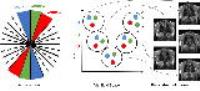
Five-Dimensional Respiratory and Cardiac Motion Compensation
Based on Strongly Undersampled MR Data
Christopher M Rank1, Sebastian Sauppe1,
Thorsten Heußer1, Andreas Wetscherek1,
and Marc Kachelrieß1
1Medical Physics in Radiology, German Cancer
Research Center (DKFZ), Heidelberg, Germany
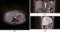
We propose a new method for 5D respiratory and cardiac
motion compensation (MoCo), which employs highly
undersampled MR data and thus requires acquisition times as
low as 2 minutes. Radial MR data of the thorax of three
free-breathing patients were acquired. Respiratory and
cardiac motion vector fields were estimated allowing for 5D
MoCo reconstructions, which employ 100% of the measured raw
data for reconstruction of each combination of respiratory
and cardiac phase. These 5D MoCo reconstructions clearly
resolve different combinations of respiratory and cardiac
phases while achieving high temporal and spatial resolution
as well as low noise and artifact levels.
|
| |
17:24
|
0787.
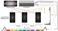 |
Respiratory and Cardiac Dual Soft-Gated 4D Cardiovascular MRI
Ziwu Zhou1, Fei Han1, Takegawa Yoshida1,
Kim-Lien Nguyen1, Paul Finn1, and Peng
Hu1
1Radiological Sciences, University of California,
Los Angeles, Los Angeles, CA, United States
In this study, we proposed a respiratory and cardiac dual
soft-gated technique that efficiently suppresses respiratory
motion and resolves cardiac motion in 4D cardiovascular MRI.
Comparing with existing methods that exploited data
redundancy in respiratory and cardiac dimensions using joint
reconstruction, proposed method weights data consistency
according to the degree of motion corruption. A big
advantage of this approach is its short reconstruction time
and low computation burden, making it feasible for practical
usage.
|
| |
17:36
|
0788.
 |
Quantification and Artifact Reduction from Simple Modeling of
DESS Signals
Bragi Sveinsson1, Garry Gold1, and
Brian Hargreaves1
1Stanford University, Stanford, CA, United States
The double-echo in steady-state (DESS) sequence offers both
3D anatomical imaging and 3D quantitative mapping
(SNR-efficient 3D maps of T2 and apparent diffusion
coefficent) in various applications, such as breast imaging
or knee cartilage imaging. The complicated signal behavior
remains a challenge for quantitative imaging, and strong
spoiling can lead to motion artifacts. Here, we introduce
simplified methods for modeling DESS signals, enabling more
accurate T2 measurements and better motion artifact
reduction.
|
| |
17:48
|
0789.
 |
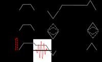
Fully self-gated motion compensated cine reconstruction from
free-breathing ungated 2D radial cardiac MRI data
André Fischer1,2, Anne Menini1,
Aurelien Bustin1,3, Kevin M Johnson4,
Christopher J Francois5, and Anja C.S. Brau2
1GE Global Research, Garching bei München,
Germany, 2Cardiac
Center of Excellence, GE Healthcare, Garching bei München,
Germany, 3Computer
Science, Technical University Munich, München, Germany,4Medical
Physics, University of Wisconsin, Madison, WI, United
States, 5Radiology,
University of Wisconsin, Madison, WI, United States
Cardiac MRI is affected by both cardiac and respiratory
motion. While ECG-gated imaging in breath hold is the
clinical method of choice, free-breathing methods are needed
in patients with limited breath hold capability. This works
describes a method to obtain free-breathing cine datasets
with high SNR and high spatial resolution (1.4mm in-plane)
from a completely self-gated Golden Angle radial scan within
an 11s scan time. The motion compensated reconstruction
technique takes advantage of calibrated displacement fields
extracted from the radial data to recover motion
artifact-free cardiac phases. Beyond cine imaging,
contrast-enhanced cardiac imaging can also be expected to
benefit from this motion compensated reconstruction
strategy.
|
|