| |
16:00
|
1050.
 |
Evaluation of Renal Blood flow in subjects with Diabetic
Nephropathy using ASL Perfusion MRI - Permission Withheld
Lu-Ping Li1,2, Huan Tan1, Jon Thacker3,
Wei Li1,2, Ying Zhou4, Orly Kohn5,
Stuart Sprague2,6, and Pottumarthi V Prasad1,2
1Radiology, Northshore University HealthSystem,
Evanston, IL, United States, 2Pritzker
School of Medicine, University of Chicago, Chicago, IL,
United States, 3Biomedical
Engineering, Northwestern University, Evanston, IL, United
States, 4Center
for Biomedical Research & Informatics, Northshore University
HealthSystem, Evanston, IL, United States, 5Medicine,
University of Chicago, Chicago, IL, United States,6Medicine,
Northshore University HealthSystem, Evanston, IL, United
States
Renal blood flow is thought to be reduced in subjects of
diabetic nephropathy (DN). However, there is limited amount
of quantitative data on renal blood flow in patients with
DN. In this study, ASL MRI data was acquired in 28 patients
with diabetes and stage-3 CKD along with 30 healthy
controls. Renal blood flow was found to be significantly
lower in subjects with DN with a large Cohen’s d value.
Renal blood flow also showed a significant correlation with
eGFR and age was not found to be a significant confounder in
this relationship.
|
| |
16:12
|
1051.
 |
Are Renal Lipids Increased in Overweight Diabetic Patients? A MR
Spectroscopy and Dixon Fat/Water Imaging Study 
Gaëlle Diserens1, Waldo Valenzuela2,
Maryam Seif1, Laila Mani3, Daniel
Fuster3, Christoph Stettler4, Bruno
Vogt3, Mauricio Reyes2, Chris Boesch1,
and Peter Vermathen1
1Depts Clinical Research and Radiology,
University of Bern, Bern, Switzerland, 2Institute
for Surgical Technology and Biomechanics, University of
Bern, Bern, Switzerland, 3Dept.
of Nephrology, Hypertension and Clinical Pharmacology,
University of Bern, Bern, Switzerland, 4Dept.
of Endocrinology, Diabetes and Clinical Nutrition,
University of Bern, Bern, Switzerland
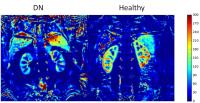
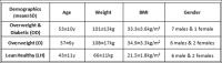
Renal ectopic lipid accumulation may lead to kidney
dysfunction. The study purpose was to determine (1) renal
ectopic lipid content in overweight type-2 diabetic patients
compared to (a) overweight non-diabetic patients and (b)
lean volunteers by 1H-MRS and (2) renal sinus fat content by
DIXON-MRI in the same three patient groups. This study
demonstrates that renal ectopic lipids appear to be not
higher in overweight diabetic patients compared to
overweight non-diabetic subjects, while ectopic lipids are
higher in both groups compared to healthy subjects.
Significantly higher renal sinus bulk lipids were detected
for overweight diabetic patients compared to BMI-matched
non-diabetics.
|
| |
16:24
 |
1052.
 |
Simultaneous quantification of intragastric secretion and fat
distribution
Dian Liu1, Helen Louise Parker2,
Jelena Curcic1,2, Sebastian Kozerke1,
and Andreas Steingoetter1,2
1Institute for Biomedical Engineering, University
and ETH Zurich, Zurich, Switzerland, 2Division
of Gastroenterology and Hepatology, University Hospital
Zurich, Zurich, Switzerland
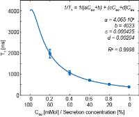
Simultaneous assessment of both intragastric secretion and
fat is important in food science but has hitherto been
hampered by the bi-exponential relaxation behavior of fat
emulsions. In combination with IDEAL, this work introduced a
fat correction for rapid T1 mapping,
which enabled the simultaneous measurement of the
intragastric distribution and temporal development of
gastric secretion and fat. Results revealed the interaction
between these two components by dilution and mixing, making
this method a promising tool to non-invasively assess the
emulsification and emptying of ingested fat.
|
| |
16:36
|
1053.
 |
Non-invasive postprandial fatty acid tracking with 1H-[13C]
Magnetic Resonance Spectroscopy in the human liver
Lucas Lindeboom1,2,3, Robin A. de Graaf4,
Christine I. Nabuurs1,2,3, Matthijs K.C.
Hesselink2, Joachim E. Wildberger1,
Patrick Schrauwen2,3, and Vera B.
Schrauwen-Hinderling1,2,3
1Radiology, Maastricht University Medical Center,
Maastricht, Netherlands, 2Human
Biology and Human Movement Sciences, Maastricht University
Medical Center, Maastricht, Netherlands, 3Top
Institute Food and Nutrition, Wageningen, Netherlands, 4Diagnostic
Radiology, Magnetic Resonance Research Center, Yale
University School of Medicine, New Haven, CT, United States
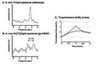
We here show that postprandial 13C
fatty acid tracking is feasible in the human liver using
ge-HSQC. Experiments in two human volunteers revealed that
intake of 5 or 7 grams of 13C-labeled
fatty acids resulted in two- or threefold increase in
hepatic 13C-enrichment
after 3 hours. It is estimated that 3% of the oral load is
stored in the liver at this time point. The ge-HSQC sequence
can be used to reveal the contribution of dietary fat to the
development of hepatic steatosis.
|
| |
16:48
|
1054.
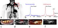 |
Detection of human brown adipose tissue by MRI with
hyperpolarized Xe-129 gas and validation by FDG-PET/MRI
Rosa Tamara Branca1,2, Le Zhang3,4,
Alex Burant1,4, Laurence Katz5, and
Andrew McCallister1,4
1Physics and Astronomy, University of North
Carolina at Chapel Hill, Chapel Hill, NC, United States, 2Biomedical
Research Imaging Center, Chapel Hill, NC, United States, 3Material
Science, University of North Carolina at Chapel Hill, Chapel
Hill, NC, United States, 4Biomedical
Research Imaging Center, University of North Carolina at
Chapel Hill, Chapel Hill, NC, United States, 5Emergency
Medicine, University of North Carolina at Chapel Hill,
Chapel Hill, NC, United States
Despite histological evidence that all humans have brown
adipose tissue, the detection of this tissue in overweighs
and obese subjects has proven to be a challenge. A recent
study showed that MRI by hyperpolarized xenon gas (HP129Xe)
enables the detection of this tissue in both lean and obese
animal phenotype, with enhanced sensitivity in the latter
with respect to the gold standard, FDG-PET. Here we
demonstrate that HP129Xe gas MRI can also be used to detect
human BAT with better sensitivity than FDG-PET.
|
| |
17:00
|
1055.
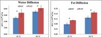 |
Diffusion Spectroscopy of White and Brown Adipose Tissues
Sanjay Kumar Verma1, Kaz Nagashima1,
Swee Shean Lee1, Tian Xianfeng1,
Jadegoud Yaligar1, Venkatesh Gopalan1,
Bhanu Prakash KN1, and S. Sendhil Velan1
1Laboratory of Molecular Imaging, Singapore
Bioimaging Consortium, Singapore
There are two types of fat tissues, white adipose tissue
(WAT) and brown adipose tissue (BAT), which essentially
perform opposite functions in whole body energy metabolism.
There is a large interest in development of MR Imaging
techniques that will be suitable for separating white and
brown fat. In this work we have implemented diffusion NMR
spectroscopy to differentiate these two types of tissues.
Water diffused faster than the fat in both WAT and BAT. Fat
diffusion was faster in WAT compared to BAT. Our findings
also suggest restricted behavior of fat molecules in BAT and
not in WAT.
|
| |
17:12
|
1056.
 |
Deep subcutaneous adipose tissue lipid unsaturation associates
with intramyocellular lipid content
Jesper Lundbom1,2, Alessandra Bierwagen1,2,
Kálmán Bodis1,2, Jaakko Kaprio3,4,5,
Aila Rissanen6,7, Nina Lundbom8,
Michael Roden1,2,9, and Kirsi Pietiläinen4,6,10
1German Diabetes Center, Leibniz Center for
Diabetes Research, Düsseldorf, Germany, 2German
Center for Diabetes Research (DZD e.V.), Partner Düsseldorf,
Düsseldorf, Germany, 3Finnish
Twin Cohort Study, Department of Public Health, Hjelt
Institute, Helsinki, Finland, 4FIMM,
Institute for Molecular Medicine, University of Helsinki,
Helsinki, Finland, 5National
Institute for Health and Welfare, Helsinki, Finland,6Obesity
Research Unit, Diabetes and Obesity, University of Helsinki,
Helsinki, Finland, 7Department
of Psychiatry, Helsinki University Central Hospital,
Helsinki, Finland, 8HUS
Medical Imaging Center, University of Helsinki, Helsinki,
Finland, 9Department
of Endocrinology and Diabetology, Medical Faculty,
Heinrich-Heine University, Düsseldorf, Germany, 10Endocrinology,
Abdominal Center, Helsinki University Central Hospital,
Helsinki, Finland
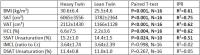
The present study uses non-invasive MRS to examine whether
MZ twins discordant for BMI display depot specific
differences in adipose tissue unsaturation (DSAT and SSAT),
and how the unsaturation relates to body fat distribution
and ectopic fat. The main finding of the twin study is that
DSAT lipid unsaturation associates with intramyocellular
lipid content, which was further confirmed in a general
population study and for the repeated sampling of one
volunteer. These results highlight the role of fatty acid
composition in adipose tissue - skeletal muscle crosstalk.
|
| |
17:24
|
1057.
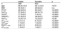 |
MR-derived indices for identification of quantity and
distribution of adipose tissue – age- and gender related
differences in a cohort at increased risk for metabolic diseases
Jürgen Machann1, Malte Niklas Bongers2,
Andreas Fritsche3, Norbert Stefan3,
Hans-Ulrich Häring3, Konstantin Nikolaou4,
and Fritz Schick2
1Section on Experimental Radiology, Department of
Diagnostic and Interventional Radiology, IDM of the
Helmholtz Center Munich at the University Tübingen, German
Center for Diabetes Research (DZD), Tuebingen, Germany, 2Section
on Experimental Radiology, Department of Diagnostic and
Interventional Radiology, University Hospital Tuebingen,
Tuebingen, Germany, 3Department
of Endocrinology and Diabetology, Angiology, Nephrology and
Clinical Chemistry, IDM of the Helmholtz Center Munich at
the University Tübingen, German Center for Diabetes Research
(DZD), Tuebingen, Germany, 4Department
of Diagnostic and Interventional Radiology, University
Hospital Tuebingen, Tuebingen, Germany
MR-based phenotyping is of increasing interest for
cross-sectional and interventional studies on large cohorts.
Quantification of adipose tissue (AT) compartments – e.g. by
T1-weighted MRI – has mainly been performed by giving the
absolute amounts in litres. However, this does not directly
reflect the distribution and quantity (e.g. for people with
different size). Thus, the percentage of AT compartments are
given as percent of total AT and new fat indices, corrected
for height (comparable to BMI) are introduced and age- and
gender related differences are determined in a large cohort
of people at increased risk for metabolic diseases.
|
| |
17:36
|
1058.
 |
Hepatic lipid alterations monitored by 1H-MRS in
vivo in the
ontogeny of obesity-related metabolic dysregulation.
Ana Francisca Soares1, João M. N. Duarte1,
Blanca Lizarbe1, and Rolf Gruetter1,2,3,4
1Laboratory of Functional and Metabolic Imaging
(LIFMET), Swiss Federal Institute of Technology Lausanne
(EPFL), Lausanne, Switzerland, 2Center
for Biomedical Imaging (CIBM), Lausanne, Switzerland,3Department
of Radiology, University of Geneva (UNIGE), Geneva,
Switzerland, 4Department
of Radiology, University of Lausanne (Unil), Lausanne,
Switzerland
Obesity is associated with a loss of metabolic control,
largely driven by alterations in whole-body lipid
distribution. Impaired insulin action leads to hepatic lipid
accumulation and, conversely, high levels of liver lipids
also cause insulin resistance. We followed the loss of
glucose homeostasis in mice fed a high-fat diet for 18
weeks. In parallel, we assessed their hepatic lipids by 1H-MRS in
vivo. In this model, glucose intolerance preceded
hepatic lipid accumulation that then contributed to
aggravate the phenotype. Moreover, fasting-induced hepatic
lipid dynamics was hampered with high-fat diet feeding.
|
| |
17:48
|
1059.
 |
TOFI – Thin Outside, Fat Inside – identifying non-obese subjects
at high risk for metabolic diseases based on MRI and MRS
Jürgen Machann1, Malte Niklas Bongers2,
Norbert Stefan3, Andreas Fritsche3,
Konstantin Nikolaou4, Hans-Ulrich Häring3,
and Fritz Schick5
1Section on Experimental Radiology, IDM of the
Helmholtz Center Munich at the University Tübingen, German
Center for Diabetes Research (DZD), Tuebingen, Germany, 2Department
of Diagnostic and Interventional Radiology, Section on
Experimental Radiology, Tuebingen, Germany, 3Department
of Endocrinology and Diabetology, Angiology, Nephrology and
Clinical Chemistry, IDM of the Helmholtz Center Munich at
the University Tübingen, German Center for Diabetes Research
(DZD), Tuebingen, Germany, 4Department
of Diagnostic and Interventional Radiology, University
Hospital Tübingen, Tuebingen, Germany, 5Section
on Experimental Radiology, University Hospital Tübingen,
Tuebingen, Germany
Axial T1-weighted MRI and volume selective 1H-MRS
were performed in a cohort of almost 500 non-obese subjects
at increased risk for metabolic diseases. Adipose (AT) and
lean tissue (LT) compartments from different body regions
were quantified and are expressed as percentage of the
entire volume in order to display tissue distribution and to
differentiate metabolically healthy (insulin sensitive, IS)
and unhealthy (insulin resistant, IR) subgroups.
Additionally, intrahepatic lipids (IHL) were quantified. It
could be shown that IS subjects are characterized by lower
percentage of AT in abdominal regions but higher amounts in
the extremities whereas IHL are almost doubled in IR
subjects.
|
|