| |
16:00
|
0526.
 |
Magnetic Resonance Imaging of the Pancreas in a Transgenic Mouse
Model of Pancreatic Carcinogenesis 
Conny F. Waschkies1,2, Theresia F. Reding1,
Gitta Maria Seleznik1, Udo Ungethuem1,
and Rolf Graf1
1Division of Visceral and Transplantation
Surgery, University Hospital Zurich, Zurich, Switzerland, 2Institute
for Biomedical Engineering, ETH and University Zurich,
Zurich, Switzerland
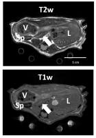
Few preclinical studies rely on MRI to monitor pancreatic
tissue changes in commensurate animal models, mostly due to
inherently low conspicuity of the rodent pancreas.
Pancreatic inflammation is a risk factor for pancreatic
ductal adenocarcinoma development, and its initiation is
linked to activating mutations in KRAS oncogene, known as
the KPC mouse model. In the present study we demonstrate the
potential of preclinical MRI to visualize the murine
pancreas and its changes associated with cellular
transformations in this mouse model of pancreatic
carcinogenesis.
|
| |
16:12
|
0527.
 |
Pilot Study of Rapid MR Pancreas Screening for Patients with
BRCA Mutation Undergoing Screening Breast MRI – Preliminary Data
Mitchell C Raeside1, Andrea Agostini1,
Richard K.G. Do1, Amita Shukla-Dave1,2,
David Aramburu-Nunez2, Ramesh Paudyal2,
Olga Smelianskaia1, Monika Khan1,
David Kelsen3, and Lorenzo Mannelli1
1Radiology, Memorial Sloan-Kettering Cancer
Center, New York, NY, United States, 2Medical
Physics, Memorial Sloan-Kettering Cancer Center, New York,
NY, United States, 3Medicine,
Memorial Sloan-Kettering Cancer Center, New York, NY, United
States
The purpose of this study was to develop and optimize a
rapid MR pancreas screening protocol to be performed in
conjunction with breast MRI screening in BRCA-positive
individuals. 15 patients underwent a rapid pancreatic
screening at the conclusion of their breast MRI
examination. Images were acquired with the patient in the
prone position, with the breast coil still in place, but
using the built-in body coil on a 3T magnet, and evaluated
for image quality (including SNR and CNR), and detection of
pancreatic lesions. Rapid MR protocol for pancreatic cancer
screening is feasible and provides diagnostic quality
images.
|
| |
16:24
|
0528.
 |
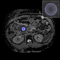
Contrast Agent Uptake Analysis at 3T for Pancreatic Cancer
Douglas Arthur Charles Kelley1, Eric Collisson2,
Benjamin M Yeh3, Michael Ohliger3, and
Zhen Wang3
1Neuro Applications and Workflow, GE Healthcare,
Corte Madera, CA, United States, 2Medicine,
University of California, San Francisco, San Francisco, CA,
United States, 3Radiology,
University of California, San Francisco, San Francisco, CA,
United States
Pancreatic cancer is highly desmoplastic and slowly takes up
extracellular gadolinium based contrast agents during MR
imaging. Quantitative estimation of gadolinium based
contrast uptake in pancreas cancers may help assess the
tumor stroma, which is implicated in tumor aggressiveness
and treatment response. However, tissue motion and
sensitivity inhomogeneity on abdominal MR scans present
complications for quantitative analysis of contrast uptake.
A new MR PET system with higher performance gradient and RF
capabilities coupled with data-driven analysis methods allow
robust estimation of contrast agent concentration in
pancreatic cancer patients.
|
| |
16:36
|
0529.
 |
Differentiating Pancreatic Cancer from Mass-Forming Focal
Pancreatitis with a Novel Inhomogeneity Index Based on ADC Map
Analysis
Chao Ma1, Li Liu1, Jing Li1,
Li Wang1, Xu Fang1, Jianxun Qu2,
Shi-yue Chen1, and Jianping Lu1
1Department of Radiology, Changhai Hospital of
Shanghai, Shanghai, China, People's Republic of, 2MR
Research China, GE Healthcare, Beijing, China, People's
Republic of
Differentiating mass-forming focal pancreatitis (FP) and
pancreatic ductal adenocarcinoma (PDAC) is of great
importance and yet remains a challenge in clinical practice.
In this work, we propose a novel method to address the
challenge with a new parameter (inhomogeneity index) based
on the ADC map analysis with different region of interest
(ROI) size.
|
| |
16:48
|
0530.
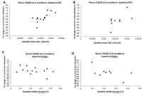 |
Prediction of response by DCE-MRI and DW-MRI for intrahepatic
cholangiocarcinomas treated with locoregional and systemic
chemotherapy: a preliminary analysis
Kristen L Zakian1, Richard K Do2,
Taryn Boucher2, Mithat Gonen3, Andrea
Cercek4, William R Jarnagin5, and
Nancy Kemeny4
1Medical Physics, Memorial Sloan-Kettering Cancer
Center, New York, NY, United States, 2Radiology,
Memorial Sloan-Kettering Cancer Center, New York, NY, United
States, 3Epidemiology
and Biostatistics, Memorial Sloan-Kettering Cancer Center,
New York, NY, United States, 4Medicine,
Memorial Sloan-Kettering Cancer Center, New York, NY, United
States, 5Surgery,
Memorial Sloan-Kettering Cancer Center, New York, NY, United
States
Intrahepatic cholangiocarcinoma (ICC) is the second most
common primary liver malignancy and has few treatment
options. A previous study suggested that MRI may help
identify patients likely to benefit from hepatic arterial
infusion pump therapy with floxuridine (HAI-FUDR). The
purpose of this prospective study was to investigate the
ability of pre-treatment and early-in-treatment DCE and
DW-MRI to predict ICC response to combined HAI-FUDR and
systemic chemotherapy given in a Phase 2 clinical trial. Our
preliminary analysis suggests that DW-MRI may predict
response of unresectable ICC using data acquired at baseline
or at 1 month after treatment start.
|
| |
17:00
|
0531.
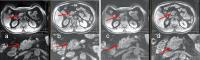 |
An Indenting Abdominal Array for 2-Fold SNR Improvement in
Pancreatic MRI
Scott B. King1, Jarod Matwiy1, Calvin
Bewsky1, Hung-Yu Lin1, and Masoom A.
Haider2,3
1Medical Devices, National Research Council
Canada, Winnipeg, MB, Canada, 2Dept
of Medical Imaging, Sunnybrook Health Sciences Center,
Toronto, ON, Canada, 3Department
of Medical Imaging, University of Toronto, Toronto, ON,
Canada
The pancreas is difficult to image because of its central
location deep in the abdomen, often with overlapping
artifact in parallel MRI reconstruction through the mid
abdomen and drop off of signal. In this new concept for
improved SNR and parallel MRI in pancreas MRI, a surface
array is pushed/indented into the abdomen, bringing smaller
array elements closer to the pancreas. Compared to the
benchmark array, the Indenting Array demonstrated >2x SNR
and 40% improved R=3 A-P parallel MRI with g-factor = 1
within the pancreas. This new “indenting” array design
could have a significant impact on pancreas diagnostic MRI.
|
| |
17:12
|
0532.
|
Repeatability of MRI and MRS pancreatic proton density fat
fraction (PDFF) quantification methods
Alexandra N Schlein1, Yesenia Covarrubias1,
Adrija Mamidipalli1, Jonathan Hooker1,
Michael S Middleton1, Rohit Loomba2,
Tanya Wolfson3, Claude B Sirlin1, and
Gavin Hamilton1
1Radiology, UCSD, San Diego, CA, United States, 2Hepatology,
UCSD, San Diego, CA, United States, 3Computational
and Applied Statistics Laboratory UCSD, San Diego, CA,
United States
Advanced MR techniques have been developed to estimate
proton density fat fraction (PDFF) of pancreatic fat. The
purpose of this study is to assess intra- and
inter-examination repeatability of 1H MRS and multi-echo MRI
to estimate pancreatic PDFF. Subjects were scanned with both
MRI and MRS three times: twice without subject repositioning
and then once more after having subjects get off and back on
the table. The results suggest that MRI is more repeatable
than MRS, especially when subjects are repositioned between
acquisitions, which more closely simulates the conditions in
which these techniques might be applied clinically and in
research.
|
| |
17:24
|
0533.
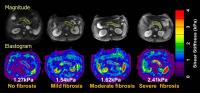 |
Quantitative analysis of diagnosing pancreatic fibrosis using
Magnetic resonance elastography
Yu Shi1, qiyong guo1, He An1,
Kevin J Glaser2, and Ehman L Richard3
1Department of radiology, Shengjing hospital of
China medical university, shenyang, China, People's Republic
of, 2Rochester,
MN, United States, 3Department
of radiology, Mayo Clinic, Rochester, MN, United States
An accurate diagnosis of pancreatic fibrosis is important in
clinical work. MR elastography (MRE) can be used for staging
the degrees of pancreatic fibrosis that reflects the
severity of chronic pancreatitis. Our work proved that both
fibrosis (P< 0.001) and inflammation (P=
0.014) contribute to higher stiffness of pancreatic
parenchyma, excluding fat deposition (P= 0.082).
The sensitivity and specificity was 100% and 86% for
diagnosing ≥F2, and 100% and 100% for diagnosing =F3
fibrosis stage, respectively.
|
| |
17:36
|
0534.
 |
Impact of Inter-lobular Fat on the Repeatability of Pancreatic
Fat Fraction Measurement by MRI.
Adam Jaster1, Ivan Pedrosa1,2, Robert
E. Lenkinski1,2, Ildiko Lingvay3,4,
and Takeshi Yokoo1,2
1Radiology, UT Southwestern Medical Center,
Dallas, TX, United States, 2Advanced
Imaging Research Center, UT Southwestern Medical Center,
Dallas, TX, United States, 3Internal
Medicine, UT Southwestern Medical Center, Dallas, TX, United
States, 4Clinical
Sciences, UT Southwestern Medical Center, Dallas, TX, United
States
In pancreatic steatosis, fat accumulates within
intra-lobular (parenchyma) and inter-lobular (adipose)
tissue. Regions of interest (ROIs) placed in pancreas
include heterogeneous population of pixels of lower-fat
intra-lobular tissue, higher-fat inter-lobular tissue, and
admixture of the two by partial volume effects. In this
study of 21 subjects with insulin-dependent type 2 diabetes,
we investigated the impact of inter-lobular fat on the
repeatability of pancreatic fat fraction (FF) measurement by
multiecho gradient-echo MRI. We found that the mean FF
measurement within segmented pancreatic ROI is highly
repeatable with intraclass correlation of 0.965 after
exclusion of high-fat (FF≥50%) pixels contaminated by
inter-lobular fat.
|
| |
17:48
|
0535.
 |
Longitudinal change of pancreatic proton density fat fraction
(PDFF) and its correlates during weight loss in initially obese
adults
Yesenia Covarrubias1, Alexandra N Schlein1,
William M Haufe1, Catherine A Hooker1,
Adrija Mamidipalli1, Tanya Wolfson2,
Garth Jacobson3, Santiago Horgan3,
Jeffrey B Schwimmer4, Scott B Reeder5,
and Claude B Sirlin1
1Liver Imaging Group, Department of Radiology,
University of California, San Diego, School of Medicine, San
Diego, CA, United States, 2Computational
and Applied Statistics Laboratory (CASL), SDSC, University
of California, San Diego, La Jolla, CA, United States, 3Department
of Surgery, University of California, San Diego, La Jolla,
CA, United States, 4Division
of Gastroenterology, Hepatology, and Nutrition & Department
of Pediatrics, University of California, San Diego, School
of Medicine, San Diego, CA, United States, 5Departments
of Radiology, Medical Physics, Biomedical Engineering,
Medicine, Emergency Medicine, University of Wisconsin,
Madison, WI, United States
This pilot, prospective, longitudinal study in 9 obese
adults explored the relationship between weight loss and
longitudinal change in MRI-determined pancreatic proton
density fat fraction (PDFF), as well as the relationships
between rates of change in pancreatic PDFF, hepatic PDFF,
and anthropometric measures. Pancreatic PDFF decreased in
every subject from a mean of 15.5% at the first study visit
to a mean of 8.6% at the last study visit (p=0.006). Further
research in larger cohorts is needed to confirm our findings
and to understand the clinical and biological relevance of
pancreatic PDFF reduction.
|
|