| |
16:00
 |
0469.
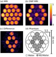 |
Nanodiamond Imaging with Room Temperature Dynamic Nuclear
Polarization 
David E J Waddington1,2,3, Mathieu Sarracanie2,3,4,
Huiliang Zhang3,5, Torsten Gaebel1,
David R Glenn3,5, Ewa Rej1, Najat
Salameh2,3,4, Ronald L Walsworth3,5,
David J Reilly1, and Matthew S Rosen2,3,4
1School of Physics, University of Sydney, Sydney,
Australia, 2A.A
Martinos Center for Biomedical Imaging, Massachusetts
General Hospital, Charlestown, MA, United States, 3Department
of Physics, Harvard University, Cambridge, MA, United
States, 4Harvard
Medical School, Boston, MA, United States, 5Harvard-Smithsonian
Center for Astrophysics, Cambridge, MA, United States
Overhauser-enhanced MRI (OMRI) is a double resonance
technique that has been developed to image free radicals in
vivo. Here, we use an ultra-low field MRI scanner
with a highly efficient b-SSFP OMRI protocol to image
synthetic nanodiamonds (NDs) in water at room temperature.
Surprisingly, we find that high contrast can be generated
via the Overhauser effect due to paramagnetic impurities in
the ND. Given the already established application of ND as
a biocompatible platform for drug delivery, these results
are encouraging for applications based on the non-invasive
tracking of nanoparticles using MRI.
|
| |
16:12
|
0470.
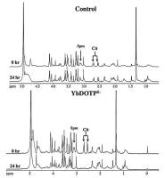 |
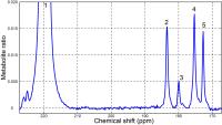
Imaging Spermine using LnDOTP5-Towards a Noninvasive Staging of
Prostate Cancer
Abiola Olatunde1, Taylor Fuss1,
Phillip Zhe Sun1, Leo L Cheng1, and
Peter Caravan1
1Massachusetts General Hospital, Boston, MA,
United States
Prostate cancer (PCa) is the most frequently diagnosed
malignancy in men worldwide. Previous studies have indicated
the utility of spermine as a potential biomarker for
prostate cancer; however, quantifying spermine using MRS is
difficult due to overlapping chemical shifts of spermine
with other metabolites. We used LnDOTP5-, an
anionic lanthanide macrocyclic complex, to form a stable
ternary complex with positively-charged spermine to
selectively shift spermine MR resonances. Here we report the
affinity of different LnDOTP5- complexes
for spermine and the effect of complex formation on spermine
MR resonances in both D2O and serum solutions and
intact human prostate tissue.
|
| |
16:24
 |
0471.
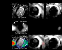 |
Molecular imaging of inflammation and extracellular matrix
remodelling after myocardial infarction
Isabel Ramos1,2, Markus Henningsson1,
Maryam Nezafat1, Begoña Lavin1,2,
Pierre Gebhardt1, Andrea Protti1,2,
Sara Lacerda1,2, Silvia Lorrio1,2,
Alkystis Phinikaridou1,2, Ulrich Flögel3,
Ajay M. Shah2, and René M. Botnar1,2
1Imaging Sciences and Biomedical Engineering,
King's College London, London, United Kingdom, 2Cardiovascular
Division, The British Heart Foundation Centre of Excellence,
King's College London, London, United Kingdom, 3Department
of Molecular Cardiology, Heinrich Heine University
Düsseldorf, Düsseldorf, Germany
Optimal post-MI healing relies on a suitable degree of
inflammation and its timely resolution, which is directly
related to a well-orchestrated degradation and deposition of
extracellular matrix (ECM) proteins, leading to cardiac
remodeling. Here we explored the merits of multinuclear 1H/19F
MRI for the simultaneous assessment of cardiac inflammation
and subsequent remodelling in a murine model of MI. To
investigate inflammatory cell recruitment into injured
myocardium, a 19F
containing nanoparticle that is avidly taken up by
macrophages was used1. To evaluate changes of
elastin content in the ECM post-MI, a small molecular weight
gadolinium-based elastin-specific MR contrast agent was
investigated2.
|
| |
16:36
|
0472.
|
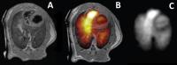
Correlation of Hyperpolarized 13C-Lactate Measurements and Ex
Vivo NMR using a [3-13C]Pyruvate Injection
Casey Y. Lee1,2, Justin Y. C. Lau1,2,
Albert P. Chen3, Yi-Ping Gu2, and
Charles H. Cunningham1,2
1Medical Biophysics, University of Toronto,
Toronto, ON, Canada, 2Physical
Sciences, Sunnybrook Research Institute, Toronto, ON,
Canada, 3GE
Healthcare, Toronto, ON, Canada
Lactate has been proposed as a potential marker to
non-invasively predict cancer progression and monitor
response to the therapy. Previously, hyperpolarized [1-13C]pyruvate
have been used to study the metabolic properties of tumor
through measuring the rapid conversion of pyruvate to
lactate. However, the fate of the 13C-lactate,
following the hyperpolarized experiment, has been less
understood due to the fast, irreversible decay of the
hyperpolarized signal. In this work, lactate concentrations
(total, 13C1-
and 13C3-lactate)
has been estimated in rat tumor extracts following the
injection of hyperpolarized [1-13C]pyruvate and
non-hyperpolarized [3-13C]pyruvate in rats.
|
| |
16:48
|
0473.
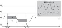 |
Velocity-Selective Tip-Back Excitation for Hyperpolarized [13C]
Urea Cardiac Perfusion Imaging
Maximilian Fuetterer1, Julia Busch1,
Constantin von Deuster1,2, Christian Binter1,
Nikola Cesarovic3, Miriam Lipiski3,
Christian Torben Stoeck1,2, and Sebastian Kozerke1,2
1Institute for Biomedical Engineering, University
and ETH Zurich, Zurich, Switzerland, 2Division
of Imaging Sciences and Biomedical Engineering, King's
College London, London, United Kingdom, 3Division
of Surgical Research, University Hospital Zurich, Zurich,
Switzerland
A velocity-selective excitation scheme with bipolar
slice-select gradients for hyperpolarized cardiac perfusion
imaging is presented. Using the approach, an excitation
ratio of >5 of myocardial signal to left-ventricular blood
pool signal can be achieved based on differences in blood
and tissue velocities. Thereby increased myocardial signal
and reduced left-ventricular signal spilling is obtained.
Dynamic perfusion images acquired with hyperpolarized [13C]urea
in pigs show higher SNR and less signal leakage in the
myocardium relative to a conventional excitation approach.
|
| |
17:00
|
0474.
 |
Investigating in vivo cardiac ketone bodies metabolism using
hyperpolarized 13C acetoacetate
Way Cherng Chen1, Xing Qi Teo1, and
Teck Hock Philip Lee1
1Laboratory of metabolic imaging, Singapore
Bioimaging Consortium, Singapore, Singapore
The use of hyperpolarized 3-13C acetoacetate to probe in
vivo cardiac ketone bodies metabolism was investigated.
Preliminary results showed the successful detection of 1-13C
citrate and 1-13C acetylcarnitine after hyperpolarized
acetoacetate delivery. Specifically, a significant increase
in citrate with a corresponding decrease in acetylcarnitine
was observed in the rat heart in vivo after 24hrs of
fasting.
|
| |
17:12
|
0475.
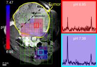 |
Dynamic Nuclear Polarization of Biocompatible 13C-Enriched
Carbonates for In vivo pH Imaging
David E Korenchan1,2, Robert Flavell1,
Renuka Sriram1, Celine Baligand1, Kiel
Neumann1, Subramaniam Sukumar1, Daniel
B Vigneron1,2, Henry VanBrocklin1,
David M Wilson1, and John Kurhanewicz1,2
1Radiology and Biomedical Imaging, University of
California at San Francisco, San Francisco, CA, United
States, 2Bioengineering,
University of California at Berkeley, Berkeley, CA, United
States
Although large gains in hyperpolarized 13C-bicarbonate
signal are obtainable for extracellular pH imaging, toxicity
becomes a concern for clinical implementation of current
methods. We report an approach in which a precursor
molecule, 1,2-glycerol carbonate, is hyperpolarized and
decomposed to form bicarbonate, CO2, and glycerol
using base-catalyzed hydrolysis. This technique enables
concentrations and polarizations similar to those previously
reported, and its application to pH imaging, both in phantom
experiments and in
vivo in a
mouse model of prostate cancer, is demonstrated.
|
| |
17:24
|
0476.
 |
Towards High Resolution Chemical Shift Imaging of the Lungs
using Hyperpolarized Carbon-13
Mehrdad Pourfathi1,2, Stephen J. Kadlecek1,
Harrilla Profka1, Sarmad M. Siddiqui1,3,
Heather Gatens1, and Rahim R. Rizi1
1Radiology, University of Pennsylvania,
Philadelphia, PA, United States, 2Electrical
and Systems Engineering, University of Pennsylvania,
Philadelphia, PA, United States, 3Bioengineering,
University of Pennsylvania, Philadelphia, PA, United States
We present the utility of a under-sampled single-shot turbo
spin-echo (TSE) sequence for high resolution T2 mapping
and imaging of the lungs using hyperpolarized carbon-13
agents. We then demonstrate the possibility of using this
sequence selectivity excite different carbon-13 species via
a minimum-phase frequency-selective excitation pulse.
|
| |
17:36
|
0477.
|
Heteronuclear cross- relaxation and polarization transfer
effects enable spectroscopic measurements of enzymatic activity
by hyperpolarized proton NMR.
Piotr Dzien*1, Anne Fages*2, Kevin
Michael Brindle3, Markus Schwaiger1,
and Lucio Frydman2
1Nuklearmedizinische Klinik und Poliklinik
Klinikum rechts der Isar der TUM, Technische Universität
München, Munich, Germany, 2Chemical
Physics, Weizmann Institute of Science, Rehovot, Israel, 3CRUK
Cambridge Institute, University of Cambridge, Cambridge,
United Kingdom
Disolution DNP increases the sensitivity of 13C MR
sufficiently to allow real time measurements of 13C-
labelled substrates and products of their metabolism in
vivo. While advantages could also result from hyperpolarized
observations based on 1H MR, the fast relaxation times of 1H
resonances prevent in vivo applications of this kind. Here
we demonstrate, in vitro, that a substantial enhancement of
the 1H resonance of [1-1H, 2,2,2-2H3,1-13C] acetaldehyde,
produced in situ by solutions containing purified yeast
Pyruvate Decarboxylase (yPDC) from 13C - hyperpolarized
[U-2H3,2-13C] pyruvate, can be achieved. This enhancement
can arise from either spontaneous or INEPT-driven 13C --> 1H
polarization transfers.
|
| |
17:48
|
0478.
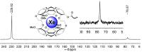 |
Functionalized Cryptophane-129Xe MRI Biosensor for Biothiols
Detection through Thiol-addition Reaction
Shengjun Yang1, Weiping Jiang1, Qing
Luo1, Qianni Guo1, and Xin Zhou1
1Wuhan Institute of Physics and Mathematics,
Chinese Academy of Sciences, Wuhan, China, People's Republic
of
Biothiols such as cysteine, homocysteine and glutathione
play an important role in regulating the vital functions of
living organisms. Here, we report a biosensor for biothiol
detection and imaging using nuclear spin resonance of 129Xe.
The 129Xe biosensor consists of cryptophane-A cage
encapsulating xenon atom and acrylate group. The latter
serves as a reactive site to covalently bond biothiols
through thiol-addition reaction. The selectivity of the
biosensor enables discrimination of Cys from Hcy and GSH
through the chemical reaction rate. Our results indicate
that this biosensor is a promising strategy for the
real-time imaging of biothiol distributions.
|
|