| |
16:30
|
0207.
 |
Optimal data acquisition for application to the continuous time
random walk diffusion model - Permission Withheld
Thomas Richard Barrick1, Andrew Mott1,
Diggory North1, and Franklyn Arron Howe1
1Neuroscience Research Centre, St George's,
University of London, London, United Kingdom
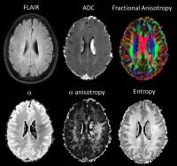
This study aims to optimise diffusion-weighted MRI (DW-MRI)
acquisition for applications involving the continuous time
random walk (CTRW) diffusion model. Minimum acquisition time
and effects of inversion recovery are considered. Optimisation
indicates a 6 minute 4 b-value DW-MRI acquisition is
sufficient for diffusion tensor data. Inversion recovery
significantly reduces the variability in calculated α, β and
ADC due to effects of CSF in grey matter and periventricular
white matter. Analysis of water diffusion in brain with the
CTRW model may reveal more subtle effects of neuronal damage
than conventional DWI.
|
| |
16:42
|
0208.
 |
The Effects of Navigator Distortion Level on Interleaved EPI DWI
Reconstruction: A Comparison between Image and K-space Based
Method 
Erpeng Dai1, Xiaodong Ma1, Zhe Zhang1,
Chun Yuan1,2, and Hua Guo1
1Center for Biomedical Imaging Research,
Department of Biomedical Engineering, School of Medicine,
Tsinghua University, Beijing, China, People's Republic of, 2Vascular
Imaging Laboratory, Department of Radiology, University of
Washington, Seattle, WA, United States
One of the challenges for interleaved EPI (iEPI) DWI is the
phase inconsistency among different shots. Several methods,
performed either in the image or k-space domain, have been
proposed to solve this problem with extra acquired navigator
data. However, the navigator is usually acquired with a
lower bandwidth in the phase encoding direction than the
image echo, which can cause different distortion levels. In
this study, the effects of such distortion for the image or
k-space based reconstruction are investigated. It has been
shown that the k-space based method is more tolerant to
the navigator distortion.
|
| |
16:54
|
0209.
 |
Experimental detection of imaginary signals in diffusion pore
imaging using double diffusion encoding - Permission Withheld
Kerstin Demberg1, Frederik Bernd Laun1,
Johannes Windschuh1, Reiner Umathum1,
Peter Bachert1, and Tristan Anselm Kuder1
1Medical Physics in Radiology, German Cancer
Research Center (DKFZ), Heidelberg, Germany
By diffusion pore imaging, the average shape of arbitrary
closed pores in an imaging volume element can be detected
employing a long-narrow gradient profile. Alternative
approaches use short gradient pulses only. Until now,
however, diffusion pore imaging of non-point-symmetrically
shaped pores has not been demonstrated using short gradient
pulses only. In this abstract, we present a first
experimental verification using double diffusion encoded
experiments. Non-point-symmetric pores result in
non-vanishing imaginary parts in the double diffusion
encoded signal. Thus the phase of the form factor can be
estimated with an iterative approach. This allows for
unambiguous pore image reconstruction.
|
| |
17:06
|
0210.
 |
Virtual Coil Reconstruction for 3D Diffusion-Weighted Multi-Shot
MRI using a Single Reference Shot.
Eric Y. Pierre1, Jacques-Donald Tournier1,2,
and Alan Connelly1
1Florey Institute of Neuroscience and Mental
Health, Melbourne, Australia, 2Centre
for the Developing Brain, King's College London, London,
United Kingdom
We introduce an efficient Mult-Shot Diffusion-Weighted (DW)
3D-GRASE acquisition and reconstruction technique to produce
DW image volumes free of motion-induced phase artifacts,
without relying on explicit measurement or inference of the
phase information. The method replaces navigators
measurements by a single reference scan for the whole
acquisition. Virtual Coil concepts for Parallel Imaging
techniques are used to map the multi-shot data onto a
k-space signal with consistent phase information.
|
| |
17:18
 |
0211.
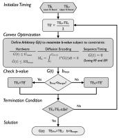 |
Convex Optimized Diffusion Encoding (CODE) Gradient Waveforms
for Minimum TE and Bulk Motion Compensated Diffusion Weighted
MRI
Eric Aliotta1,2, Holden H Wu1,2, and
Daniel B Ennis1,2
1Radiological Sciences, UCLA, Los Angeles, CA,
United States, 2Biomedical
Physics IDP, UCLA, Los Angeles, CA, United States
Spin-Echo EPI Diffusion Weighted MRI (SE-EPI DWI) typically
uses a diffusion encoding gradient waveform with two
identical gradients on either side of the 180° pulse which,
in combination with the temporal footprint of the EPI
readout results in sequence dead time. This dead time can be
used for additional diffusion encoding which can, in turn,
reduce TE and/or be used to null gradient moments for bulk
motion compensated diffusion encoding. Convex Optimized
Diffusion Encoding (CODE) was developed to minimize TE for
DWI with and without motion compensation, implemented on a
clinical scanner and tested in volunteers.
|
| |
17:30
|
0212.
 |
Detection of Microscopic Diffusion Anisotropy in Human Brain
Cortical Gray Matter in Vivo with Double Diffusion Encoding
Marco Lawrenz1 and
Juergen Finsterbusch1
1Systems Neuroscience, University Medical Center
Hamburg-Eppendorf, Hamburg, Germany
Double diffusion encoding experiments with two weighting
periods applied successively in the same acquisition offer
access to microscopic tissue properties. Rotationally
invariant measures of the so-called microscopic diffusion
anisotropy as a marker for cell or compartment shape have
reliably been determined in brain white matter. In this
study, it is demonstrated that microscopic diffusion
anisotropy can also be detected in cortical gray matter in
vivo and measures of it can be determined extending first
evidences presented recently. However, an inversion recovery
pulse is required to null white matter signals and avoid
partial volume effects.
|
| |
17:42
|
0213.
 |
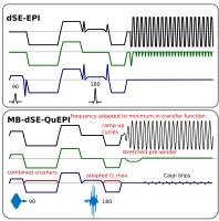
High-resolution diffusion imaging at 7T using 3D multi-slab EPI
Wenchuan Wu1, Peter J Koopmans1,
Robert Frost1, Myung-Ho In2, Oliver
Speck3, and Karla L Miller1
1FMRIB, Nuffield Department of Clinical
Neurosciences, University of Oxford, Oxford, United Kingdom, 2Department
of Neurologic Surgery, Mayo Clinic, Rochester, MN, United
States, 3Department
of Biomedical Magnetic Resonance, Otto-von-Guericke
University, Magdeburg, Germany
In this work, we combined 3D multi-slab imaging (optimal SNR
efficiency for spin-echo sequence) and 7T (higher SNR) to
enhance diffusion imaging. With the newly developed
Slice-FLEET technique and NPEN correction, we successfully
achieved robust high resolution diffusion MRI at 7T with
high SNR.
|
| |
17:54
|
0214.
 |
Efficient quiet multiband accelerated HARDI fetal Diffusion
Jana Maria Hutter1, J-Donald Tournier1,
Anthony N Price1, Lucilio Cordero Grande1,
Emer Judith Hughes1, Kelly Pegoretti1,
Laura McCabe1, Mary Rutherford1, and
Joseph V Hajnal1
1Centre for the developing brain, King's College
London, London, United Kingdom
Fetal diffusion MRI analysis is often limited by the
ability of the conventional ssEPI to allow an efficient,
high-resolution acquisition, able to produce multi-shell
high angular resolution dMRI data as required by advanced
analysis tools. This abstract presents a novel, multiband
accelerated, sinusoidal, quiet and efficient ssEPI
acquisition. The first results on 3 fetuses with 54
directions show promising data quality and significantly
decreased scan time.
|
| |
18:06
|
0215.
 |
Microscopic Anisotropy of the Rat Spinal Cord In vivo with DW
PRESS
Matthew Budde1 and
Nathan Skinner1
1Neurosurgery, Medical College of Wisconsin,
Milwaukee, WI, United States
Diffusion weighted imaging of the spinal cord has seen
promising applications to diagnosis and prognosis, yet it is
limited by technical challenges. This work presents the
implementation of diffusion weighted spectroscopy of the
water signal in the rat spinal cord in vivo with the goal of
reducing acquisition times and post processing requirements
to promote wider clinical feasibility.
|
| |
18:18
|
0216.
 |
Diffusion-weighted MRI using undersampled radial STEAM with
iterative image reconstruction
Andreas Merrem1, Jakob Klosowski1,
Sabine Hofer1, Klaus-Dietmar Merboldt1,
and Jens Frahm1
1Biomedizinische NMR Forschungs GmbH,
Max-Planck-Institut für Biophysikalische Chemie, Göttingen,
Germany
Single-shot STEAM MRI is a method for black-blood
diffusion-weighted imaging where the use of
radiofrequency-refocussed echoes leads to no image
distortions, no susceptibility artifacts, and no violations
of the Carr-Purcell-Meiboom-Gill condition. Despite these
favorable properties, clinical applications have been
limited by a low signal-to-noise ratio. Here, we demonstrate
the development of highly undersampled radial
diffusion-weighted single-shot STEAM MRI with iterative
reconstruction to achieve acceptable signal-to-noise for
studies of the human brain.
|
|