| |
13:30
 |
0920.
 |
Intravoxel Incoherent Motion MRI in a 3-Dimensional
Microvascular Flow Phantom 
Moritz Schneider1, Thomas Gaaß1,2,
Julien Dinkel1,2, Michael Ingrisch1,
Maximilian F Reiser1, and Olaf Dietrich1
1Institute for Clinical Radiology, Ludwig-Maximilians-University
Hospital Munich, Munich, Germany, 2Comprehensive
Pneumology Center, German Center for Lung Research, Munich,
Germany
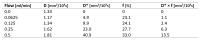
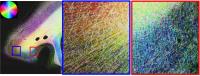
In this study we present intravoxel incoherent motion (IVIM)
measurements in a flow phantom consisting of a 3-dimensional
capillary network made from melt-spun, sacrificial sugar
structures embedded in a synthetic resin. IVIM parameters
were determined at varying water flow rates. The
pseudodiffusion D* (associated with flow velocity) as well
as the product D*×f (which constitutes a measure of flow)
show proportionality to the applied flow rates. These
results demonstrate that the presented flow phantom is ideal
to assess the applicability of IVIM measurements and
influence factors such as flow rates, capillary diameter or
acquisition parameters.
|
| |
13:42
|
0921.
 |
Single MR spectral peak diffusion phantom with wide ADC range
based on acetone, H2O and manganese chloride
Xiaoke Wang1, Scott B Reeder1,2,3,4,5,
and Diego Hernando2
1Biomedical Engineering, University of
Wisconsin-Madison, Madison, WI, United States, 2Radiology,
University of Wisconsin-Madison, Madison, WI, United States, 3Medical
Physics, University of Wisconsin-Madison, Madison, WI,
United States, 4Medicine,
University of Wisconsin-Madison, Madison, WI, United States, 5Emergency
Medicine, University of Wisconsin-Madison, Madison, WI,
United States
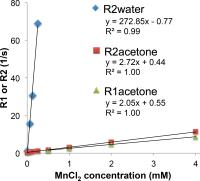
Practical diffusion phantoms are urgently needed for
technique development, protocol harmonization and quality
assurance of quantitative diffusion MRI. Ideally, a
diffusion phantom should have a single-peak NMR spectrum,
Gaussian diffusion, with a wide range of tunable apparent
diffusion coefficients (ADC). In this work, we developed and
validated a novel diffusion phantom based on acetone-water
mixtures doped with MnCl2. This phantom exhibits
the desired signal behavior, where water modulates the ADC
of acetone, and MnCl2 both
eliminates water signal (through T2 shortening) and shortens
the T1 of acetone.
|
| |
13:54
|
0922.
 |
Validation of Diffusion Tensor MRI with Structure Tensor
Synchrotron Imaging
Irvin Teh1, Darryl McClymont1,
Marie-Christine Zdora2,3, Valentina Davidoiu4,
Hannah J Whittington1, Christoph Rau2,
Irene Zanette2, and Jürgen E Schneider1
1Division of Cardiovascular Medicine, Radcliffe
Department of Medicine, University of Oxford, Oxford, United
Kingdom, 2Diamond
Light Source, Didcot, United Kingdom, 3Department
of Physics and Astronomy, University College London, London,
United Kingdom, 4Department
of Imaging Sciences and Biomedical Engineering, King's
College London, London, United Kingdom
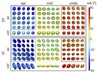
Diffusion tensor imaging (DTI) is widely used to assess
tissue microstructure, but is limited in resolution and
cannot resolve multiple fibre populations within a voxel.
Existing methods for validating DTI are limited in either
resolution or coverage. 2D histological methods are
additionally destructive and prone to tissue distortion. In
contrast, synchrotron imaging strikes an excellent balance
between resolution and coverage. Here, we demonstrate for
the first time, the prospect of validating DTI with
structure tensor analysis of synchrotron imaging data.
Tensors reconstructed with DTI and structure tensor
synchrotron imaging were consistent across the left
ventricular wall of the heart.
|
| |
14:06
 |
0923.
 |
Neuroplasticity changes in rat brain following targeted
irradiation assessed by diffusion MRI tractography validated by
histology and behavioral tests - Permission Withheld
Julie Constanzo1, Matthieu Dumont2,
Luc Tremblay1, Philippe Sarret3,
Jean-Michel Longpré3, Karyn Kirby3,
Sameh Geha4, Laurence Masson-Côté1,
Benoit Paquette1, and Maxime Descoteaux2
1Nuclear Medicine and Radiobiology, Sherbrooke
University, Sherbrooke, QC, Canada, 2Computing
Science, Sherbrooke University, Sherbrooke, QC, Canada, 3Pharmacology
and biophysics, Sherbrooke University, Sherbrooke, QC,
Canada, 4Pathology,
Sherbrooke University, Sherbrooke, QC, Canada
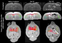
Despite its high efficiency for treating brain tumors and
metastases, stereotactic radiosurgery (SRS) may lead to
brain swelling, necrosis, and neuronal dysfunction, thus
inducing delayed adverse effects such as cognitive decline
and stroke-like symptoms. Altogether, our results revealed
that SRS treatment induces region-specific plasticity (i.e.
structural and function changes), as demonstrated by
neuronal matrix remodeling using diffusion MRI and
appropriate HARDI reconstruction, corresponding to
histopathological modifications and changes in behavioral
responses.
|
| |
14:18
|
0924.
 |
Comparing Diffusion MRI with the Fiber Architecture and Tract
Density of Gyral Blades
Kurt Schilling1, Vaibhav Janve1, Yurui
Gao1, Iwona Stepniewska1, Bennett
Landman1, and Adam Anderson1
1Vanderbilt University, Nashville, TN, United
States
It has been reported that diffusion tractography has a
tendency for streamlines to terminate preferentially on
gyral crowns rather than on sulcal walls or fundi. Rather
than anatomical reality, it has been suggested that this is
a bias associated with tractography. To better understand
this issue, we compare histology to diffusion MRI of the
same specimen. We measure the trajectories and density of
axons crossing the gray matter/white matter boundary and
compare to diffusion tensor measures and deterministic
tractography. The results of this study lead to a better
understanding of gyral anatomy and potential limitations of
fiber tractography.
|
| |
14:30
|
0925.
 |
Post-mortem inference of the inner connectivity of the human
hippocampus using ultra-high field diffusion MRI at 11.7T
Justine Beaujoin1,2,3, Fawzi Boumezbeur1,2,3,
Jérémy Bernard1,2,3, Markus Axer4,
Jean-François Mangin2,3,5,6, and Cyril Poupon1,2,3,6
1CEA NeuroSpin / UNIRS, Gif-sur-Yvette, France, 2Université
Paris-Saclay, Orsay, France, 3FLI
/ Noeud Paris-Sud, Orsay, France, 4Forschungszentrum
Jülich, INM1, Jülich, Germany, 5CEA
NeuroSpin / UNATI, Gif-sur-Yvette, France, 6http://cati-neuroimaging.com/,
Gif-sur-Yvette, France
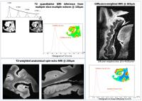
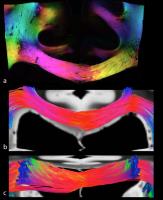
In this work, we demonstrate that post-mortem ultra-high
field (11.7T) / ultra-high gradients (760mT/m)
diffusion-weighted MRI allows to finely map the inner
connectivity of the human hippocampus and we show that the
polysynaptic intra-hippocampal pathway can be accurately
reconstructed using fiber tractography techniques at very
high spatial/angular resolutions.
|
| |
14:42
|
0926.
 |
Post-mortem diffusion MRI of cervical spine and nerves roots
Wieke Haakma1,2,3, Lidy Kuster2,
Martijn Froeling1, Lars Uhrenholt2,
Michael Pedersen3,4, Jeroen Hendrikse1,
Alexander Leemans5, and Lene Warner Thorup Boel2
1Radiology, University Medical Center Utrecht,
Utrecht, Netherlands, 2Forensic
Medicine, Aarhus University, Aarhus, Denmark, 3Comparative
Medicine Lab, Department of Clinical Medicine, Aarhus
University, Aarhus, Denmark, 4MR
Research Center, Department of Clinical Medicine, Aarhus
University, Aarhus, Denmark, 5Image
Sciences Institute, University Medical Center Utrecht,
Utrecht, Netherlands
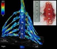
In this work we examined the architecture and diffusion
measures of the cervical spine and nerves in non-fixated
post-mortem subjects. We were able to display the
architectural configuration of the cervical nerves at the
level of C4-C8 and we computed reference values for the
diffusion measures in these nerves. We showed with great
detail the ventral and dorsal nerve roots with fiber
tractography. Microscopic examination revealed normal
anatomy. We expect that post-mortem diffusion MRI will be
valuable for understanding of pathological mechanisms
underlying degenerative neurological diseases, as it is
possible to compare any findings directly to histological
examinations.
|
| |
14:54
|
0927.
 |
Microstructure models for diffusion MRI in breast cancer and
surrounding stroma: an ex vivo study
Colleen Bailey1, Bernard Siow2,
Eleftheria Panagiotaki1, John H Hipwell1,
Sarah E Pinder3, Daniel C Alexander1,
and David J Hawkes1
1Centre for Medical Image Computing, University
College London, London, United Kingdom, 2Centre
for Advanced Biomedical Imaging, University College London,
London, United Kingdom, 3Breast
Research Pathology, King's College London and Guy's
Hospital, London, United Kingdom
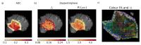
A variety of one- and two-compartment models were fitted to
rich diffusion data sets from ex vivo breast tissue samples
containing tumour. Two compartment models with restriction
explained the data better than conventional ADC and
bi-exponential models, as determined by the Akaike
Information Criterion. In four of seven samples, anisotropy
was also observed, although parametric maps of the primary
eigenvector direction show that regions of coherence are
small (~1 mm diameter).
|
| |
15:06
|
0928.
 |
Validation of quantitative MRI metrics using full slice
histology with automatic axon segmentation
Tanguy Duval1, Blanche Perraud1,
Manh-Tung Vuong1, Nibardo Lopez Rios1,2,
Nikola Stikov1,3, and Julien Cohen-Adad1,4
1Polytechnique Montréal, Montréal, QC, Canada, 2Medical
Biophysics Center, Oriente University, Santiago de Cuba,
Cuba, 3Montreal
Heart Institute, Montréal, QC, Canada, 4Functional
Neuroimaging Unit, CRIUGM, Université de Montréal, Montréal,
QC, Canada
In this work we propose to validate and compare AxCaliber/ActiveAx/Noddi/MTV
in the spinal cord using full slice histology with
axon/myelin segmentation. High resolution data (150µm/px)
were acquired on an ex vivo spinal cord and compared voxel
by voxel with histology. We found that q-space metrics were
precise enough to distinguish between various fiber
distributions. A correlation coefficient of r=0.62 was found
between AxCaliber and histology for axon diameter metric.
Also, good agreement were found between the different
q-space models and with MTV.
|
| |
15:18
|
0929.
 |
Validating tractography of high resolution post-mortem human
brain at 7T with polarized light imaging
Sean Foxley1, Jeroen Mollink1, Saad
Jbabdi1, Stuart Clare1, Moises
Hernandez Fernandez1, Connor Scott2,
Olaf Ansorge2, and Karla Miller1
1FMRIB Centre, University of Oxford, Oxford,
United Kingdom, 2Nuffield
Department of Clinical Neurosciences, University of Oxford,
Oxford, United Kingdom
In this work we present voxel-wise orientation estimates
from diffusion-weighted steady state free precession MRI
data of post-mortem human brain, acquired with three
resolutions at 7T. Data were acquired with 0.5mm, 1mm, and
2mm isotropic resolution over 90 directions. These
resolutions were chosen because 1mm and 2mm are typical of in
vivo DTI.
Deterministic tractography was produced in various regions
using the highest resolution dataset. Orientation maps
demonstrate small structures that are less apparent in lower
resolution data. Orientation estimates and tractography
results were validated with polarized light microscopy
imaging.
|
|