| |
10:00
|
0557.
 |
Quantitative Determination of Pediatric Myelination Using Fast
Bound-Pool Fraction Imaging 
Hunter R Underhill1,2 and
Gary Hedlund2
1Pediatrics, University of Utah, Salt Lake City,
UT, United States, 2Radiology,
University of Utah, Salt Lake City, UT, United States
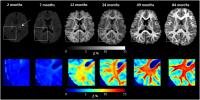
Fast bound-pool fraction imaging (FBFI) is a quantitative
MRI technique validated with histology to measure
whole-brain, voxel-based myelin density. In this study, FBFI
was translated to a whole-body 3T clinical scanner using
only standard preset sequences without modifications to
measure myelin density in the developing pediatric brain via
a time-efficient methodology (<7 min). We found that FBFI
effectively quantifies myelin density during normal
development. Progressive myelination identified in the
posterior white matter corresponded strongly to a bounded
exponential growth curve. Quantification of myelin density
with FBFI in pediatric patients may improve detection of
delayed or altered myelination.
|
| |
10:12
|
0558.
 |
Longitudinal Probing Infant Brain Connectomes Using Graph Theory
Longchuan Li1,2, Sarah Shultz1,
Xiaoping Hu2, Ami Klin1, and Warren
Jones1
1Marcus Autism Center, Emory University, Atlanta,
GA, United States, 2Biomedical
Imaging Technology Center, Emory University, Atlanta, GA,
United States
We used diffusion tractography and network theory to examine
the organizational development of the brain in typical
infants in their first 6 months of life. Data were
longitudinally sampled at randomized time points between
birth and 6 months and collected on a Siemens 3T TIM Trio
system with 32-channel coil using multiband techniques. We
found that network-based metrics may reveal unique
information in the organizational principles of the brain
and its development that is impossible with conventional
methods focusing on specific pathways and regions,
demonstrating the usefulness of the approach in studying
early typical brain development and its disruptions.
|
| |
10:24
|
0559.
 |
Toward routine assessment of cerebral blood flow in neonates and
infants: a phase-contrast MRI study
Peiying Liu1, Ying Qi2, Zixuan Lin1,
Xuna Zhao3, Qiyong Guo2, Xiaoming Wang2,
and Hanzhang Lu1
1Department of Radiology, Johns Hopkins
University School of Medicine, Baltimore, MD, United States, 2Shengjing
Hospital of China Medical University, Shenyang, China,
People's Republic of, 3Philips
Healthcare, Beijing, China, People's Republic of
Knowledge of CBF in neonates or infants may provide valuable
information in many pathological conditions. When applied to
very young children, CBF mapping using
arterial-spin-labeling (ASL) MRI suffers from low SNR and
poor quantification, whereas phase-contrast (PC) MRI may
provide reliable estimation of global CBF. Therefore, this
study aim to 1) provide a set of age-specific PC-MRI
protocols for CBF quantification in children under 1.5 years
old; 2) establish typical arterial flow velocity in children
at this age which could guide future ASL efforts in labeling
pulse optimization; 3) report how CBF changes during this
early stage of life.
|
| |
10:36
 |
0560.
 |
Global and regional cortical connectivity maturation index
(CCMI) of developmental human brain with quantification of
short-range association tracts
Minhui Ouyang1, Tina Jeon1, Jennifer
Muller1, Virendra Mishra2, Haixiao Du3,
Yu Wang3, Yun Peng4, Bo Hong5,
and Hao Huang1,6
1Department of Radiology, Children's Hospital of
Philadelphia, Philadelphia, PA, United States, 2Cleveland
Clinic Lou Ruvo Center for Brain Health, Las Vegas, NV,
United States, 3Department
of Electronic Engineering, Tsinghua University, Beijing,
China, People's Republic of, 4Department
of Radiology, Beijing Children's Hospital, Capital Medical
University, Beijing, China, People's Republic of, 5Department
of Biomedical Engineering, School of Medicine, Tsinghua
University, Beijing, China, People's Republic of, 6Department
of Radiology, Perelman School of Medicine, University of
Pennsylvania, Philadelphia, PA, United States
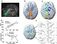
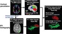
Disturbance of precisely balanced strengthening of certain
axons and pruning of others in developmental human brains is
associated with mental disorders such as autism and
schizophrenia. To characterize this balance, we defined a
cortical connectivity maturation index (CCMI) derived from
short-range association tracts traced with diffusion MRI
tractography. The brain CCMI values were measured with
diffusion MRI and T1-weighted datasets of 21
healthy subjects with age of 2-25 years. CCMI in all
cortical regions decreased in early developmental stage and
increased later, yet with distinctive trajectories. The
observed CCMI dynamics may be underlaid by heterogeneous
pruning among cortical regions.
|
| |
10:48
|
0561.
 |
Parental Education and Childhood Brain and Behavioral
Development
Sean Deoni1,2, Holly Dirks2, Jonathan
O'Muircheartaigh3, and Douglas C Dean4
1CHILD Lab, Children's Hospital, Colorado,
Aurora, CO, United States, 2Advanced
Baby Imaging Lab, Brown University, Providence, RI, United
States, 3Neuroimaging,
King's College, London, London, United Kingdom, 4Waisman
Lab for Brain Imaging and Behavior, University of Wisconsin
Madison, Madison, WI, United States
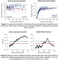
It is well established that family socioeconomic status
(SES), related to parental education level, occupation, and
income, is associated with differences in offspring
educational outcomes and cognitive abilities. However,
while brain imaging studies in older children have revealed
altered brain structure associated with SES, the influence
of SES on infant and childhood brain development remain
unclear. Here we investigated longitudinal trajectories of
brain and cognitive development in a large cohort of
typically-developing children from 2 months to 6 years of
age. Results reveal diverging developmental trends
associated with parental education (PE) level even when
controlling for common confounds.
|
| |
11:00
|
0562.
 |
Age-related Magnetic Susceptibility in the Deep Gray Nuclei from
1 month to 6 Years: Comparison between Quantitative
Susceptibility and R2* Mapping
Ning Ning1, Peng Wu2, Xianjun Li3,
Yajie Hu3, Weishan Zhang1, Lei Zhang1,
Sung-Min Gho4, Dong-Hyun Kim4, Hua Guo2,
and Jian Yang1,3
1Department of Diagnostic Radiology, the First
Affiliated Hospital of Xi’an Jiaotong University, Xi'an,
China, People's Republic of, 2Department
of Biomedical Engineering, Tsinghua University, Beijing,
China, People's Republic of, 3Department
of Biomedical Engineering, School of Life Science and
Technology, Xi'an Jiaotong University, Xi'an, China,
People's Republic of, 4Department
of Electrical and Electronic Engineering, Yonsei University,
Seoul, Korea, Republic of
To observe the age-related susceptibility changes in the
deep gray nuclei and assess the superiority of the
quantitative susceptibility mapping(QSM) and effective
transverse relaxation rate(R2*) for quantifying the iron
deposits in children. 87 subjects(1M-6Y) were enrolled. The
susceptibility in QSM and R2* values exhibited positive
correlations with age and the reference iron concentrations
calculated using an empirical equation. The correlation of
the susceptibility with the iron is higher than the R2* with
it. QSM may provide a more promising and reliable tool for
assessment of iron content in children’s deep gray nuclei,
even in the regions with lower iron content.
|
| |
11:12
|
0563.
 |
To smell or not to smell: does the newborn habituate to
sustained odorant stimulation?
Frédéric Grouiller1, Alexandra Adam-Darqué2,
Russia Ha-Vinh Leuchter2, Petra S Hüppi2,
and François Lazeyras1
1Department of Radiology and Medical Informatics,
University of Geneva, Geneva, Switzerland, 2Division
of Development and Growth, Department of Pediatrics,
University of Geneva, Geneva, Switzerland
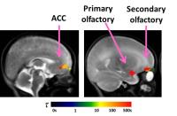
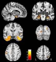
The aim of this study is to better characterize the
habituation effect of sustained odorant stimulation and to
investigate if this effect is already present in newborns.
Olfactory fMRI was acquired in adults and newborns using a
20s block design. After modelling habituation, activations
in the primary and secondary olfactory cortices were
observed in adults and newborns. Habituation effect to
sustained odorant stimulation was strong in adults but
unseen in the newborns. This study shows that the olfactory
cortex of newborns is highly functional soon after birth and
that the habituation effect is not observed in newborns
compared to adults.
|
| |
11:24
|
0564.
 |
Structural neuroimages revealed limited parental care affect
development of gray matter rather than white matter in
left-behind children
Yuan Xiao1,2, Lili Yang2, Lu Liu1,
Xin Gao1, Bo Tao1, Min Wu1,
Yuchuan Fu2, Meimei Du2, Zhihan Yan2,
and Su Lui1,2
1Department of Radiology, HMRRC, West China
Hospital of Sichuan University, Chengdu, China, People's
Republic of, 2Department
of Radiology, The Second Affiliated Hospital & Yuying
Children’s Hospital of Wenzhou Medical University, Wenzhou,
China, People's Republic of
This study provided the first empirical evidence of larger
gray matter volume in left-behind children than comparison
children who lived in the nuclear family, especially in
emotional circuit, suggesting the early parental care would
affect the brain development of gray matter rather than
white matter.
|
| |
11:36
 |
0565.
 |
Local shape analysis of the thalamus in extremely preterm born
young adults
Eliza Orasanu1, Andrew Melbourne1,
Zach Eaton-Rosen1, David Atkinson2,
Joshua Lawan3, Joanne Beckmann4, Neil
Marlow4, and Sebastien Ourselin1
1Translational Imaging Group, Centre for Medical
Image Computing, University College London, London, United
Kingdom, 2University
College London, London, United Kingdom, 3University
College Hospital, London, United Kingdom, 4Institute
for Women's Health, University College London, London,
United Kingdom
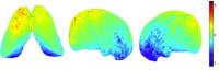
Alterations of thalamic structures may cause disruptions in
thalamic-cortical-thalamic circuitry and affect cognition.
In this work we present a local shape analysis of the
thalamus in extremely preterm born young adults when
compared to their term born peers. We perform a groupwise
shape analysis after spectral matching registration. After
correcting for gender and thalamic volume, it resulted that
the anterior and superior thalamic regions, connected to
regions responsible for executive function, working memory,
language and verbal memory, show most shape variations.
|
| |
11:48
|
0566.
 |
Segmentation of the fetal brain cortical plate using
diffusion-weighted imaging cues - Permission Withheld
Rosita Shishegar1,2, Shreya Rana3,
Mary Tolcos3, David W. Walker3, and
Leigh A. Johnston1,4
1Dept. Electrical & Electronic Engineering,
University of Melbourne, Melbourne, Australia, 2NICTA
Victoria Research Laboratory, Melbourne, Australia, 3The
Ritchie Centre, Hudson Institute of Medical Research, Monash
University, Melbourne, Australia, 4Florey
Institute of Neuroscience and Mental Health, Melbourne,
Australia
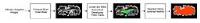
Segmentation of the developing cortical plate from MRI data
of the fetal brain is highly challenging due to partial
volume effects, low contrast and heterogeneous maturation
caused by ongoing myelination processes. We present a new
atlas-free method for segmenting the boundary between the
cortical plate and subplate in fetal brains, by exploiting
diffusion-weighted imaging cues. The accuracy of the
segmentation algorithm is demonstrated by application to
fetal sheep brain MRI data.
|
|