| |
08:00
 |
1088.
 |
Learning a Variational Model for Compressed Sensing MRI
Reconstruction 
Kerstin Hammernik1, Florian Knoll2,
Daniel K Sodickson2, and Thomas Pock1,3
1Institute for Computer Graphics and Vision, Graz
University of Technology, Graz, Austria, 2Center
for Biomedical Imaging and Center for Advanced Imaging
Innovation and Research (CAI2R), Department of Radiology,
NYU School of Medicine, New York, NY, United States, 3Safety
& Security Department, AIT Austrian Institute of Technology
GmbH, Vienna, Austria
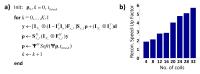
Compressed sensing techniques allow MRI reconstruction from
undersampled k-space data. However, most reconstruction
methods suffer from high computational costs, selection of
adequate regularizers and are limited to low acceleration
factors for non-dynamic 2D imaging protocols. In this work,
we propose a novel and efficient approach to overcome these
limitations by learning a sequence of optimal regularizers
that removes typical undersampling artifacts while keeping
important details in the imaged objects and preserving the
natural appearance of anatomical structures. We test our
approach on patient data and show that we achieve superior
results than commonly used reconstruction methods.
|
| |
08:12
|
1089.
 |
SENSE-LORAKS: Phase-Constrained Parallel MRI without Phase
Calibration
Tae Hyung Kim1, Kawin Setsompop2, and
Justin P. Haldar1
1Electrical Engineering, University of Southern
California, Los Angeles, CA, United States, 2Radiology,
Harvard Medical School, Boston, MA, United States
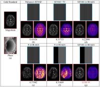
We introduce a novel framework called SENSE-LORAKS for
partial Fourier phase-constrained parallel MRI
reconstruction. SENSE-LORAKS combines classical SENSE data
modeling with advanced regularization based on the novel
low-rank modeling of local k-space neighorhoods (LORAKS)
framework. Unlike conventional phase-constrained SENSE
techniques, SENSE-LORAKS enables use of phase constraints
without requiring a prior estimate of the image phase or a
fully sampled region of k-space that could be used for phase
autocalibration. Compared to previous SENSE-based and
LORAKS-based reconstruction approaches, SENSE-LORAKS is
compatible with a much wider range of sampling trajectories,
which can be leveraged to achieve much higher acceleration
rates.
|
| |
08:24
|
1090.
 |
k-t ESPIRiT:
Efficient Auto-Calibrated Parallel Imaging Reconstruction by
Exploiting k-t Space
Correlations
Claudio Santelli1, Adrian Huber1, and
Sebastian Kozerke1
1Institute for Biomedical Engineering, University
and ETH Zurich, Zurich, Switzerland
Using eigen-decomposition of a modified k-t SPIRiT
operator, computationally optimized reconstruction formally
translating into auto-calibrated SENSE-like reconstruction
of a coil-combined x-f image
(k-tESPIRiT) is proposed. 2D and 3D in-vivo
experiments show equivalence of k-t SPIRiT
and k-t ESPIRiT,
and significant reconstruction time speed-up's of the
proposed relative to the standard technique.
|
| |
08:36
|
1091.
 |
Motion-Resolved Golden-Angle Radial Sparse MRI Using
Variable-Density Stack-of-Stars Sampling
Li Feng1, Tiejun Zhao2, Hersh
Chandarana1, Daniel K Sodickson1, and
Ricardo Otazo1
1Center for Advanced Imaging Innovation and
Research (CAI2R), New York University School of Medicine,
New York, NY, United States, 2Siemens
Medical Solutions, New York, NY, United States
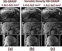
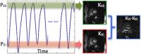
This work proposes a 3D free-breathing MRI technique called
variable-density XD-GRASP, which employs stack-of-stars
sampling with variable-density kz-undersampling
and motion-resolved sparse reconstruction. The new sampling
scheme combines the advantages of conventional
stack-of-stars sampling and kooshball-type 3D radial
sampling, enabling 3D continuous MRI with flexible slice
resolution, robust fat suppression and low sensitivity to
eddy currents. The performance of variable-density XD-GRASP
is demonstrated for free-breathing liver MRI.
|
| |
08:48
|
1092.
 |
Non-Iterative Motion-Error Regularized Reconstruction for
Efficient Respiratory Gating with Auto-Calibrating Parallel
Imaging
Peng Lai1, Joseph Yitan Cheng2,
Shreyas S Vasanawala2, and Anja C.S Brau3
1Global MR Applications and Workflow, GE
Healthcare, Menlo Park, CA, United States, 2Radiology,
Stanford University, Stanford, CA, United States, 3Global
MR Applications and Workflow, GE Healthcare, Munich, Germany
Respiratory gating (RG) is commonly used for free-breathing
3D MRI. Conventional RG based on acceptance/rejection
performs hard-threshholding on acquired data and suffers
from either increased motion corruption with a large
acceptance window or long scan time/increased undersampling
artifacts with a small window. This work developed a
non-iterative respiratory soft-threshholding method by
incorporating the motion-induced error into autocalibrating
parallel imaging (ac-PI). The proposed method showed more
effective motion suppression on free-breathing 3D cine than
conventional respiratory gating on the same datasets. This
method can be generalized to suppress other types of motion
with full acquisition or ac-PI as well.
|
| |
09:00
 |
1093.
 |
Towards a Parameter-Free ESPIRiT: Soft-Weighting for Robust Coil
Sensitivity Estimation
Siddharth Srinivasan Iyer1, Frank Ong1,
and Michael Lustig1
1Electrical Engineering and Computer Sciences,
University of California, Berkeley, Berkeley, CA, United
States
ESPIRiT is a robust, auto-calibrating approach to parallel
MR image reconstruction that estimates the subspace of
sensitivity maps using an eigenvalue-based method. While it
is robust to a range of parameter choices, having parameters
that result in a tight subspace yields the best performance.
We propose an automatic, parameter free method that
appropriately weights the subspace using a shrinkage
operator derived from Stein's Unbiased Risk Estimate. We
demonstrate the efficacy of our technique by showing
superior map estimation without user intervention in
simulation and in-vivo data compared to the current default
method of subspace estimation.
|
| |
09:12
|
1094.
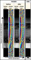 |
Self-Calibrated Phase-Correction for Superresolution of RASER at
7 T
Ute Goerke1
1CMRR/Radiology, University of Minnesota,
Minneapolis, MN, United States
RASER (rapid acquisition with sequential excitation and
refocusing) is an ultrafast imaging technique based on
spatiotemporal encoding (SPEN). The excitation with a
chirp-pulse with a low bandwidth-time product (R-value)
introduces blurring in the SPEN dimension. Superresolution
(SR) which removes the blurring fails as a result of the
spatially varying B1-phase
produced by radio-frequency coils at ultrahigh fields. A
novel iterative phase-correction of the SR-algorithm is
presented. It is shown that the spatial resolution and the
SNR of blurred RASER images acquired at 7 T are
significantly improved employing phase-corrected SR.
|
| |
09:24
|
1095.
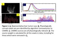 |
A convex source separation and reconstruction methodology for
filtering dynamic contrast enhancement MRI data - Permission Withheld
Sudhanya Chatterjee1, Dattesh D Shanbhag1,
Venkata Veerendranadh Chebrolu1, Uday Patil1,
Sandeep N Gupta2, Moonjung Hwang 3,
Jeong Hee Yoon4, Jeong Min Lee4, and
Rakesh Mullick1
1GE Global Research, Bangalore, India, 2GE
Global Research, Niskayuna, NY, United States, 3GE
Healthcare, Seoul, Korea, Republic of, 4Seoul
National University Hospital, Seoul, Korea, Republic of
Main aim of this research is to investigate a source
separation based approach to remove noise from true signal,
while maintaining original tissue enhancement signature. It
is based on the hypothesis that there exists overlapping
temporal information in the DCE-MRI data, which if
identified, can be used for filtering noise out of the true
concentration data. We demonstrate the utility of source
separation and subsequent weight estimation methodology to
filter “noise” from DCE concentration data and impact on the
pK model parameters in liver DCE-MRI.
|
| |
09:36
|
1096.
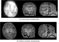 |
3D motion corrected SENSE reconstruction for multishot
multislice MRI
Lucilio Cordero-Grande1, Emer Hughes1,
Anthony Price1, Jana Hutter1, A. David
Edwards1, and Joseph V. Hajnal1
1Centre for the Developing Brain, King's College
London, London, United Kingdom
A framework for retrospectively motion corrected
reconstruction of multislice multishot MRI in the presence
of 3D rigid motion is developed. The method is able to cope
both with within-plane and through-plane motion by
estimating the motion states corresponding to the acquired
shots and slices. It has been applied to 478 T1 and T2
newborn brain studies, including many severely motion
corrupted examples, for which consistent structures have
been recovered in more than 96% of cases. Due to its
robustness and flexibility, our technique has wide potential
application for both clinical and research examinations.
|
| |
09:48
|
1097.
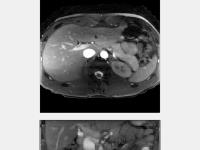 |
4D radial fat-suppressed alternating-TR bSSFP MRI with
compressed sensing reconstruction for abdominal imaging during
free breathing.
Jasper Schoormans1, Oliver Gurney-Champion1,
Remy Klaassen2, Jurgen H. Runge1,
Sonia I. Gonçalves3, Bram F. Coolen1,
Abdallah G. Motaal1, Hanneke W.M. van Laarhoven2,
Jaap Stoker1, Aart J. Nederveen1, and
Gustav J. Strijkers4
1Department of Radiology, AMC, Amsterdam,
Netherlands, 2Department
of Medical Oncology, AMC, Amsterdam, Netherlands, 3Institute
for Biomedical Imaging and Life Sciences, University of
Coimbra, Coimbra, Portugal, 4Department
of Biomedical Engineering and Physics, AMC, Amsterdam,
Netherlands
We developed a 4D radial fat-suppressed alternating-TR bSSFP
sequence with T2-like contrast for abdominal free-breathing
imaging of pancreatic cancer patients. The sequence was
tested in healthy volunteers and patients with pancreatic
cancer and provided images of the abdomen during different
respiratory motion states of diagnostic quality.
|
|