| |
10:45
|
0052.
 |
Reduced functional segregation between the default mode network
and the executive control network in healthy older adults: a
longitudinal study 
Kwun Kei Ng1, June C. Lo1, Michael W.L.
Chee1, and Juan Zhou1,2
1Duke-NUS Graduate Medical School, Singapore,
Singapore, 2Clinical
Imaging Research Centre, the Agency for Science, Technology
and Research and National University of Singapore,
Singapore, Singapore
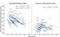
The effects of age on functional connectivity (FC) of
intrinsic connectivity networks (ICNs) have largely been
derived from cross sectional studies. Far less is known
about longitudinal changes in FC and how they relate to
ageing-related cognitive decline. We found progressive loss
of functional specialization with ageing evidenced by a
decline in intra-network FC within the executive control (ECN)
and default mode networks (DMN). In contrast, longitudinal
change in FC between ECN and DMN followed a u-shaped
trajectory whereby functional segregation between these two
networks initially increased over time and later decreased
as participants aged. The rate of loss in ECN-DMN functional
segregation was associated with decline in processing speed.
|
| |
10:57
 |
0053.
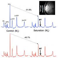 |
Aging Effect on Creatine Kinase Enzyme Activity in Resting Human
Brain: An In Vivo 31P-MT Study at 7T - Permission Withheld
Byeong-Yeul Lee1, Xiao-Hong Zhu1, and
Wei Chen1
1Center for Magnetic Resonance Research,
Radiology, University of Minnesota, Minneapolis, MN, United
States
In this work, we investigated the aging effect on the enzyme
activity of creatine kinase (CK) in healthy human visual
cortex at resting state using a newly developed in
vivo 31P
magnetization transfer (31P-MT) method at 7T. Our
results show that there was a strong aging dependence of the
CK enzyme activity in the resting brain, implying a
significant decline of brain energy metabolism in elderly
people. In vivo 31P-MT
technique should provide a valuable tool for clinical
research aiming to study aging-related neurodegenerative
diseases such as Alzheimer’s disease, and potentially for
other metabolic disorders/diseases.
|
| |
11:09
|
0054.
 |
Cerebral venous oxygenation as a potential marker to
differentiate normal aging from neurodegeneration
Zixuan Lin1, Marilyn Albert2, Peiying
Liu3, Anja Soldan2, Abhay Moghekar3,
Shin-Lei Peng4, Michael Miller1, Peter
van Zijl3, and Hanzhang Lu3
1Biomedical Engineering, Johns Hopkins
University, Baltimore, MD, United States, 2Department
of Neurology, Johns Hopkins University School of Medicine,
Baltimore, MD, United States, 3Department
of Radiology, Johns Hopkins University School of Medicine,
Baltimore, MD, United States, 4Department
of Radiology, China Medical University, Taichung, Taiwan
Decreased cerebral venous oxygenation (Yv) has been
considered as a compensation for aging which is diminished
in neurodegeneration. We substantiated this hypothesis by
examining the relationship between Yv and several
Alzheimer-specific hallmarks on 65 normal elderly subjects.
We demonstrated that Yv is higher in ApoE4 carriers who have
increased risks of AD and that higher Yv is associated with
poorer cognitive performance, indicating that assessment of
Yv with non-invasive MRI methods may present a potential
simple opportunity to identify the transition point from
normal to pathological aging.
|
| |
11:21
|
0055.
 |
Hippocampal subfield diffusivity changes in healthy ageing
Daniel J Cox1,2, Hamied A Haroon2,
Daniela Montaldi1, and Laura M Parkes2
1School of Psychological Sciences, University of
Manchester, Manchester, United Kingdom, 2Centre
for Imaging Sciences, University of Manchester, Manchester,
United Kingdom
Alterations to hippocampal microstructure may precede gross
volumetric changes in ageing, and these changes may occur
preferentially in different hippocampal subfields. We
investigated both established (FA and mADC) and novel (DOC)
measurements of diffusion in these regions, in addition to
volume, in order to determine where age-related changes
occurred. The results showed changes across the majority of
subfields for mADC and FA, but only in left CA 2/3 for DOC
measures 1, 3 and >3. We suggest this could be related to
differential degradation of particular cellular structures
in these regions.
|
| |
11:33
|
0056.
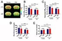 |
Early Shifts of Brain Metabolism by Caloric Restriction Preserve
White Matter Integrity and Long-term Memory in Aging Mice
Janet Guo1, Ailing Lin1,2, and Vikas
Bakshi1
1Department of Pharmacology & Nutritional
Sciences, University of Kentucky, Lexington, KY, United
States, 2Department
of Biomedical Engineering, Lexington, KY, United States
Caloric restriction (CR) has been shown to increase
healthspan in various species; however, its effects on
preserving brain functions in aging remain largely
unexplored. We used multimodal neuroimaging (PET/MRI/MRS)
and behavioral testing to determine in vivo brain glucose
metabolism, energy metabolites, and white matter structural
integrity in young and old mice fed with either control or
40% CR diet. Blood glucose and ketone bodies were measured.
Our findings suggest CR could slow brain aging, partly due
to early shift of energy metabolism caused by lower caloric
intake. These results provide rationale for CR-induced
sustenance of brain health with extended longevity.
|
| |
11:45
|
0057.
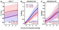 |
Age-dependent changes in the BOLD Cerebrovascular Reactivity
Curve in Response to Progressive Hypercapnia
Alex Bhogal1, Jill B de Vis1, Jeroen
C.W. Siero1, Esben T Petersen2, Peter
R. Luijten1, Jeroen Hendrikse1,
Marielle E.P. Philippens3, and Hans Hoogduin4
1Radiology, UMC Utrecht, Utrecht, Netherlands, 2Danish
Research Centre for Magnetic Resonance, Centre for
Functional and Diagnostic Imaging and Research, Copenhagen
University Hospital Hvidovre, Copenhagen, Denmark, 3Radiotherapy,
UMC Utrecht, Utrecht, Netherlands, 4Utrecht,
Netherlands
Characterizing healthy, age-related changes in the BOLD-CVR
response can provide a reference point from which to
distinguish abnormal CVR from the otherwise normal effects
of ageing. In this study, we examine age-dependent
differences in grey and white matter BOLD-CVR response to
progressive hypercapnia between young and elderly subjects.
|
| |
11:57
 |
0058.
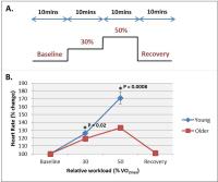 |
Assessment of cerebral response to exercise: effects of ageing
and cardiorespiratory fitness
Andrew Hale1, Penny Gowland1, Paul
Greenhaff2, and Susan Francis1
1Sir Peter Mansfield Imaging Centre, University
of Nottingham, Nottingham, United Kingdom, 2Faculty
of Medicine & Health Sciences, Queens Medical Centre,
University of Nottingham, Nottingham, United Kingdom
Although there is a general relationship between age and
brain function, habitual physical activity levels may also
impact on brain health. We performed a MR study involving
low and moderate intensity supine exercise in healthy young
and older subjects. We assess the effect of exercise on CBF
response in large arteries, regional perfusion and BOLD, and
the relationship of grey matter volume with physical fitness
and ageing. On exercise there was a clear CBF, perfusion and
BOLD response to exercise in young volunteers, whilst a
reduced CBF, perfusion and BOLD response to exercise was
found in the older volunteers.
|
| |
12:09
|
0059.
 |
Consistent detection of age-dependent variations of the
longitudinal relaxation time in cortical brain regions
investigated by MP2RAGE at 9.4T: influence of correcting for a
non-uniform transmit field
Gisela E Hagberg1,2, Jonas Bause1,
Thomas Ethofer2,3, Philipp Ehses1,
Thomas Dresler3, G Shajan1, Rolf
Pohmann1, Cornelia Herbert3, Andreas
Fallgatter3, Christoph Laske3, Marina
Pavlova2, and Klaus Scheffler1,2
1High Field Magnetic Resonance, Max Planck
Institute for Biological Cybernetics, Tübingen, Germany, 2Biomedical
Magnetic Resonance, University Hospital Tübingen, Tübingen,
Germany, 3General
Psychiatry&Psychotherapy, University Hospital Tübingen,
Tübingen, Germany
Accurate and precise determination of T1 values is of
central importance in clinical studies and for tissue
segmentation based on the myeloarchitecture that transcends
T1. Here we investigate whether well-described age-dependent
changes can be detected by high field T1 relaxometry, and
how different transmit field correction methods influence
the results. We found that the intrinsic bias correction of
the MP2RAGE technique is not sufficient to achieve reliable
quantification of T1 at ultra high magnetic fields. But,
provided that a correction for transmit field inhomogeneity
is performed, T1 maps that consistently reveal age-related
changes can be generated. The technique holds promise for
investigation of local myeloarchitectonics for
neuroscientific and clinical studies.
|
| |
12:21
|
0060.
 |
Changes in white matter structural connectivity and cortical
functional connectivity over the healthy adult lifespan
Adrian Tsang1,2,3, Catherine Lebel1,4,
Signe Bray1,4, Brad Goodyear1,2,3,
Roberto C. Sotero1, Cheryl McCreary1,3,
and Richard Frayne1,2,3
1Department of Radiology, University of Calgary,
Calgary, AB, Canada, 2Hotchkiss
Brain Institute, Calgary, AB, Canada, 3Seaman
Family MR Research Centre, Calgary, AB, Canada, 4Child
and Adolescent Imaging Research Program, Alberta Children's
Hospital Research Institute, Calgary, AB, Canada
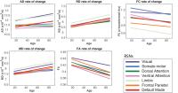
This study investigates how both structural and functional
connectivity (SC and FC) changes in the adult lifespan as
well as to explore the relationship between measures that
are commonly used for SC and FC in the context of normal
aging. A multi-modal analysis using DTI and resting-state
fMRI data was performed from 183 healthy participants aged
18 – 87 years. We found that fractional anisotropy (FA) and
FC showed similar rate of change and correlation strengths
with age in the 7 resting-state networks explored. However
none of the SC measures showed significant correlations with
FC measure.
|
| |
12:33
|
0061.
 |
Diagnostic accuracy of MRS for Hereditary Neurodegeneration at
3T and 7T
Uzay E Emir1,2, Tianmeng Lyu3, Dinesh
K Deelchand2, James M Joers2, Diane
Hutter2, Christopher M Gomez4, Khalaf
O Bushara5, Lynn E Eberly3, and Gulin
Oz2
1FMRIB Centre, University of Oxford, Oxford,
United Kingdom, 2Center
for Magnetic Resonance Research, Department of Radiology,
University of Minnesota, Minneapolis, MN, United States, 3Division
of Biostatistics, School of Public Health, University of
Minnesota, Minneapolis, MN, United States, 4Department
of Neurology, University of Chicago, Chicago, IL, United
States, 5Department
of Neurology, Medical School, University of Minnesota,
Minneapolis, MN, United States
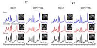
To evaluate diagnostic accuracy of state-of-the-art MRS in
early neurodegenerative disease, we measured neurochemical
profiles in the vermis, cerebellar hemisphere and brainstem
of genetically confirmed subjects with spinocerebellar
ataxia type 1 and controls by 3T and 7T 1H MRS.
Concentrations of major metabolites obtained at 3T and 7T
were strongly correlated. While 3T showed great potential by
enabling detection of abnormal metabolite levels even in the
presymptomatic stage, the increased sensitivity at 7T
enabled group separation with higher significance and
identification of subtle neurochemical alterations in early
symptomatic disease stage more robustly than at 3T.
|
|