| |
10:00
|
0617.
 |
Improving Respiratory Phase-resolved 3D Body Imaging Using
Iterative Motion Correction and Average (MoCoAve) 
Xiaoming Bi1, Jianing Pang2, Wensha
Yang2, Matthias Fenchel3, Zixin Deng2,
Yuhua Chen4, Richard Tuli2, Debiao Li2,
Gerhard Laub1, and Zhaoyang Fan2
1Siemens Healthcare, Los Angeles, CA, United
States, 2Cedars-Sinai
Medical Center, Los Angeles, CA, United States, 3Siemens
Healthcare GmbH, Erlangen, Germany, 4University
of Pennsylvania, Philadelphia, PA, United States
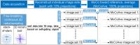
4D (respiratory phase-resolved 3D) MRI has been increasingly
used for the planning of radiotherapy and minimally invasive
surgery. Recently developed self-gating methods showed great
potential in 4D MRI by providing high imaging efficiency and
isotropic spatial resolution. However, images of individual
phases may suffer from decreased SNR and increased streaking
artifact since only a subset of data were used for
reconstruction. A motion correction and average (MoCoAve)
framework was developed in this work to address such
limitations. Preliminary results from patients showed that
the proposed method can significantly improve SNR and image
quality without compromising motion information.
|
| |
10:12
|
0618.
 |
Variable Density Compressed Sensing Single Shot Fast Spin Echo
Valentina Taviani1, Daniel V. Litwiller2,
Jonathan I. Tamir3, Andreas M. Loening1,
Brian A. Hargreaves1, and Shreyas S. Vasanawala1
1Stanford University, Stanford, CA, United
States, 2Global
MR Applications and Workflow, GE Healthcare, New York, NY,
United States, 3University
of California Berkeley, Berkeley, CA, United States
Variable density (VD) sampling was implemented into an
extended echo train single shot fast spin echo (SSFSE) pulse
sequence. Compressed sensing (CS) reconstuction was used.
With respect to regular undersampling and ARC (Autocalibrated
Reconstruction for Cartesian imaging), VD CS SSFSE allows
higher acceleration factors, which translates in increased
flexibility in the choice of echo times for full-Fourier
imaging (shorter minimum TEs) and faster acquisitions
(shorter breath-holds).
|
| |
10:24
|
0619.
 |
Free-breathing non-contrast enhanced 3D radial
respiratory-motion resolved pancreatic MRI at 3T using sparse
iterative reconstruction
Jessica AM Bastiaansen1, Jerome Yerly1,2,
Jean-Baptiste Ledoux2, Ruud B van Heeswijk1,2,
Davide Piccini3, and Matthias Stuber1,2
1Department of Radiology, University hospital
(CHUV) and University of Lausanne (UNIL), Lausanne,
Switzerland, 2Center
for Biomedical Imaging, Lausanne, Switzerland, 3Advanced
Clinical Imaging Technology, Siemens Healthcare, Lausanne,
Switzerland
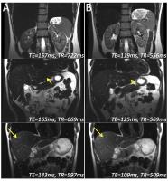
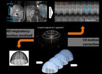
Pancreatic MRI is commonly performed during breath-held or
navigator-gated acquisitions. The long breath-holds needed
for high spatial resolution are not always feasible in
patients and residual respiratory motion may still occur.
Additionally, in some implementations, the navigator leads
to a local signal void that may obscure parts of the anatomy
of interest. Here we used a free-breathing self-navigated 3D
radial gradient-recalled-echo (GRE) imaging sequence, and
compared the 1D motion correction as performed on the
scanner versus a motion-resolved 4D sparse iterative
reconstruction. We show that non-contrast enhanced
pancreatic MRI can be performed at 3T during free-breathing,
while motion-resolved sparse reconstruction can efficiently
minimize the adverse effects of respiratory motion.
|
| |
10:36
|
0620.
 |
Radial Volumetric Interpolated Breath-hold Examination of the
Liver: Clinical Impact of Self-gated 3D Isotropic
Contrast-enhanced Late-Phase MR Imaging - Permission Withheld
Jakob Weiss1, Jana Taron1, Ahmed E.
Othman1, Robert Grimm2, Petros
Martirosian1, Christina Schraml1,
Konstantin Nikolaou1, and Mike Notohamiprodjo1
1Diagnostic and Interventional Radiology,
University of Tuebingen, Tuebingen, Germany, 2Siemens
Healthcare, Erlangen, Germany
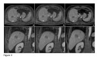
To evaluate clinical performance of contrast-enhanced
3D-isotropic radial volumetric interpolated breath-hold
examination (VIBE) for late-phase MR imaging of the liver. A
prototype retrospective self-gating algorithm for more
motion-robust data acquisition was implemented and compared
to standard Cartesian VIBE. Utilization of self-gating VIBE
provides significantly improved image quality, especially in
coronal reformations and Gd-EOB-DTPA-enhanced late-phase
scans. Moreover, in 11% only radial VIBE provided diagnostic
image quality, thus having a direct implication on patient
care. Therefore, self-gated radial VIBE seems a valuable
approach to improve diagnostic accuracy in late-phase MR
imaging of the liver.
|
| |
10:48
|
0621.
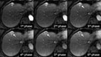 |
Improved detection of capsular enhancement in hepatocellular
carcinoma using multiphasic hepatic arterial imaging and
DIfferential Sub-sampling with Cartesian Ordering (DISCO) in
gadoxetic acid-enhanced magnetic resonance imaging
Shintaro Ichikawa1, Utaroh Motosugi1,
Tetsuya Wakayama2, Takashi Kakegawa1,
Hiroshi Kumagai1, and Hiroshi Onishi1
1University of Yamanashi, Yamanashi, Japan, 2GE
Healthcare Japan, Tokyo, Japan
DIfferential Subsampling with Cartesian Ordering (DISCO) is
a new high spatiotemporal resolution, dynamic
contrast-enhanced magnetic resonance imaging (MRI)
technique. We evaluated the usefulness of multiple (n=6)
hepatic arterial phases (HAPs) with DISCO in gadoxetic
acid-enhanced dynamic MRI for detecting capsular enhancement
in hepatocellular carcinoma (HCC). Such capsular enhancement
is detected more frequently by combining portal venous phase
(PVP) images and multiphasic hepatic arterial images with
DISCO. Combining DISCO with PVP improved the liver imaging
reporting and data system (LI-RADS) v2014 score from LR4 to
LR5.
|
| |
11:00
|
0622.
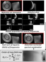 |
Multispectral Body Diffusion-Weighted Imaging
Valentina Taviani1, Shreyas S. Vasanawala1,
and Brian A. Hargreaves1
1Stanford University, Stanford, CA, United States
A diffusion-weighted (DW) imaging method was developed to
mitigate off-resonance-induced distortion and signal loss,
which are problematic for body applications. A 2D RF pulse
is used in place of the conventional spectral-spatial
excitation used for DW spin echo echo-planar imaging. In the
presence of off-resonance, a narrow band of frequencies is
excited due to the different bandwidths between excitation
and refocusing pulses. By progressively shifting the center
frequency, the whole range of off-resonance can be excited
and a composite image, corrected for off-resonance-induced
distortion, can be reconstructed by estimating the field map
from the spectral information.
|
| |
11:12
|
0623.
 |
3D whole liver black blood imaging: a 3 min solution consisting
of respiratory triggering and free breathing imaging techniques
Li Jiang1, Chenguang Zhao1, Andy Jiang1,
Ming Yang1, Wengu Su1, Allan Jin1,
Ping Yang1, Stephon Xu1, and Feng
Huang1
1Philips Healthcare (Suzhou), Suzhou, Jiangsu,
China, People's Republic of
Liver black blood imaging helps to detect and characterize
focal liver lesions and thus is highly desirable clinically.
The commonly used low b-value DWI sequence is limited due to
inherent limitations of EPI, such as low spatial resolution
and motion artifacts including blurring and ghosting. We
proposed a 3D whole liver black blood imaging solution
within 3 min. By combining with existing black blood
preparation, a respiratory triggered VISTA sequence and a
free breathing imaging technique utilizing GROWL
reconstruction were proposed. Six healthy volunteers with
stable and irregular respiration were scanned to further
validate the feasibility of our proposed solution.
|
| |
11:24
 |
0624.
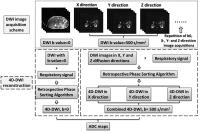 |
Potential Improvement in Apparent Diffusion Coefficient (ADC)
Measurement by Respiratory Correlated Four Dimensional
Diffusion-Weighted MRI (4D-DWI): Initial Investigation on
Digital Phantoms and Human Subjects
Yilin Liu1, Fang-Fang Yin2, Brian Gary
Czito2, Mustafa R. Bashir 3,
Manisha Palta 2,
Xiaodong Zhong 4,
Brian M. Dale 5,
and Jing Cai2
1Medical Physics Graduate Program, Duke
University Medical Center, Durham, NC, United States, 2Radiation
Oncology, Duke University Medical Center, Durham, NC, United
States, 3Department
of Radiology, Center for Advanced Magnetic Resonance
Development, Duke University Medical Center, Durham, NC,
United States, 4MR
R&D Collaborations, Siemens Healthcare, Atlanta, GA, United
States, 5MR
R&D Collaborations, Siemens Healthcare, Cary, NC, United
States
Diffusion-weighted imaging (DWI) has been shown to have
superior tumor-to-tissue contrast for cancer detection in
abdominal region. However, the respiratory motion may induce
severe imaging errors or artifacts for DWI images. This
study aims at developing and evaluating a respiratory
correlated 4D-DWI technique using a retrospective sorting
method for imaging respiratory motion on human subjects.
Comparing to free breathing DWI, 4D-DWI can lead to more
accurate measurement of ADC. This has a great potential to
improve the visualization and delineation of cancer tumors
for radiotherapy.
|
| |
11:36
|
0625.
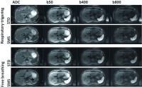 |
Simultaneous multislice accelerated diffusion-weighted imaging
of the liver: comparison of different breathing schemes with
standard sequences as reference
Christina Schraml1, Jana Taron1, Nina
F Schwenzer1, Holger Schmidt1, Thomas
Kuestner2, Michael Erb3, Mike
Notohamiprodjo1, Konstantin Nikolaou1,
Fritz Schick4, and Petros Martirosian4
1Diagnostic and Interventional Radiology,
Department of Radiology, University Hospital Tuebingen,
Tuebingen, Germany, 2Institute
of Signal Processing and System Theory, University of
Stuttgart, Stuttgart, Germany, 3Department
of Biomedical Magnetic Resonance, University Hospital
Tuebingen, Tuebingen, Germany, 4Section
on Experimental Radiology, Department of Radiology,
University Hospital Tuebingen, Tuebingen, Germany
SMS-acceleration allows for considerable scan time reduction
in hepatic DWI without substantial drawbacks in image
quality both using respiratory-triggering and free-breathing
acquisitions. In the present study set-up, ADC measured in
SMS-DWI were lower than in standard DWI which should be
considered when using absolute ADC for clinical reading. The
demonstrated high image quality of SMS-DWI obtained in FB
indicates great potential for scan time reduction in DWI for
abdominal and whole-body applications.
|
| |
11:48
|
0626.
 |
Intravoxel incoherent motion diffusion-weighted imaging of
hepatocellular carcinoma: is there a correlation with flow and
perfusion metrics obtained with dynamic contrast-enhanced MRI?
Stefanie Hectors1, Mathilde Wagner1,
Cecilia Besa1, Hadrien Dyvorne1,
Octavia Bane1, M. Isabel Fiel2, Hongfa
Zhu2, and Bachir Taouli1,3
1Translational and Molecular Imaging Institute,
Icahn School of Medicine at Mount Sinai, New York, NY,
United States, 2Department
of Pathology, Icahn School of Medicine at Mount Sinai, New
York, NY, United States, 3Department
of Radiology, Icahn School of Medicine at Mount Sinai, New
York, NY, United States
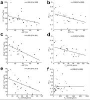
We assessed the correlation between intravoxel incoherent
diffusion-weighted imaging (IVIM-DWI) and dynamic
contrast-enhanced MRI (DCE-MRI) in hepatocellular carcinoma
(HCC) and liver parenchyma. DCE-MRI-derived arterial
fraction and arterial flow were significantly negatively
correlated with IVIM-DWI-derived perfusion fraction and
pseudodiffusion in the liver, while IVIM-DWI parameters did
not correlate with DCE-MRI parameters in HCC. These results
indicate that IVIM-DWI and DCE-MRI provide non-redundant
information in HCC.
|
|