|
|
|
Plasma # |
 |
0363.
 |
1 |
Bone Quantitative Susceptibility Mapping using tissue specific
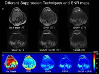
R2* and multi-peak fat spectrum to model ultra-short TE gradient
echo signal 
Alexey V. Dimov1,2, Zhe Liu1,2, Pascal
Spincemaille2, and Yi Wang1,2
1Department of Biomedical Engineering, Cornell
University, Ithaca, NY, United States, 2Radiology
Department, Weill Cornell Medical College, New York, NY,
United States
Bone quantitative susceptibility mapping (QSM) using
standard IDEAL fat water/signal model often suffers from
erroneous labeling of water component. We propose a new
field estimation approach incorporating the negligible T2*
decay of fat compared to bone water signal, and modeling fat
with multiple spectral peaks. This tissue specific R2*
multi-peak signal allows robust field mapping from radial
ultra-short TE gradient echo data, enabling in vivo bone QSM
with consistent high quality.
|
|
0364.
 |
2 |
Reproducibility and regional variations of an optimized gagCEST
protocol for the in vivo evaluation of knee cartilage at 7 Tesla
Markus Matthias Schreiner1,2, Stefan Zbyn2,
Benjamin Schmitt3, Stephan Domayer1,
Reinhard Windhager1, Siegfried Trattnig2,
and Vladimir Mlynarik2
1Department of Orthopaedic Surgery, Medical
University of Vienna, Vienna, Austria, 2Department
of Biomedical Imaging and Imag-Guided Therapy, High Field MR
Centre, Medical University of Vienna, Vienna, Austria, 3Siemens
Healthcare Pty Ltd, Macquarie Park, Australia
Early onset osteoarthritis is associated with
ultrastructural and compositional changes of cartilage, in
particular with a loss of glycosaminoglycans (GAGs) and
disorganization of the collagen matrix. Both changes remain
elusive to morphological MRI. GagCEST is a promising tool
for the evaluation of glycosaminoglycan content in articular
cartilage. However, it is affected by many variables, thus
rendering its application challenging. The implementation of
a novel saturation scheme combined with optimized fixation
seems to improve the robustness of the technique as
indicated by increased reproducibility. Our optimized
protocol seems to be sensitive to regional differences in
the GAG content.
|
|
0365.
 |
3 |
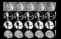
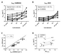
Muscle functional oxidative capacity varies along the length of
healthy tibialis anterior
Andreas Boss1, Linda Heskamp1, Mark
Jacobus van Uden1, Lauren Jean Bains2,3,
Vincent Breukels1, and Arend Heerschap1
1Radiology and Nuclear Medicine, Radboud
university medical center, Nijmegen, Netherlands, 2Donders
Institute for Brain, Cognition and Behaviour, Radboud
University, Nijmegen, Netherlands,3Donders Centre
for Cognitive Neuroimaging, Radboud University, Nijmegen,
Netherlands
Traditional PCr recovery experiments are performed in a
non-localized way, while skeletal muscle is not homogeneous.
In this study we performed localized 31P-MRS
using a ladder-shaped 31P-phased
array receive coil optimized for the tibialis anterior and
found a pronounced variation in the rate of PCr recovery
after isometric exercise along the length of this muscle in
healthy volunteers. In addition, we observed similar
regional differences in the time-to-peak signal intensity of
muscle functional MRI obtained after exercise in the same
volunteers. The reasons for this strong functional gradient
along the tibialis anterior remain, however, to be
elucidated.
|
|
0366.
 |
4 |
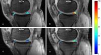
Assessment of meniscus with adiabatic $$$T_{1\rho}$$$ and
$$$T_{2\rho}$$$ in asymptomatic subjects and patients with early
osteoarthritis: Oulu knee osteoarthritis study
Abdul Wahed Kajabi1,2,3, Victor Casula2,3,
Arttu Peuna2,3,4, Simo Saarakkala2,5,
Eveliina Lammentausta3,4, Ali Guermazi6,
and Miika T. Nieminen2,3,4
1Department of Biomedical Engineering, University
of Oulu, Oulu, Finland, 2Research
Unit of Medical Imaging, Physics and Technology, University
of Oulu and Oulu University Hospital, Oulu, Finland,3Medical
Research Center, University of Oulu and Oulu University
Hospital, Oulu, Finland, 4Department
of Diagnostic Radiology, Oulu University Hospital, Oulu,
Finland, 5Department
of Medical Technology, Institute of Biomedicine, University
of Oulu, Oulu, Finland, 6Department
of Radiology, Boston University School of Medicine, MA, MA,
United States
Evaluation of meniscal degeneration in asymptomatic subjects
and patients with early osteoarthritis (KL = 1,2) was
performed using adiabatic $$$T_{1\rho}$$$ and
$$$T_{2\rho}$$$ ($$$AdT_{1\rho}$$$ and $$$AdT_{2\rho}$$$,
respectively) measurements in sagittal plane. Menisci of all
subjects were also evaluated using semiquantitative MRI OA
Knee Score (MOAKS). The results show that the length of
$$$AdT_{1\rho}$$$ and $$$AdT_{2\rho}$$$ is directly related
to clinical symptoms and the severity of meniscal
degeneration. $$$AdT_{1\rho}$$$ and $$$AdT_{2\rho}$$$ may
provide a non-invasive means of detecting and monitoring
degenerative changes in the meniscus.
|
|
0367.
 |
5 |
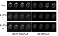
Diffusion Tensor Imaging of Human Achilles Tendon by Stimulated
Echo RESOLVE (ste-RESOLVE)
Xiang He1, Kenneth Wengler2, Alex C
Sacher3, Marco Antonio Oriundo Verastegui1,
Alyssa Simeone4, Mingqian Huang1,
Elaine Gould1, and Mark Schweitzer1
1Department of Radiology, Stonybrook University
School of Medicine, Stony Brook, NY, United States, 2Department
of Biomedical Engineering, Stonybrook University School of
Medicine, Stony Brook, NY, United States, 3SUNY
Binghamton University, Binghamton, NY, United States, 4New
York Medical College, Valhalla, NY, United States
Diffusion tensor imaging (DTI) is sensitive to the
injury-induced changes on the tendons microstructure.
However, conventional spin-echo based DTI techniques often
lead to poor tendon MR signal and difficulty on diffusion
quantification, mainly due to the short tendon T2/T2*
relaxation time constant. In this study, a novel method of
combining stimulated-echo based DTI and readout-segmented
multi-shot EPI (ste-RESOLVE) has been developed and
evaluated. TE value can be as low as 20 ms for b value of
800 s/mm2, enabling robust investigation of
Achilles tendon microscopic tissue integrity on clinical MR
scanners.
|
|
0368.
 |
6 |
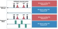
MR NeuroAngiography: Simultaneous Acquisition of Brachial Plexus
MR Neurography and Subclavian MR Angiography Using phase-cycling
Motion-Sensitized Driven-Equilibrium (pcMSDE)
Masami Yoneyama1, Hajime Tanji2,
Tomoya Yamaki2, Daisuke Takahashi2,
Makoto Obara1, Tomoyuki Okuaki3, and
Marc Van Cauteren3
1Philips Electronics Japan, Tokyo, Japan, 2Kita-Fukushima
Medical Center, Fukushima, Japan, 3Philips
Healthcare Asia Pacific, Tokyo, Japan
Simultaneous acquisition of both MR angiography and MR
neurography would be extremely helpful for diagnosing
thoracic outlet syndrome. This study proposed a novel
sequence, motion-sensitized driven-equilibrium (MSDE)
prepared phase-cycling gradient echo (pcMSDE), for achieving
simultaneous depiction of both MR angiography and MR
neurography. By using this sequence, MR neurography images
were obtained by MSDE (with motion sensitized gradient
(MSG)) scan. MR angiography images were obtained by
subtraction between “b0” scan (without MSG) and MSDE (with
MPG) images. Additionally, this sequence could
simultaneously offer the anatomical proton-density images
and “self-fusion” images (MR NeuroAngiography) by using MR
neurography and MR angiography. This sequence has great
potential to help the diagnosis for any type of TOS. Further
clinical investigation is needed.
|
|
0369.
 |
7 |
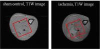
Detection of Alterations in Intramyocellular Lipid and Creatine
Diffusivities during Muscle Ischemia by Diffusion Weighted MRS
Anna M. WANG1,2 and
Ed X. Wu1,2
1Laboratory of Biomedical Imaging and Signal
Processing, The University of Hong Kong, Hong Kong, China,
People's Republic of, 2Department
of Electrical and Electronic Engineering, The University of
Hong Kong, Hong Kong, China, People's Republic of
We measured the apparent diffusion coefficients (ADCs), as
well as the relative concentrations of both intramyocellular
lipid (IMCL) and creatine in the rat muscle ischemia model.
Comparing with the metabolite concentration changes, the
IMCL and creatine ADCs had largely increased during muscle
ischemia and the IMCL ADC increase was more drastic than
creatine. The IMCL ADC, measured by diffusion weighted MRS,
had shown the potential to probe the alterations in lipid
droplet size and lipid metabolism in skeletal muscles.
|
 |
0370.
 |
8 |
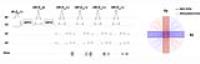
Clinically Viable Diffusion-Weighted Imaging Near Metal using
2D-MSI PROPELLER DUO
Suryanarayanan Sivaram Kaushik1, Ajeet Gaddipati2,
Brian Hargreaves3, Dawei Gui4, Robert
Peters2, Tugan Muftuler5, and Kevin
Koch1
1Radiology, Medical College of Wisconsin,
Milwaukee, WI, United States, 2GE
Healthcare, Waukesha, WI, United States, 3Radiology,
Stanford University, Stanford, CA, United States, 4GE
Healthcare, Waukesh, WI, United States, 5Neurosurgery,
Medical College of Wisconsin, Milwaukee, WI, United States
While FSE-based multi-spectral imaging (MSI) sequences help
overcome the artifacts caused by metallic hardware,
diffusion-weighted imaging remains a challenge. The non-CPMG
artifacts caused by adding diffusion lobes to an FSE train
can be mitigated by modulating the phase of the refocusing
pulses. Another solution involves splitting the contribution
made by the spin and stimulated echoes (DUO acquisition).
Here, we combine a 2D version of MSI with a PROPELLER-DUO
sequence to obtain clinically-feasible, artifact-minimized,
diffusion-weighted images in subjects that have cancerous
lesions in close proximity to metallic hardware.
|
|
0371.
 |
9 |
Evaluation of Different Fat Suppression Techniques for Clinical
Knee MRI at 7.0 Tesla
Michael Wyss1, Andrei Manoliu2, Georg
Spinner1, Magda Marcon2, Roger
Luechinger1, Daniel Nanz2, Klaas P.
Pruessmann1, and Gustav Andreisek2
1Institute for Biomedical Engineering, University
of Zurich and ETH Zurich, Zurich, Switzerland, 2Institute
of Diagnostic and Interventional Radiology, University
Hospital Zurich and University of Zurich, Zurich,
Switzerland
Reliable fat suppression is challenging but mandatory for
clinical 7.0T imaging. Purpose of this study was to evaluate
different fat suppression techniques for clinical 7.0T knee
MRI. Eight volunteers were imaged at 7.0T (Achieva, Philips)
using a dedicated 28-channel TX-knee coil (QED) and axial
PDw-TSE sequences without fat suppression, with SPIR, with
SPAIR, with SSGR and with the combination of SSGR+SPIR.
|
|
0372.
 |
10 |
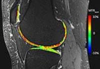
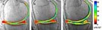
3-D cones UTE-T2* maps show early cartilage degeneration 2 years
after ACL reconstruction
Ashley Anne Williams1, Matthew R Titchenal1,
and Constance R Chu1
1Orthopaedic Surgery, Stanford University,
Stanford, CA, United States
3-D cones UTE-T2* maps were examined in 22 subjects with
reconstructed anterior cruciate ligaments (ACLR) and 16
uninjured controls for evidence of alterations to the
subsurface cartilage matrix suggestive of cartilage at risk
for early OA 2 years after surgery. Elevated UTE-T2* values
in regions of deep tibiofemoral cartilage and in
side-to-side UTE-T2* differences were detected. UTE-T2*
values correlated to standard T2 values in tibial and
posterolateral femoral regions. Together, these findings
suggest that UTE-T2* mapping detects “pre-osteoarthritic”
subsurface cartilage matrix changes that may occur following
ACLR and thus can help to identify subjects at risk of
developing OA.
|
|
0373.
 |
11 |
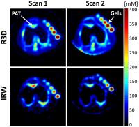
Longitudinal sodium MRI of cartilage in patients with knee
osteoarthritis: Baseline vs. 16 months follow-up
Guillaume Madelin1, Ding Xia1, Gregory
Chang1, Svetlana Krasnokutsky2, Steven
B Abramson2, and Ravinder R Regatte1
1Department of Radiology, New York University
Langone Medical Center, New York, NY, United States, 2Department
of Rheumatology, New York University Langone Medical Center,
New York, NY, United States
In this longitudinal study, we measured the sodium
concentration in knee cartilage in 12 patients with
osteoarthritis (OA) with quantitative 23Na
MRI at 7 T. Sodium measurements were performed at baseline
and 16 months follow-up (on average), with and without fluid
suppression by inversion recovery (IR). We show that only
fluid-suppressed measurements show a significant decrease of
mean [Na+] in different regions of cartilage over
16 months follow-up in OA patients. Quantitative 23Na
IR-MRI could therefore be a useful imaging biomarker to
monitor cartilage degradation over time, and help assess the
efficiency of potential disease modifying OA drugs.
|
|
0374.
 |
12 |
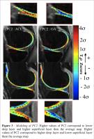
PCA-T1? Voxel-Based Relaxometry of the Articular Cartilage: a
Comparison of Biochemical Pattern Changes in Knees with
Osteoarthritis and ACL Injury - Permission Withheld
Valentina Pedoia1, Colin Russell1,
Allison Randolph V1, Keiko Amano1,
Xiaojuan Li1, and Sharmila Majumdar1
1University of California, San Francisco, San
Francisco, CA, United States
MR quantitative T1ρ mapping has been extensively used to
probe articular biochemical changes. While several studies
are still limited to analyzing average T1ρ values, there is
growing interest in the analysis of local patterns of T1ρ
maps. A novel algorithm for locally studying knee relaxation
times using Voxel-Based Relaxometry (VBR) was recently
proposed. In this study we propose to couple VBR and
Principal Component Analysis in order to analyze local
pattern changes in OA and ACL patients. Specific features,
behind the expected average elevation of T1ρ values, are
observed able to distinguish between OA, ACL and Controls
subjects.
|
|
0375.
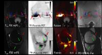 |
13 |
Correlation of Bone Pathology on MRI with 18F-fluoride PET
Uptake in Subchondral Bone
Feliks Kogan1, Audrey Fan1, Emily
McWalter1, Edwin Oei2, Andrew Quon1,
and Garry Gold1
1Radiology, Stanford University, Stanford, CA,
United States, 2Radiology,
Erasmus Medical Center, Rotterdam, Netherlands
Osteoarthritis (OA) is a debilitating disease that affects
27 million?Americans, causing pain, stiffness and loss of
mobility. Simultaneous PET-MR imaging provides an
opportunity to combine metabolic information regarding bone
remodeling with high resolution images on MR. This work
demonstrates that simultaneous 18F-fluoride PET/MR may
provide additional metabolic information regarding bone
pathology seen on conventional MR. This will allow for a
better understanding of the role of bone degeneration in OA
disease processes. Additionally, 18F-fluoride PET/MR may
detect knee abnormalities unseen on MRI alone and is a
promising tool for detection of early metabolic changes in
OA.
|
|
0376.
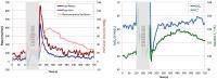 |
14 |
Quantitative assessment of muscle metabolism and dynamics of
oxygen consumption with vPIVOT
Erin Kristine Englund1, Zachary Bart Rodgers1,
Michael C Langham2, Emile R Mohler3,
Thomas F Floyd4, and Felix W Wehrli2
1Department of Bioengineering, University of
Pennsylvania, Philadelphia, PA, United States, 2Department
of Radiology, University of Pennsylvania, Philadelphia, PA,
United States, 3Department
of Medicine, University of Pennsylvania, Philadelphia, PA,
United States, 4Department
of Anesthesiology, Stony Brook University, Stony Brook, NY,
United States
A method to simultaneously measure blood flow, perfusion,
venous oxygen saturation, and muscle T2* using a
3-slice interleaved PASL, multi-echo GRE sequence is
presented. The method, termed Velocity and Perfusion,
Intravascular Venous Oxygen saturation and T2* (vPIVOT)
was assessed in five subjects during a series of
ischemia-reperfusion paradigms. Results indicate that vPIVOT
faithfully measures all four parameters at 4-second temporal
resolution. Dynamic measurement of these parameters was
completed following a bout of dynamic plantar flexion
contractions. vPIVOT
allows for quantification of muscle oxygen consumption and
evaluation of macro/microvascular flow dynamics, and may be
useful for the development of biophysical models.
|
|
0377.
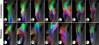 |
15 |
Synchronous Magnetic Resonance Imaging of Muscle Contraction
induced by Electrical Stimulation
Xeni Deligianni1,2, Michele Pansini3,
Meritxell Garcia4, Anna Hirschmann4,
Arno Schmidt-Trucksäss5, Oliver Bieri1,
and Francesco Santini1,2
1Department of Radiology, Division of
Radiological Physics, University of Basel Hospital, Basel,
Switzerland, 2Department
of Biomedical Engineering, University of Basel, Basel,
Switzerland, 3Radiology,
Kantonsspital Basel-Landschaft, Brudeholz, Switzerland, 4Department
of Radiology, University of Basel Hospital, Basel,
Switzerland, 5Department
of Sports Medicine, University of Basel, Basel, Switzerland
Magnetic Resonance Imaging can be used to provide structural
and functional muscle information either from oxygenation or
contraction imaging. Contraction imaging can be based on
real-time imaging or on voluntary movements. However,
synchronization of the acquisition is challenging. Here, we
present a new method for accurate, quantitative measurement
of muscle contraction using a commercially available
electrical muscle stimulator. This allows the direct
assessment of the reaction time of muscle fibers,
contraction speed, displacement, and strain providing
complementary information to electromyography. MR images of
the vastus lateralis muscle of five healthy volunteers were
acquired at 3 Tesla field strength during
electro-stimulation.
|
|