|
|
|
Plasma # |
 |
0715.
 |
1 |
Abdominal and Body Imaging Using a 16 Channel Dipole RF Array at
7.0 T 
Celal Oezerdem1, Till Huelnhagen1,
Lukas Winter1, and Thoralf Niendorf1,2
1Berlin Ultrahigh Field Facility (B.U.F.F), Max
Delbrück Center for Molecular Medicine in the Helmholtz
Association (MDC), Berlin, Germany, 2Experimental
and Clinical Research Center, a joint cooperation between
the Charité Medical Faculty and the Max Delbrück Center for
Molecular Medicine in the Helmholtz Association, Berlin,
Germany
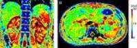
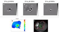
This pilot study demonstrates the feasibility of abdominal
imaging and parametric T2* mapping
of the liver and kidney at 7.0T by employing a 16 channel
electrical dipole RF array. The large field of view and
rather uniform excitation field enabled by the proposed bow
tie antenna array affords comprehensive anatomic coverage
and enhanced spatial resolution. Our initial results suggest
that high spatial resolution anatomic and functional UHF-MR
can be of benefit for clinical liver and kidney imaging.
|
|
0716.
 |
2 |
Free-Breathing 3D Abdominal Magnetic Resonance Fingerprinting
Using Navigators
Yong Chen1, Bhairav Mehta1, Jesse
Hamilton2, Dan Ma1, Nicole Seiberlich2,
Mark Griswold1, and Vikas Gulani1
1Department of Radiology, Case Western Reserve
University, Cleveland, OH, United States, 2Department
of Biomedical Engineering, Case Western Reserve University,
Cleveland, OH, United States
In this study, a free-breathing quantitative abdominal
imaging method was developed using the MRF technique in
combined with navigators, which allows simultaneous and
volumetric quantification of multiple tissue properties in
abdomen.
|
 |
0717.
 |
3 |
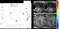
Multiple Linear Regression for Predicting Fibrosis in the Kidney
using T1 Mapping and ‘RESOLVE’ Diffusion-Weighted MRI
Iris FRIEDLI1, Lindsey Alexandra CROWE1,
Lena BERCHTOLD2, Solange MOLL3, Karine
HADAYA2, Thomas DE PERROT1,
Pierre-Yves MARTIN2, Sophie DE SEIGNEUX2,
and Jean-Paul VALLEE1
1Department of Radiology, Geneva University
Hospitals, Geneva, Switzerland, 2Department
of Nephrology, Geneva University Hospitals, Geneva,
Switzerland, 3Department
of Pathology, Geneva University Hospitals, Geneva,
Switzerland
Multi-parametric studies are beginning to emerge in renal
disease assessment. However these studies investigated each
MR parameter independently and compare the MR sequences but
do not combine multiple parameters in a single statistic. In
this multi-parametric 3T MR study, the sensitivity of T1
mapping and Readout Segmentation Of Long Variable Echo train
(RESOLVE) DWI parameters was first independently evaluated
and compared against interstitial fibrosis of 31 Chronic
Kidney Disease patients undergoing renal biopsy. The two MR
parameters were then associated in a single statistic with
the hypothesis that used together they can improve the
non-invasive detection of interstitial fibrosis.
|
|
0718.
 |
4 |
Towards Quantitative Renal MR Blood Oximetry by Combined
Monitoring of T2*, T2 and Blood Volume Fraction
Andreas Pohlmann1, Karen Arakelyan1,2,
Leili Riazy1, Till Huelnhagen1,
Stefanie Kox1, Kathleen Cantow2, Sonia
Waiczies1, Bert Flemming2, Erdmann
Seeliger2, and Thoralf Niendorf1
1Berlin Ultrahigh Field Facility, Max Delbrueck
Center for Molecular Medicine, Berlin, Germany, 2Institute
of Physiology, Charite Universitaetsmedizin, Berlin, Germany
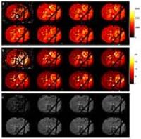
Acute kidney injuries are often characterized by tissue
oxygen hypoxia. T2*-mapping permits probing renal
oxygenation but provides a surrogate rather than a
quantitative measure of oxygen saturation. The link between
pO2 and
T2* is influenced by changes in blood volume
fraction (BVf). Monitoring BVf in combination with recently
developed quantitative BOLD approaches could permit
unambiguous interpretation of renal T2*. To test
the feasibility of this new approach we monitored renal T2*/T2 during
baseline and short periods of venous occlusion. This was
performed in the same animal under naïve conditions and
again with USPIO to permit estimation of BVf and SO2.
|
|
0719.
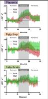 |
5 |
BOLD MRI of human placenta and fetuses under maternal
hyperoxygenation in growth restricted twin pregnancies
Jie Luo1,2, Esra Abaci Turk1,2,
Carolina Bibbo3, Borjan Gagoski1, Mark
Vangel4, Clare M Tempany-Afdhal5,
Norberto Malpica6, Arvind Palanisamy7,
Elfar Adalsteinsson2,8,9, Julian N Robinson3,
and Patricia Ellen Grant1
1Fetal-Neonatal Neuroimaging & Developmental
Science Center, Boston Children's Hospital, Harvard Medical
School, Boston, MA, United States, 2Madrid-MIT
M+Vision Consortium in RLE, Massachusetts Institute of
Technology, Cambridge, MA, United States, 3Maternal
and Fetal Medicine, Brigham and Women's Hospital, Boston,
MA, United States, 4Department
of Radiology, Harvard Medical School, Boston, MA, United
States, 5Department
of Radiology, Brigham and Women's Hospital, Boston, MA,
United States, 6Medical
Image Analysis and Biometry Laboratory, Universidad Rey Juan
Carlos, Madrid, Spain, 7Division
of Obstetric Anesthesia, Brigham and Women's Hospital,
Boston, MA, United States, 8Department
of Electrical Engineering and Computer Science,
Massachusetts Institute of Technology, Cambridge, MA, United
States, 9Harvard-
MIT Health Sciences and Technology, Massachusetts Institute
of Technology, Cambridge, MA, United States
Adequate oxygen transport across the placenta from mother to
fetus is critical for fetal growth and development. In this
pilot study, BOLD MRI with maternal hyperoxygenation show
great potential in differentiating IUGR fetuses from
controls. Not only the placentae show significant difference
in rate of oxygen uptake, fetal organs also have distinct
response to exposure to hyperoxia. Differences between fetal
brain and liver responses to hyperoxygenation are observed
in some cases, which might suggest variations in fetal
hemodynamic autoregulation.
|
|
0720.
 |
6 |
Ingestion of carbohydrate solutions of glucose-fructose versus
glucose-alone during a prolonged exercise in individuals with
type 1 diabetes
Tania Buehler1, Lia Bally2, Ayse Sila
Dokumaci1, Christoph Stettler2, and
Chris Boesch1
1Depts. Radiology and Clinical Research,
University of Bern, Bern, Switzerland, 2Division
of Endocrinology, Diabetes and Clinical Nutrition,
Inselspital Bern, Bern, Switzerland
In comparison to healthy subjects, there is scarce data on
the influence of different carbohydrate-types on the
metabolism in exercising individuals with type 1 diabetes
mellitus (T1DM). Based on 13C-MRS,
blood sampling, stable isotopes, and indirect calorimetry
the impact of glucose-fructose and glucose-alone was
investigated in T1DM subjects without prior insulin
reduction. Glucose-fructose ingestion showed a shift in fuel
metabolism towards increased fat oxidation and potential
glycogen sparing effects. Despite the negative reputation of
fructose it seems to be a more efficient fuel in exercising
T1DM subjects, since blood glucose levels are not
immediately elevated due to its different metabolization.
|
|
0721.
 |
7 |
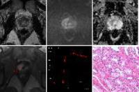
Pancreatic disease in obesity: observations on fat content,
diffusion, T2* relaxometric and mechanical properties in the rat
ex vivo
Philippe Garteiser1, Sabrina Doblas1,
Jean-Baptiste Cavin1, André Bado1,
Vinciane Rebours1,2, Maude Le Gall1,
Anne Couvelard1,3, and Bernard E Van Beers1,4
1Center For Research on Inflammation, Inserm
U1149, Paris, France, 2Pancreatology
Unit, AP-HP, Beaujon Hospital, Clichy, France, 3Pathology
department, AP-HP, Bichat Hospital, Paris, France,4Radiology
department, AP-HP, Beaujon Hospital, Clichy, France
Multiparametric assessment of pancreas in the obese rat was
used to evaluate alterations linked to obesity-mediated
inflammation. Mechanical properties and T2* values are
significantly affected by disease, and reflect accurately
the histological features of the obese pancreas.
|
|
0722.
 |
8 |
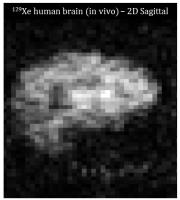
MR of hyperpolarized Xe-129 dissolved in the human brain at 1.5
T and 3.0 T
Madhwesha Rao1, Neil J Stewart1,
Graham Norquay1, Paul D Griffiths1,
and Jim M Wild1
1Academic unit of Radiology, University of
Sheffield, Sheffield, United Kingdom
Xenon is an inert noble gas which can be safely inhaled. In
the lungs, it diffuses into the bloodstream and is then
transported to distal organs (brain, kidneys and liver). In
this study, we have directly imaged the uptake of
hyerpolarized 129Xe
in the human brain in vivo. Thus demonstrated the
feasibility as a safe and non-invasive contrast agent for
functional imaging of the brain in diagnosing diseases
related to cerebral perfusion such as brain ischemia. In
addition, using tracer kinetic analysis we provide
quantitative measurement for the intrinsic physiological
characteristic of the blood brain barrier.
|
|
0723.
 |
9 |
Pulmonary Thin-Section MRI with Ultrashort TE: Capability for
Lung Nodule Screening and Subtype Classification as Compared
with Low- and Standard-Dose CTs - Permission Withheld
Yoshiharu Ohno1,2, Yuji Kishida2,
Shinichiro Seki2, Hisanobu Koyama2,
Takeshi Yoshikawa1,2, Daisuke Takenaka3,
Masao Yui4, Aiming Lu5, Mitsue
Miyazaki5, Katsusuke Kyotani6, and
Kazuro Sugimura2
1Advanced Biomedical Imaging Research Center,
Kobe University Graduate School of Medicine, Kobe, Japan, 2Radiology,
Kobe University Graduate School of Medicine, Kobe, Japan, 3Radiology,
Hyogo Cancer Center, Akashi, Japan, 4Toshiba
Medical Systems Corporation, Otawara, Japan, 5Toshiba
Medical Research Institute USA, Vernon Hills, IL, United
States, 6Center
for Radiology and Radiation Oncology, Kobe University
Hospital, Kobe, Japan
MRI with ultrashort TE (UTE) has been suggested as
useful for morphological assessment of lung as well as CT.
However, no reports have been found to study the capability
of thin-section MRI with UTE for pulmonary nodule detection
and nodule type assessment as compared with thin-section
CTs. We hypothesized that pulmonary MRI with UTE has a
similar potential for nodule detection and nodule type
evaluation as compared with thin-section CT. The purpose of
this study was to compare the capability of pulmonary MRI
with UTE for nodule detection and nodule type assessment
with low- and standard-dose CTs.
|
|
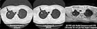
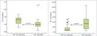
0724.
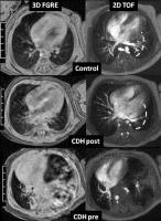 |
10 |
Quantitative Assessment of Pulmonary Blood Flow in Infants with
Congenital Diaphragmatic Hernia by CINE Phase Contrast MRI
Jean A Tkach1, Ryan A Moore2, Nara S
Higano1,3,4, Laura L Walkup1,3,
Mantosh S Rattan5, Paul S Kingma6,
Michael D Taylor2, and Jason C Woods1,3,4
1Imaging Research Center, Department of
Radiology, Cincinnati Children's Hospital Medical Center,
Cincinnati, OH, United States, 2The
Heart Institute, Cincinnati Children's Hospital Medical
Center, Cincinnati, OH, United States, 3Center
for Pulmonary Imaging Research, Division of Pulmonary
Medicine, Cincinnati Children's Hospital Medical Center,
Cincinnati, OH, United States, 4Department
of Physics, Washington University, St. Louis, MO, United
States, 5Department
of Radiology, Cincinnati Children's Hospital Medical Center,
Cincinnati, OH, United States, 6Division
of Neonatology and Pulmonary Biology, Cincinnati Children's
Hospital Medical Center, Cincinnati, OH, United States
Pulmonary arterial hypertension (PAH) is common in
congenital diaphragmatic hernia (CDH) and is a major
contributor to morbidity and mortality. Echocardiography and
cardiac catheterization are the current standards for
evaluating pulmonary hemodynamics in CDH infants, but both
have significant limitations and/or risks. Phase contrast
(PC) MRI can provide quantitative information about velocity
and flow longitudinally, with minimal risk. We demonstrate
the feasibility of applying PC MRI in the neonatal ICU
(NICU) to obtain a quantitative assessment of pulmonary
blood flow in CDH infants with the long-term goal to
establish imaging biomarkers to predict PAH and assess
therapeutic response.
|
|
0725.
 |
11 |
Pretreatment intravoxel incoherent motion diffusion-weighted
imaging for predicting the response of locally advanced rectal
cancer to neoadjuvant chemoradiation therapy
Hongliang Sun1, Yanyan Xu1, Kaining
Shi2, and Wu Wang1
1Radiology, China-Japan Friendship hospital,
Beijing, China, People's Republic of, 2Philips
Healthcare China, Beijing, China, People's Republic of
Neoadjuvant chemoradiation therapy (CRT) followed by surgery
has been established as the standard for locally advanced
rectal cancer[1]. The treatment response after CRT is
normally evaluated by MRI. However, MRI morphology
techniques suffer from limitations in the interpretation of
fibrotic scar tissue and inflammation. Diffusion weighted
MRI has shown its potentially beneficial role for response
evaluation, but with conflicting results[2]. Intravoxel
incoherent motion (IVIM) which enable quantitative
parameters that separately reflect tissue diffusivity and
tissue microcapillary perfusion[3-4]. However, the
pretreatment tumor IVIM MRI parameters predicting treatment
response were not clarified.
|
|
0726.
 |
12 |
Prostate cancer detection with multi-parametric MRI : PI-RADS
version 1 versus version 2
Zhaoyan Feng1, Xiangde Min1, and Liang
Wang1
1Tongji Hospital, Tongji Medical College,
Huazhong University of Science and Technology, Wuhan, China,
People's Republic of
The new PI-RADS version 2 classification (PI-RADS v2) was
proposed together with the European Society of Urogenital
Radiology (ESUR) and the American College of Radiology (ACR)
in December 2014. In contrast to PI-RADS v1, the v2 regulate
how to classify final PI-RADS score. for routine clinical
use, test of the validity of v2, including its sensitivity
and specificity for prostate cancer (PCa) detection should
raise concerns, and literature of them less. So, the purpose
of our study was to compare the diagnostic performance of v1
and v2 for the detection of PCa.
|
|
0727.
 |
13 |
Radiomic features on T2w MRI to predict tumor invasiveness for
pre-operative planning in colorectal cancer: preliminary results
Jacob Antunes1, Scott Steele2, Conor
Delaney2, Joseph Willis3, Justin Brady4,
Rajmohan Paspulati5, Anant Madabhushi1,
and Satish Viswanath1
1Department of Biomedical Engineering, Case
Western Reserve University, Cleveland, OH, United States, 2Department
of Colon and Rectal Surgery, University Hospitals Case
Medical Center, Cleveland, OH, United States, 3Department
of Anatomic Pathology, University Hospitals Case Medical
Center, Cleveland, OH, United States, 4Department
of General Surgery, University Hospitals Case Medical
Center, Cleveland, OH, United States, 5Department
of Radiology, University Hospitals Case Medical Center,
Cleveland, OH, United States
Pre-operative planning in colorectal cancer is highly
dependent on extent of tumor into the mesorectum, but tumor
margin is currently only assessed on excised pathology.
Radiomic features may capture subtle microarchitectural
changes on a restaging MRI, enabling characterization of
tumor extent prior to surgery, even when residual disease
may not be visually discernible. We present preliminary
results for identifying radiomic features which discriminate
invasive from noninvasive tumor on a 3 Tesla restaging T2w
MRI in colorectal cancer. In a cohort of 24 patients,
multi-scale gradient (Gabor) radiomic features demonstrated
high accuracy in segregating patients with invasive
colorectal cancer.
|
|
0728.
 |
14 |
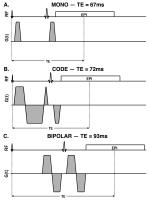
Motion Compensated Diffusion-Weighted MRI in the Liver with
Convex Optimized Diffusion Encoding (CODE)
Eric Aliotta1,2, Holden H Wu1,2, and
Daniel B Ennis1,2
1Radiological Sciences, UCLA, Los Angeles, CA,
United States, 2Biomedical
Physics IDP, UCLA, Los Angeles, CA, United States
Bulk motion artifacts in liver DWI can be substantially
reduced with first moment nulled diffusion encoding.
However, the bipolar diffusion encoding gradient waveforms
generally used for this purpose extend TE and limit SNR. We
have developed a Convex Optimized Diffusion Encoding (CODE)
framework to design time-optimal, motion compensated
diffusion encoding gradients that remove sequence dead times
and minimize TE. CODE gradients were designed and
implemented for liver DWI on a 3.0T clinical scanner, then
evaluated in healthy volunteers and patients. Bulk motion
artifacts were significantly reduced and ADC maps were
improved compared to conventional monopolar encoding.
|
|
0729.
 |
15 |
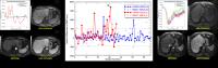
Quantitative Analysis of Arterial Phase Transient Respiratory
Motions Induced by Two Contrast Agents for Dynamic Liver MR
Imaging
Yuxi Pang1, Dariya Malyarenko1,
Matthew Davenport1, Hero Hussain1, and
Thomas Chenevert1
1Department of Radiology, UNIVERSITY OF MICHIGAN,
ANN ARBOR, MI, United States
This work is to analyze the respiratory waveforms from
dynamic liver MR images related to the motion artifacts in
arterial phase images induced by the contrast-media
administration. The discriminative metrics were defined to
quantify the likelihood of the acutely and temporally
impaired breath-holding by the subjects who received
gadoxetate disodium and gadobenate dimeglumine contrast
agents. Our preliminary results show that the indicative
metrics derived from recorded respiratory waveforms
objectively confirm prior reported observations that
gadoxetate disodium has a significantly higher likelihood of
inducing acute transient breath-holding difficulties that
adversely affect arterial phase image quality.
|
|