|
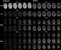
|
|
Plasma # |
 |
0273.
 |
1 |
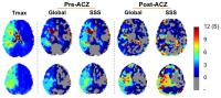
Simultaneous evaluation of hemodynamic and functional
connectivity in patients with chronic steno-occlusive disease of
the cerebrovascular system: A study using BOLD with
acetazolamide 
Junjie Wu1, Seena Dehkharghani1, Tyler
Gleason1, Fadi Nahab2, and Deqiang Qiu1
1Department of Radiology and Imaging Sciences,
Emory University, Atlanta, GA, United States, 2Department
of Neurology, Emory University, Atlanta, GA, United States
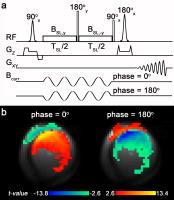
In this paper we applied a temporal-shift analysis
of the BOLD signal to delineate regions with abnormal
perfusion in patients with chronic steno-occlusive disease
of the cerebrovascular system. We proposed an improved
method of analysis based on an iterative approach for the
temporal shift analysis. We further explored the effects of
acetazolamide, a vasodilator, on the assessment of
hemodynamic compromise using temporal-shift analysis and
functional connectivity.
|
|
0274.
 |
2 |
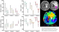
Electrical Conductivity Characteristics of Glioma: Noninvasive
Assessment by MRI and Its Validity
Khin Khin Tha1,2, Ulrich Katscher3,
Shigeru Yamaguchi4, Shunsuke Terasaka4,
Toru Yamamoto5, Kohsuke Kudo2,6, and
Hiroki Shirato1,2
1Department of Radiobiology and Medical
Engineering, Hokkaido University Graduate School of
Medicine, Sapporo, Japan, 2Global
Institution for Quantum Medical Science and Engineering,
Hokkaido University, Sapporo, Japan, 3Research
Laboratories, Hamburg, Germany, 4Department
of Neurosurgery, Hokkaido University Graduate School of
Medicine, Sapporo, Japan, 5Graduate
School of Health Sciences, Sapporo, Japan, 6Hokkaido
University Hospital, Sapporo, Japan
Electric Properties Tomography was performed in 24 glioma
patients, and the electrical conductivity characteristics of
glioma were determined noninvasively. Diagnostic performance
of electrical conductivity in distinguishing glioma grades
was also evaluated. Validity of noninvasive electrical
conductivity measurement was proved by correlating with the
conductivity values measured ex vivo by a dielectric probe.
|
 |
0275.
 |
3 |
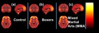
Quantifying differences in the cerebral blood flow (CBF) between
controls, professional boxers and Mixed Martial Arts (MMA)
fighters using arterial spin labeling (ASL) MRI
Virendra R Mishra1, Karthik Sreenivasan1,
Xiaowei Zhuang1, Zhengshi Yang1, Sarah
Banks1, Dietmar Cordes1, and Charles
Bernick1
1Cleveland Clinic Lou Ruvo Center for Brain
Health, Las Vegas, NV, United States
The professional fighters brain health study (PFBHS) is a
longitudinal study of active professional fighters with
age-matched healthy controls using multimodal MRI methods.
Using ASL-MRI, we report for the first time that cerebral
blood flow (CBF) is significantly lower in boxers and
mixed-martial-arts fighters (MMA) than age-matched healthy
controls. Most of the clusters were located in the
fronto-temporal lobe, cerebellum and thalamus. No
significant difference in perfusion between boxers and MMA
suggests that type of combat sports have an indiscernible
effect on CBF, further suggesting that perfusion may not
account for different patterns of cognitive decline observed
later in the life of these athletes.
|
|
0276.
 |
4 |
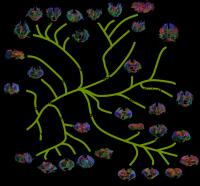
The Evolution of the Mammalian Connectome -
Video Not Available
Yossi Yovel1, Omri Zomet1, Arieli
Bonzach2, Assaf Marom1, and Yaniv
Assaf1
1Tel Aviv University, Tel Aviv, Israel, 2Beit
Dagan Veterinary institute, Beit Dagan, Israel
Despite its importance, little is known on the evolution of
the mammalian brain. Previous work suggests that body size
and behavioral function are intertwined in their influence
on the evolution of the brain. Most previous studies focused
on examining gray matter. Because the underlying white
matter connectome facilitates the connections between gray
matter areas, it must have simultaneously evolved to support
gray matter evolution. In this work we used a wide
comparative approach relying on diffusion MRI based
fiber-tracking to reconstruct whole-brain structural
connectomes and explore its evolution.
|
|
0277.
 |
5 |
Neurite Orientation Dispersion and Density Imaging (NODDI) in
Young Onset Alzheimer's Disease and Its Syndromic Variants - Permission Withheld
Jiaying Zhang1, Catherine F Slattery2,
Ross W Paterson2, Alexander JM Foulkes2,
Laura Mancini2, David L Thomas2, Marc
Modat1, Nicolas Toussaint2, David M
Cash2, John S Thornton2, Daniel C
Alexander1, Sebastien Ourselin1, Nick
C Fox2, Jonathan M Schott2, and Hui
Zhang1
1Department of Computer Science and Centre for
medical image computing, University College London, London,
United Kingdom, 2Department
of Neurodegenerative disease, Institute of Neurology,
University College London, London, United Kingdom
Alzheimer's disease (AD) is now increasingly considered as a
disorder of brain networks. Therefore, it is important to
quantify the integrity of white matter (WM) connections in
AD populations. Previous DTI studies have shown WM breakdown
in patients with young onset AD (YOAD), but DTI parameters
are not specific to any tissue property. Here we
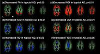
investigated WM changes using NODDI and DTI in YOAD patients
using TBSS and explored whether unique patterns of WM
changes exist in YOAD subtypes. We found NODDI was more
sensitive than DTI and demonstrated different profiles of WM
damage in YOAD syndromic subgroups.
|
|
0278.
 |
6 |
Developmental processes on the neonatal brain revealed by white
matter tract integrity metrics derived from diffusion kurtosis
imaging
Xianjun Li1,2, Jie Gao1, Yumiao Zhang1,
Yanyan Li1, Huan Li1, Mingxi Wan2,
and Jian Yang1,2
1Radiology Department of the First Affiliated
Hospital, Xi'an Jiaotong University, Xi'an, China, People's
Republic of, 2Department
of Biomedical Engineering, the Key Laboratory of Biomedical
Information Engineering of the Ministry of Education, School
of Life Science and Technology, Xi'an Jiaotong University,
Xi'an, China, People's Republic of
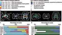
To distinguish axon-related and myelin-related developmental
processes, we tried to find a strategy for assessing white
matter developmental processes by using white matter tract
integrity (WMTI) metrics derived from diffusion kurtosis
imaging (DKI). The method was used on 41 neonates. The
proposed strategy provided more processes than conventional
diffusion tensor imaging (DTI) method. Five change patterns
were found for WMTI metrics, while 2 patterns for DTI
metrics. WMTI metrics derived from DKI could provide more
detailed developmental processes on neonatal white matter.
|
|
0279.
 |
7 |
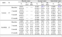
A serial microcompartment-specific T2* relaxation study of white
matter lesions in multiple sclerosis at 7T
Xiaozhen Li1,2, Peter van Gelderen2,
Pascal Sati3, Jacco de Zwart2, Daniel
Reich3, and Jeff Duyn2
1Dept. NVS, Karolinska Institutet, Stockholm,
Sweden, 2Advanced
MRI Section, LFMI, NINDS, National Institutes of Health,
Bethesda, MD, United States, 3Translational
Neuroradiology Unit, NINDS, National Institutes of Health,
Bethesda, MD, United States
Multiple sclerosis (MS) is a chronic demyelinating disease
characterized by focal lesions. Recent studies suggest the
possibility of obtaining cellular microcompartment-specific
information from three-component fitting of the T2* relaxation
decay curve, allowing determination of the relative
fractions of myelin water, axonal water and interstitial
water. The microcompartment-specific T2* relaxation
values of initially enhancing lesions were followed serially
on 7T at approximately 3, 6, and 12 months. The changes over
time that we observed in enhancing lesions are consistent
with the presence of ongoing remyelination. This may lead to
a better understanding of, and prognostic ability for, this
complex disease.
|
|
0280.
 |
8 |
Real-time fMRI Neurofeedback with Simultaneous EEG in
Combat-related PTSD: Frontal EEG Asymmetry Variations as Measure
of Treatment Response - Permission Withheld
Vadim Zotev1, Raquel Phillips1, Masaya
Misaki1, Chung Ki Wong1, Brent Wurfel1,
Matthew Meyer1,2, Frank Krueger1,3,
Matthew Feldner1,4, and Jerzy Bodurka1,5
1Laureate Institute for Brain Research, Tulsa,
OK, United States, 2Laureate
Psychiatric Clinic and Hospital, Tulsa, OK, United States, 3Neuroscience
Dept., George Mason University, Fairfax, VA, United States, 4Dept.
of Psychological Science, University of Arkansas,
Fayetteville, AR, United States, 5College
of Engineering, University of Oklahoma, Tulsa, OK, United
States
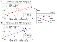
We have performed a study of emotion regulation training in
veterans with combat-related PTSD using real-time fMRI
neurofeedback (rtfMRI-nf) with simultaneous EEG. Fifteen
PTSD patients learned to upregulate their left amygdala
activity using rtfMRI-nf during a positive emotion induction
task based on retrieval of happy autobiographical memories.
Individual session-to-session variations in frontal EEG
asymmetry (FEA) changes during the rtfMRI-nf task
significantly correlated with variations in PTSD severity
(CAPS) and co-morbid depression severity (HDRS). These
results suggest that variations in task-specific FEA changes
during rtfMRI-nf training provide a sensitive measure of
individual response to treatment in PTSD patients.
|
|
0281.
 |
9 |
In-vivo detection of neuronal current using spin-lock
oscillatory excitation at 7T
Yuhui Chai1, Guoqiang Bi2, Liping Wang3,
Fuqiang Xu4, Xin Zhou4, Bensheng Qiu2,
Hao Lei4, Bing Wu5, Yang Fan5,
and Jia-Hong Gao1
1Center for MRI Research, Peking University,
Beijing, China, People's Republic of, 2University
of Science and Technology of China, Hefei, China, People's
Republic of, 3Shenzhen
Institutes of Advanced Technology, Chinese Academy of
Sciences, Shenzhen, China, People's Republic of, 4Wuhan
Institute of Physics and Mathematics, Chinese Academy of
Sciences, Wuhan, China, People's Republic of, 5GE
Healthcare, MR Research China, Beijing, China, People's
Republic of
In-vivo detection of neuronal current remains a challenging
and promising goal in fMRI. Previous work has demonstrated
its feasibility in phantom and cell culture studies, but
attempts in in-vivo studies remain few and far between. As
neuronal current is usually comprised of a series of
oscillatory waveforms rather than being a direct current, it
is most likely to be detected using oscillatory current
sensitive sequences. In this study, we explored the
potential of using the spin-lock oscillatory excitation
(SLOE) sequence to directly detect optogenetically evoked
oscillatory neuronal current in vivo for the first time.
|
|
0282.
 |
10 |
Rapid Myelin Water Imaging in Human Cervical Spinal Cord
Emil Ljungberg1, Irene Vavasour2,
Roger Tam2,3, Youngjin Yoo3, Alexander
Rauscher4, David Li2, Anthony
Traboulsee5, Alex MacKay1,2, and
Shannon Kolind5
1Physics and Astronomy, University of British
Columbia, Vancouver, BC, Canada, 2Radiology,
University of British Columbia, Vancouver, BC, Canada, 3Electrical
and Computer Engineering, University of British Columbia,
Vancouver, BC, Canada, 4Pediatrics,
University of British Columbia, Vancouver, BC, Canada, 5Medicine,
University of British Columbia, Vancouver, BC, Canada
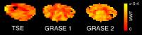
Myelin water imaging can quantify myelin in the cervical
cord in vivo. However, the established 3D Turbo Spin Echo
(TSE) approach has a lengthy scan time. We used a 3D
Gradient Spin Echo (GRASE) sequence to speed up cervical
cord myelin water acquisition by a factor of three. Average
GRASE and TSE myelin water estimates were similar (GRASE:
23±1.5%; TSE: 24±3%) and significantly correlated (R2=0.69,
p<0.001). 3D-GRASE showed good reproducibility with an
average myelin water coefficient of variation of 6%. Our
findings demonstrate that cervical cord myelin water data
can reliably be collected in clinical feasible scan times.
|
|
0283.
 |
11 |
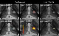
Transcranial MRI-Guided High-Intensity Focused Ultrasound for
Treatment of ?Essential Tremor: Initial Clinical Experience and
Correlation of Clinical Outcome with Lesion Size, Localization,
and Dose
Christian Federau1, Maged Goubran1,
Jason Su1, Jaimie Henderson1, Veronika
Santini1, Casey Harrison Halpern1,
Brian Rutt1, Kim Butts Pauly1, and
Pejman Ghanouni1
1Stanford University, Stanford, CA, United States
Transcranial MR-guided high-intensity focused ultrasound
ablation of the ventral division of the ventral lateral
posterior thalamic nucleus (VLpv) is a promising, minimally
invasive treatment method for essential tremor. We report
our initial clinical experience in 11 patients, and
correlate clinical outcome with lesion size, location, and
thermal dose during treatment. We found a correlation
between clinical outcome at 1 year follow-up and lesion
size (r = 0.73), as well as thermal dose in the VLpv (r =
0.65).
|
|
0284.
 |
12 |
Neuroimaging of Acute Ebola Virus Disease in a Non-Human Primate
Model
Margaret R. Lentz1, Jeffery R. Solomon2,
Srikanth Yellayi1, Richard Bennett1,
Dawn Traynor1, David Thomasson1, Anna
Honko1, Lisa Hensley1, and Peter B.
Jahrling1,3
1Integrated Research Facility, NIAID, National
Institutes of Health, Frederick, MD, United States, 2Clinical
Research Directorate/Clinical Monitoring Research Program,
Frederick National Laboratory for Cancer Research, Leidos
Biomedical Research, Inc., Frederick, MD, United States, 3Emerging
Viral Pathogens Section, NIAID, National Institutes of
Health, Frederick, MD, United States
The purpose of this study was to use MRI to assess
alterations in the brain that occur in rhesus macaques
infected with a variant of the Ebola virus (EBOV) isolated
from the most recent outbreak. EBOV was found to induce
signal alterations in susceptibility weighted imaging (SWI)
along vasculature that correlate to venous congestion and
perivascular hemorrhage. The use of SWI or other gradient
echo based methods to examine vascular changes may be of
interest when examining survivors of Ebola. Additionally,
the identification of non-invasive imaging biomarkers of
EBOV disease progression could help in development of
medical countermeasures.
|
|
0285.
 |
13 |
Structural variability in the human brain reflects functional
architecture
Gwenaelle Douaud1, Eugene Duff1,
Adrian Groves1, Thomas Nichols1,2,
Saad Jbabdi1, Christian Tamnes3, Lars
Westlye3, Andreas Engvig3, Kristine
Walhovd3, Anders Fjell3, Heidi
Johansen-Berg1, and Steve Smith1
1FMRIB Centre, University of Oxford, Oxford,
United Kingdom, 2University
of Warwick, Coventry, United Kingdom, 3University
of Oslo, Oslo, Norway
It is believed that the resting-state networks
closely relate to the underlying anatomical connectivity and
grey matter structure but cannot be understood in those
terms alone. Here, we show that a purely data-driven
approach used to co-model three complementary types of grey
matter information on a large, healthy population covering
most of the lifespan uncovers the entire repertoire of
canonical functional networks. We further demonstrate that
the modes of variation of grey matter volume across all
participants forming these structural networks spatially
co-vary with cortical area, except in primary sensory areas
where they also partially co-vary with cortical thickness.
|
|
0286.
 |
14 |
A constrained slice-dependent background suppression scheme for
simultaneous multi-slice pseudo-continuous arterial spin
labeling
Xingfeng Shao1, Yi Wang1, and Danny
J.J. Wang1
1Laboratory of FMRI Technology (LOFT), Department
of Neurology, University of California Los Angeles, Los
Angeles, CA, United States
Compared to standard two-dimensional (2D) arterial spin
labeling (ASL), simultaneous multi-slice (SMS) ASL imaging
techniques can reduce T1 relaxation effect of the label;
improve spatial coverage and resolution. However, existing
2D SMS ASL techniques are sub-optimal for the background
suppression (BS) technique since multiple SMS excitations
are required. In this study, we propose a novel constrained
slice-dependent BS scheme for 2D multi-slice
pseudo-continuous ASL (pCASL) with SMS-EPI acquisition, to
suppress background signal across a wide range of T1s. In
vivo experiment showed that the BS scheme can increase
temporal SNR of perfusion images 1.5-2 folds.
|
|
0287.
 |
15 |
Brain Catalogue and its MRI of extinct species: the example of
Thylacinus Cynocephalus
Mathieu David Santin1,2, Marc Herbin3,
and Roberto Toro4
1Centre de NeuroImagerie de Recherche - CENIR,
Paris, France, 2Inserm
U 1127, CNRS UMR 7225, Sorbonne Universités, UPMC Univ Paris
06 UMR S 1127, Institut du Cerveau et de la Moelle épinière,
ICM, Paris, France, 3Muséum
National d'Histoire Naturelle, Paris, France, 4Institut
Pasteur, Paris, France
We present here an example of one of the application of the
Brain Catalogue with an MRI of an extinct species: the
Thylacine
|
|