|
|
|
Plasma # |
 |
0631.
 |
1 |
The sensitivity of diffusion MRI in direct detection neuronal
activity: an in-vitro assessment 
Ruiliang Bai1,2, Craig Stewart3,
Dietmar Plenz3, and Peter J Basser1
1Section on Quantitative Imaging and Tissue
Science, DIBGI, NICHD, National Institutes of Health,
Bethesda, MD, United States, 2Biophysics
Program, Institute for Physical Science and Technology,
University of Maryland, College Park, MD, United States, 3Section
on Critical Brain Dynamics, LSN, NIMH, National Institutes
of Health, Bethesda, MD, United States
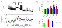
Diffusion MRI has been proposed as a noninvasive
neuroimaging method to detect neuronal activity more
directly than BOLD fMRI, yet, initial findings have proven
difficult to interpret and reproduce. Here, we study the
possible relationship between water diffusion and neuronal
activity by simultaneous intracellular calcium fluorescence
imaging and diffusion MR of organotypic rat brain cortex
cultures. Although we found that diffusion MR can follow
pathological changes during hyperexcitability, e.g., as
those seen in epilepsy or during anoxia, it does not appear
to be sensitive or specific enough to detect or follow
normal neuronal activity.
|
|
0632.
 |
2 |
Apparent diffusion coefficient correlates with gamma oscillation
of local field potentials - Permission Withheld
Tomokazu Tsurugizawa1, Yoshifumi Abe1,
and Denis Le Bihan1
1NeuroSpin, Bât 145, Commissariat à l’Energie
Atomique-Saclay Center, 91191, France, Gif-sur-Yvette,
France
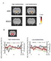
BOLD fMRI which relies on neurovascular coupling may fail
when neurovascular coupling is weakened, such as anesthesia
or alcohol intoxication. In contrast, diffusion fMRI has
been shown to be more directly linked to neuronal activation
even in the absence of neurovascular coupling. We compared
BOLD fMRI and diffusion fMRI (ADC) time-courses with local
field potentials (LFPs) in rat nucleus accumbens following
alcohol stimulation under two different doses of
medetomidine anesthesia. The ADC responses were correlated
with LFP signals while BOLD signals were not. These results
show the interest of diffusion fMRI to avoid confounds from
varying conditions of neurovascular coupling.
|
|
0633.
 |
3 |
Fast Dynamic Measurement of Functional T1 and Grey Matter
Thickness Changes During Brain Activation at 7T
Laurentius Huber1, Sean Marrett1,
Daniel A Handwerker1, Adam Thomas1,
Benjamin Gutierrez1, Dimo Ivanov2,
Benedikt A Poser2, and Peter A Bandettini1
1Section of Functional Imaging Methods, National
Institute of Mental Health, Bethesda, MD, United States, 2MBIC,
Maastricht University, Maastricht, Netherlands
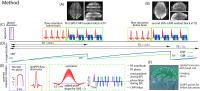
We present a fast new method for obtaining quantitative T1 maps
with high spatial (1 mm) and temporal resolutions (3 s).
This method can be useful to investigate morphological
dynamics of brain GM, e.g. during brain activity changes,
plasticity changes, or pathology. The robustness of the
developed method is demonstrated with a finger tapping fMRI
experiment. We report a functional GM T1increase
of up to 100 ms, and a GM
thickness increase by
up to 0.25 mm.
|
|
0634.
 |
4 |
Cognitive Application of Multi-Phase Passband Balanced SSFP fMRI
with 50ms Sampling rate at 7 Tesla
Zhongwei Chen1,2, Rong Xue1, Jing An3,
Kaibao Sun1,2, Zhentao Zuo1, Peng
Zhang1, and Danny JJ Wang4
1State Key Laboratory of Brain and Cognitive
Science, Institute of Biophysics, Chinese Academy of
Sciences, Beijing, China, People's Republic of, 2Graduate
School, University of Chinese Academy of Sciences, Beijing,
China, People's Republic of, 3Siemens
Shenzhen Magnetic Resonance Ltd, Shenzhen, China, People's
Republic of, 4Laboratory
of FMRI Technology (LOFT), Department of Neurology,
University of California Los Angeles, Los Angeles, CA,
United States
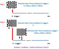
Multi-phase passband steady state free precession (SSFP)
cine fMRI can achieve a spatial resolution of a few mm3 and
a temporal sampling rate of 50ms at 7 Tesla , while
maintaining low geometric distortion and signal dropout. In
this study, the feasibility and accuracy of the technique
are demonstrated by two visual event-related functional MRI
experiments.
|
|
0635.
 |
5 |
Depth-Dependence of Visual Signals in the Human Superior
Colliculus at 9.4T: Comparison with 3T
Joana Alves Loureiro1,2, Gisela Hagberg1,
Thomas Ethofer2, Michael Erb2, Klaus
Scheffler1, and Marc Himmelbach3
1High-Field Magnetic Resonance, Max Planck
Institute for Biological Cybernetics, Tuebingen, Germany, 2BMMR,
University Hospital Tuebingen, Tuebingen, Germany, 3Division
of Neuropsychology, Centre for neurology, Tuebingen, Germany
The superior colliculus (SC) is a layered structure involved
in visual and multisensory control. Due to its small size
and location it is challenging to evaluate its function with
the conventional MR fields. In this study we compare the
depth-dependence of visual signals in SC for 9.4T and 3T
data. The highest signal was observed in the superficial
zone of the superior colliculus (for both datasets).
However, the increase in sensitivity in the blood oxygen
level dependent size allowed us to get higher response
lateralization and a significative higher depth-dependence
of visual signals in the 9.4T.
|
|
0636.
 |
6 |
Resting State Functional Connectivity is Sensitive to
Layer-specific Connectional Architecture in Cortical Columns
Yun Wang1, Jennifer Robinson1,2,3, and
Gopikrishna Deshpande1,2,3
1AU MRI Research Center, Department of Electrical
and Computer Engineering, Auburn University, Auburn, AL,
United States, 2Department
of Psychology, Auburn University, Auburn, AL, United States,3Alabama
Advanced Imaging Consortium,Auburn University and University
of Alabama Birmingham, Birmingham, AL, United States
We investigated whether resting-state functional
connectivity (FC) is sensitive to cortical layer-specific
connectional differences using high resolution resting-state
fMRI data obtained from healthy humans at 7T. Based on rat
tracing studies, we hypothesized that FC between the
thalamus and cortical layer I must be significantly greater
than between the thalamus and other layers. Our results
support this hypothesis. Further, there were no global
connectivity differences between layers, ruling out
artifactual influences from vasculature. This also opens the
future possibility of microscopic investigations of the
brain connectome using ultra-high field fMRI and will likely
move the field away from blobology.
|
|
0637.
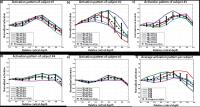 |
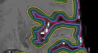
7 |
Deconvolving the laminar gradient echo activation profiles with
the spatial PSF: an approach to revealing underlying activation
patterns
Irati Markuerkiaga1 and
David G. Norris1
1Donders Institute, Nijmegen, Netherlands
The specificity of GE-BOLD profiles is suspected to be
degraded by intracortical veins. In this work
experimentally obtained GE-BOLD profiles for different
subjects are deconvolved with a laminar point spread
functions obtained from a model of cortical vasculature. The
obtained underlying activation profiles are closer to the
activity profiles expected from electrophysiology for the
type of stimulus used.
|
|
0638.
 |
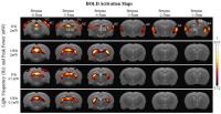
8 |
Effects of Anesthesia on White Matter BOLD Signals in Monkeys
Tung-Lin Wu1,2, Feng Wang1,3, Li Min
Chen1,3, Adam W. Anderson1,2,3,
Zhaohua Ding1,3, and John C. Gore1,2,3
1Vanderbilt University Institute of Imaging
Science, Nashville, TN, United States, 2Biomedical
Engineering, Vanderbilt University, Nashville, TN, United
States, 3Radiology
and Radiological Sciences, Vanderbilt University, Nashville,
TN, United States
We previously reported the first evidence of anisotropic
rsfMRI-BOLD signals in white matter which appear to reflect
a functional structure not previously detected. To prove
these signals have a functional basis, we performed imaging
of live squirrel monkeys under different baselines of neural
activity by altering anesthesia levels. Specifically, we
compared how different anesthesia levels modulate fractional
power and spatio-temporal correlation tensors in white
matter. Our results demonstrate that low frequency BOLD
signal fluctuations behave similarly in grey and white
matter. This indicates that anisotropic rsfMRI-BOLD signals
in white matter encode neural activity.
|
 |
0639.
 |
9 |
Cerebral vascular reactivity and baseline cerebral blood volume
contributions to the slow fluctuating baseline BOLD signal.
Jeroen C.W. Siero1, Jill B. de Vis1,
and Jeroen Hendrikse1
1Radiology, University Medical Center Utrecht,
Utrecht, Netherlands
Slow fluctuating (< 0.1 Hz) BOLD signals during baseline
conditions or ‘resting-state’ have seen interest in numerous
studies, both in healthy and disease. Here we investigate
cerebral vascular reactivity and baseline cerebral blood
volume contributions to the slow fluctuating baseline BOLD
signal.
|
|
0640.
 |
10 |
Frequency specificity of functional connectivity in rat brain
networks
Li-Ming Hsu1, Gu Hong1, Hanbing Lu1,
Elisabeth C. Caparelli1, Elliot A. Stein1,
and Yihong Yang1
1Neuroimaging Research Branch, National institute
on drug abuse, Baltimore, MD, United States
Intrinsic brain networks seen in humans, including the
default-mode network (DMN), have been demonstrated in
non-human primates and rodents using resting-state
functional fMRI (rs-fMRI). Characteristics of these brain
networks, such as frequency specificity, have been assessed
in humans, but are much less known in animal models. These
characteristics are of importance when translating findings
from preclinical models to clinical applications. The
frequency range used in a human rs-fMRI analysis is
typically ≤ 0.1 Hz; however, an appropriate frequency range
in rodents remains unclear. In this study, we investigated
the resting-state functional connectivity (rsFC) of rat
brains in three frequency ranges: 1) 0.01 – 0.1 Hz, 2) 0.1 –
0.25 Hz, and 3) 0.25 – 0.5 Hz, and compared the result with
that in human brains.
|
|
0641.
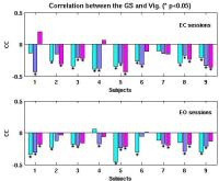 |
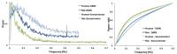
11 |
The resting state fMRI global signal is negatively correlated
with time-varying EEG vigilance
Maryam Falahpour1, Chi Wah Wong1, and
Thomas T. Liu1
1Center for Functional Magnetic Resonance
Imaging, University of California San Diego, San Diego, CA,
United States
Global signal (GS) regression is a commonly used
preprocessing approach in the analysis of resting-state fMRI
data. However GSR should be used with caution as it can not
only induce spurious anti-correlations, but may also remove
signal of neural origin. Here we used simultaneously
acquired EEG/fMRI data to study the relation between the GS
and an EEG-based measure of vigilance at rest. We found that
there is a significant negative correlation between the GS
and EEG vigilance. Our results indicate that GS has a
significant neuronal component and further emphasizes the
need to exercise caution when regressing out the GS.
|
|
0642.
 |
12 |
Detection of epileptic networks using wavelet coherence analysis
of dynamic local fMRI connectivity and simultaneous scalp EEG
Amir Omidvarnia1, David Vaughan1,2,
Mangor Pedersen1, Mira Semmelroch1,
David Abbott1, and Graeme Jackson1,2,3
1Epilepsy Imaging, The Florey Institute of
Neuroscience and Mental Health, Melbourne, Australia, 2Department
of Neurology, Austin Health, Melbourne, Australia, 3Department
of Medicine, The University of Melbourne, Melbourne,
Australia
In this study, we aimed at developing an objective method
for detecting clinically suspected epileptic networks
through possible association between interictal EEG
discharges and dynamic local fMRI connectivity in focal
epilepsy. We designed a time-frequency framework for
analysis of wavelet coherence between scalp EEG band
amplitude fluctuations (BAFs) and dynamic regional phase
synchrony (DRePS) of task-free fMRI in seven patients. The
proposed method reveals nonstationary relationship between
scalp interictal epileptic discharges (IEDs) and DRePS
within ultra-slow frequencies (~0.003 – 0.03Hz). Evaluation
of dynamic fMRI phase synchrony at rest, particularly using
data-fusion with interictal scalp EEG, may provide useful
markers of localized and transient brain connectivity
disturbance in epilepsy.
|
|
0643.
 |
13 |
Large-scale Brain Activation upon Strong Low Frequency Visual
Stimulation
Leon C. Ho1,2, Russell W. Chan1,2,
Patrick P. Gao1,2, Alex T.L. Leong1,2,
Celia M. Dong1,2, and Ed X. Wu1,2
1Laboratory of Biomedical Imaging and Signal
Processing, The University of Hong Kong, Hong Kong, China,
People's Republic of, 2Department
of Electrical and Electronic Engineering, The University of
Hong Kong, Hong Kong, China, People's Republic of
Visual inputs are primarily processed by the visual system.
However visual input also interacts with other sensory
cortices to speed up or improve sensory perception. While
the effect of different parameters of visual input to
crossmodal influences remains largely unexplored, this study
showed strong low frequency light evoked responses in
auditory cortex, secondary somatosensory cortex, cingulate
cortex and caudate putamen. The activations in those brain
regions likely propagated from the visual cortex and
influenced subcortical responses. Our current study provides
a functional understanding to cortical crossmodal processing
and its influences to subcortex upon visual stimuli of
different intensities and frequencies.
|
|
0644.
 |
14 |
Relative latency and temporal variability of BOLD fMRI signal
within human visual cortex
Jo-Fu Lotus Lin1, Jonathan R Polimeni2,
Wen-Jui Kuo3, and Fa-Hsuan Lin1
1Institute of Biomedical Engineering, National
Taiwan University, Taipei, Taiwan, 2Athinoula
A. Martinos Center, Department of Radiology, Harvard Medical
School, Massachusetts General Hospital, Charlestown, MA,
United States, 3Institute
of Neuroscience, National Yang Ming University, Taipei,
Taiwan
We used inverse imaging to spatiotemporally characterize the
relative latency and variability of the BOLD signal at human
visual cortex with 0.1 s precision. The relative BOLD
latency in the left and right visual cortex was 0.12 (s) +/-
0.33 (s). The BOLD variability in the left and right visual
cortex was 0.39 (s) +/- 0.25 (s). Local relative BOLD
latency was linearly related to local BOLD variability. The
least variability (< 0.2 s) and the earliest onset of the
BOLD signal were found at the trough of the calcarine sulcus.
|
|
0645.
 |
15 |
Globally conditioned multivariate causal influence estimates in
whole-brain functional connectivity
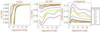
Andrea Duggento1, Luca Passamonti2,3,
Maria Guerrisi1, and Nicola Toschi1,4
1Department of biomedicine and prevention,
University of Rome "Tor Vergata", Rome, Italy, 2Institute
of Bioimaging and Molecular Physiology, National Research
Council, Catanzaro, Italy, 3Department
of Clinical Neurosciences, University of Cambridge,
Cambridge, United Kingdom, 4Department
of Radiology, Martinos Center for Biomedical Imaging and
Harvard Medical School, Boston, MA, United States
Reconstructing the direction of information flow
("causality") is crucial when studying evidence-based
network models of the brain. We use multivariate analysis to
develop a conditioning approach which measures the true
directed coupling between two signals which are also
indirectly connected through a large number of additional
interdependent sources. After validation through synthetics
noisy oscillator networks, we study data from 100 HCP
subjects, revealing a clear-cut, sparse resting-state
directed network structure and providing first-time evidence
of a concerted directional interaction between subnetworks
of the brain, with the salience network performing top-down
integration of sensory-motor and cognitive processes.
|
|