|
|
|
Plasma # |
|
0895.
 |
1 |
Vascular injury triggers a systemic response that promotes
atherosclerosis progression at a remote site of injury. 
Begona Lavin Plaza1, Alkystis Phinikaridou1,
Marcelo Andia2, Silvia Lorrio Gonzalez1,
and Rene Botnar1
1King's College London, London, United Kingdom, 2Pontificia
Universidad Catolica de Chile, Santiago de Chile, Chile
Atherothrombosis is a systemic arterial disease mainly
involving the intima of large- and medium-sized arteries
including the carotid, aorta, coronary, and peripheral
arteries. Although it has long been known that
atherosclerosis is a systemic disease, the effects of
vascular alteration distally from the site of injury and the
underlying mechanisms responsible for the systemic response
have not been elucidated. In this study, we used an
albumin-binding contrast agent to assess whether (1)
vascular injury in the abdominal aorta triggers plaque
progression in the brachiocephalic artery located distally
to the site of injury and (2) whether neutrophils can be the
link involved in this systemic response.
|
 |
0896.
 |
2 |
Translation of high-field fluorine-19 cell tracking techniques
into the clinical realm
Jeff M Gaudet1,2, Corby Fink3,4,
Matthew S Fox1, Gregory A Dekaban3,4,
and Paula J Foster1,2
1Imaging Research Laboratories, Robarts Research
Institute, London, ON, Canada, 2Medical
Biophysics, Western University, London, ON, Canada, 3Molecular
Medicine, Robarts Research Institute, London, ON, Canada, 4Microbiology
and Immunology, Western University, London, ON, Canada
Cellular MRI can be used to improve outcomes of cancer
immunotherapy by tracking the fate of these cells after
their administration. In this study, we used fluorine-19
MRI to track and quantify migration of antigen-presenting
peripheral blood mononuclear cells (PBMC). Mice were imaged
at both high-field and clinical field strengths. PBMC
migration to the node was quantified and compared under
different conditions. This study is the first to report on
fluorine-19 imaging of PBMC and demonstrates the potential
of cellular MRI to aid in the optimization of cellular
therapy.
|
|
0897.
 |
3 |
Propionate as a Probe For Myocardial Metabolism – A Biochemical
and Hyperpolarized MR Study
Mukundan Ragavan1, Xiaorong Fu2, Shawn
C Burgess2, and Matthew E Merritt1
1Department of Biochemistry & Molecular Biology,
University of Florida, Gainesville, FL, United States, 2University
of Texas Southwestern Medical Center, Dallas, TX, United
States
In this study, the utility of sodium propionate for
accentuating changes in cardiac metabolism is evaluated. The
study is performed using a murine model of cardiac
hypertrophy and employs hyperpolarized magnetic resonance
spectroscopy, mass spectrometry and a biochemical assay to
determine the cardiac redox state. Results show propionate
modulates cardiac metabolism across a range of different
concentrations.
|
|
0898.
 |
4 |
In-vivo evaluation of hypometabolism associated with muscular
dystrophy using Creatine CEST MRI
Rong-Wen Tain1,2, Ahlke Heydemann3,4,
Alessandro Scotti1,5,6, Weiguo Li7,8,
Xiaohong Joe Zhou1,5,6,9, and Kejia Cai1,5,6
1Radiology,College of Medicine, University of
Illinois, Chicago, IL, United States, 23T
Research Program, Center for MR Research, College of
Medicine, University of Illinois, Chicago, IL, United
States,3Physiology & Biophysics,College of
Medicine, University of Illinois, Chicago, IL, United
States, 4Center
for Cardiovascular Research, College of Medicine, University
of Illinois, Chicago, IL, United States,53T
Research Program, Center for MR Research, University of
Illinois, Chicago, IL, United States, 6Bioengineering,
University of Illinois, Chicago, IL, United States, 7Research
Resource Center, University of Illinois, Chicago, IL, United
States, 8Radiology,
Northwestern University, Chicago, IL, United States, 9Neurosurgery,
University of Illinois, Chicago, IL, United States
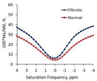
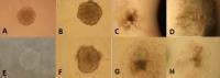
This study aims to measure hypometabolism in the muscle due
to muscular dystrophy using creatine CEST MRI. We acquired
images of the lower limbs from the diseased and wild-type
mice. Differences in the Z-spectrum and creatine CEST
contrast map were seen between fibrotic and normal muscles.
This suggested that CrCEST MRI may serve as a sensitive
imaging biomarker for metabolic changes associated with
muscular dystrophy.
|
|
0899.
 |
5 |
3D Dynamic Hyperpolarized 13C-Pyruvate MR Metabolic Imaging of
Human Prostate Cancer
Hsin-Yu Chen1, Peder E.Z. Larson1,2,
Jeremy W. Gordon2, Robert A. Bok2,
Marcus Ferrone3, Mark van Criekinge2,
Lucas Carvajal2, Peng Cao2, Ilwoo Park2,
Rahul Aggarwal4, Sarah J. Nelson1,2,
John Kurhanewicz1,2, and Daniel B. Vigneron1,2
1Graduate Program in Bioengineering, UCSF and UC
Berkeley, University of California, San Francisco, San
Francisco, CA, United States, 2Department
of Radiology and Biomedical Imaging, University of
California, San Francisco, San Francisco, CA, United States, 3Department
of Clinical Pharmacy, University of California, San
Francisco, San Francisco, CA, United States, 4Department
of Medicine, Division of Hematology/Oncology, University of
California, San Francisco, San Francisco, CA, United States
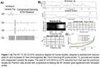
To measure the 3D spatial and temporal dynamics of
hyperpolarized [1-13C]pyruvate for patient
studies, a new compressed-sensing EPSI sequence was
developed for prostate cancer clinical research. Utilizing
multiband, variable flip angle RF excitation, this sequence
provided high temporal (2s) and spatial (0.5cm3)
resolution data detecting pyruvate uptake and its rate of
conversion to lactate. This approach provided a significant
advance over initial human HP-13C studies in
which just 1D or 2D dynamics were measured and 15s
single-timepoint 3D spectra were acquired. Following phantom
tests, patient data demonstrated high pyruvate to lactate
conversion in regions corresponding to biopsy-confirmed
prostate cancer.
|
|
0900.
 |
6 |
Positive-contrast cellular MRI of embryonic stem cells for
tissue regeneration using a highly efficient T1 MRI contrast
agent - Permission Withheld
Sadi Loai1, Inga E. Haedicke2,3, Zahra
Mirzaei1, Craig Simmons1,4, Xiao-an
Zhang2,3, and Hai-Ling Margaret Cheng1,5
1Institute of Biomaterials & Biomedical
Engineering, University of Toronto, Toronto, ON, Canada, 2Department
of Physical and Environmental Sciences, University of
Toronto Scarborough, Toronto, ON, Canada, 3Chemistry,
University of Toronto, Toronto, ON, Canada, 4Mechanical
and Industrial Engineering, University of Toronto, Toronto,
ON, Canada, 5The
Edward S. Rogers Sr. Department of Electrical & Computer
Engineering, University of Toronto, Toronto, ON, Canada
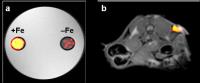
Embryonic stem (ES) cells offer promise for regenerating a
variety of tissue types. One difficult aspect to advancing
this technology is determining the fate of these cells once
they are introduced inside the body. MRI can play an
important role for non-invasive monitoring in patients, but
conventional methods based on iron oxides have limited
specificity. In this study, a novel, highly efficient T1
agent is investigated for labelling mouse ES cells. A
drastic decrease in T1 was obtained and sustained for at
least 24 hours. Viability and proliferation were unaffected,
and labelled ES cells were differentiated into beating
cardiomyocytes.
|
|
0901.
 |
7 |
Testing the Efficacy of GdDO3NI: A Novel Hypoxia-Targeting T1
Contrast Agent
Shubhangi Agarwal1, Carlos Renteria1,
Xiangxing Kong2, Yanqing Tian2, and
Vikram Kodibagkar1
1School of Biological and Health Systems
Engineering, Arizona State University, Tempe, AZ, United
States, 2Biodesign
Institute, Arizona State University, Tempe, AZ, United
States
Tumor hypoxia is a severe problem in oncology, leading to
enhanced metastatic potential and poor response to
therapies. The advent of GdDO3NI—a hypoxia-binding contrast
agent, serves to facilitate therapies by highlighting
hypoxic regions on tumors. Relaxivity studies were performed
and image registration were executed between modalities to
validate the efficacy of this novel contrast agent to
pimonidazole: the gold standard for immunohistochemical
hypoxia imaging. Results showed a strong correlation in
tumor boundaries and hypoxic fractions between modalities.
The hypoxic regions showed lower correlation than expected
however, attributed to the difference in tissue content
resulting from discrepancies in slice thickness.
|
|
0902.
 |
8 |
Tracking transplanted cells with paramagnetic fluorinated
nanoemulsions - Permission Withheld
Alexander A. Kislukhin1, Hongyan Xu1,
Stephen R. Adams2, Kazim H. Narsinh1,
Roger Y. Tsien2,3, and Eric T. Ahrens1
1Radiology, University of California San Diego,
La Jolla, CA, United States, 2Chemistry
& Biochemistry, University of California San Diego, La
Jolla, CA, United States, 3Howard
Hughes Medical Institute, La Jolla, CA, United States
Fluorine-19 magnetic resonance imaging (MRI) probes are used
to label cells for quantitative in vivo tracking of cell
therapies and visualizing inflammation. To reduce the 19F
spin-lattice relaxation time (T1) and enable
rapid imaging and improved cell detection sensitivity, we
prepared metal-binding fluorinated nanoemulsions, and then
metalated them with a panel of transition and lanthanide
ions. Iron(III) tris-β-diketonate PFPE nanoemulsion was
observed to have superior MRI properties (19F T1 as
low as 6 ms). Overall, these agents can yield a multifold
improvement in detection sensitivity over previously
employed 19F
MRI methods to track transplanted cells.
|
|
0903.
 |
9 |
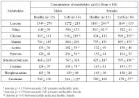
Influence of Gender and Age on the Metabolic Profile of Blood
Plasma in Celiac Disease Using Proton NMR Spectroscopy
Deepti Upadhyay1, Uma Sharma1, Govind
Makharia 2,
Prasenjit Das3, Siddharth Datta Gupta3,
and Naranamangalam R Jagannathan1
1Department of NMR & MRI Facility, All India
Institute of Medical Sciences, New Delhi, India, 2Department
of Gastroenterology and human Nutrition, All India Institute
of Medical Sciences, New Delhi, India, 3Department
of Pathology, All India Institute of Medical Sciences, New
Delhi, India
Metabonomics study on blood plasma of patients with Celiac
disease (CeD) using NMR spectroscopy revealed gender and age
specific variations. The concentrations of acetate,
pyruvate, creatine and glycine were significantly higher in
males with CeD compared to healthy males. While, levels of
β-hydroxybutyrate, glycine and alanine were significantly
elevated in females with CeD than healthy females. These
metabolic differences indicated impairment in both catabolic
and anabolic pathways of carbohydrate metabolism in CeD
patients of both genders, however, fuel preference for
energy requirement was gender specific, fatty acids were
used in males while ketone bodies were preferred in
females.
|
|
0904.
 |
10 |
Specificity and sensitivity of early predictive urinary
metabolic biomarker of radiation injury: a 1H NMR based
metabolomic study
Poonam Rana1, Ritu Tyagi1, Apurva
Watve1, Sujeet Kumar Mewar2, Uma
Sharma2, N. R. Jagannathan2, and
Subash Khushu1
1NMR Research Centre, Institute of Nuclear
Medicine and Allied Sciences (INMAS), DRDO, Delhi, India, 2Department
of NMR, All India Institute of Medical Sciences (AIIMS),
Delhi, India
Increasing radiation exposure is a big threat to population
worldwide. The present study predicts the early predictive
biomarker for radiation injury using 1H
NMR based metabolomics. The animals were exposed to 7.5 Gy
whole body radiation. The variable importance of projection
(VIP) score showed six most significant metabolites having
VIP score of >1. The partial least square discriminant
analysis (PLS-DA) based receiver-operating characteristic
(ROC) curve of all the six metabolites showed taurine with
highest area under curve (AUC) value of 0.996 and with
sensitivity (100%) and specificity (90%). It could be used
as early prognostic biomarker for radiation injury.
|
|
0905.
 |
11 |
Filtered serum-based metabolomics of prostate cancer using 1H
NMR spectroscopy - Permission Withheld
Ashish Gupta1, Deepak Kumar1, Anil
Mandhani2, and Satya Narain Sankhwar3
1metabolomics, Centre of Biomedical Research,
Lucknow, India, 2Urology,
Sanjay Gandhi Post Graduate Institute of Medical Sciences,
Lucknow, India, 3Urology,
King George’s Medical University, Lucknow, India
To address the shortcomings of clinical indexes for the
precise identification of prostate cancer (PC) and
differentiation from benign prostatic hyperplasia (BPH) and
healthy controls (HC), we applied 1H NMR spectroscopy as a
surrogate tactic for probing of PC and BPH. The study
comprises filtered sera from PC (n=75), BPH (n=70) and the
HC (n=65). NMR-measured metabolites and clinical evaluation
data were examined separately using multivariate
discriminant function analysis (DFA) to probe the signature
descriptors for each cohort. DFA reveals that filtered serum
based metabolic profiling can differentiate not only HC from
BPH and PC but also BPH from PC.
|
|
0906.
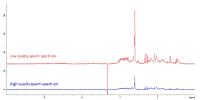 |
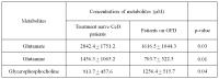
12 |
Increased metabolites in lower quality sperm suggest altered
metabolism and increased cytoplasm compared to higher quality
sperm
Sarah Calvert1, Steven Reynolds2,
Martyn Paley2, and Allan Pacey1
1Department of Oncology & Metabolism, University
of Sheffield, Sheffield, United Kingdom, 2Academic
Unit of Radiology, University of Sheffield, Sheffield,
United Kingdom
Sperm movement is necessary for reproduction and low sperm
motility can impede fertilization. There is a need for
greater understanding of the metabolic processes that drive
sperm motility. In this study, we examined differences in
sperm metabolite profiles between high and low quality sperm
in order to identify possible intracellular biomarkers of
sperm quality and motility. Sperm motility was significantly
different between the two fractions and fell either side of
the WHO lower reference limit. Low quality sperm contained
higher concentrations of choline, methyls, citrate and
lactate, indicative of increased cell membrane and altered
metabolism towards glycolysis.
|
|
0907.
 |
13 |
Assessment of changes in metabolic profile of small intestinal
mucosal biopsy of Celiac Disease patients after gluten-free
diet: An in-vitro Proton NMR Spectroscopy study
Uma Sharma1, Deepti Upadhyay1, Govind
Makharia2, Siddharth Datta Gupta3,
Prasenjit Das3, and Naranamangalam R Jagannathan1
1Department of NMR and MRI Facility, All India
Institute of Medical Sciences, New Delhi, India, 2Department
of Gastroenterology and human Nutrition, All India Institute
of Medical Sciences, New Delhi, India, 3Department
of Pathology, All India Institute of Medical Sciences, New
Delhi, India
Present in vitro proton NMR study demonstrated the metabolic
changes associated with villous abnormalities and its
recovery following gluten free diet (GFD) in patients with
Celiac disease (CeD). The concentration of glutamate and
glutamine was significantly reduced in intestinal mucosa of
CeD patients after GFD, indicating the use of these
metabolites as oxidative fuels for energy generation. The
level of glycerophosphocholine was significantly increased
after GFD in CeD patients suggesting increased turnover of
enterocytes essential for healing of intestinal mucosa in
CeD patients. The results may have implications in
determining the alternative biomarker/s for diagnosis and
treatment management of CeD.
|
|
0908.
 |
14 |
Filtered Serum Metabolomics of Myocardial Ischemia in Unstable
Angina Patients - Permission Withheld
Ashish Gupta1, Keerti Ameta2, Deepak
Ameta3, Rishi Sethi3, Deepak Kumar1,
and Abbas A Mahdi2
1metabolomics, Centre of Biomedical Research,
Lucknow, India, 2Biochemistry,
King George's Medical University, lucknow, India, 3Cardiology,
King George's Medical University, lucknow, India
This study addresses myocardial ischemia in patients
presenting with unstable angina using 1H NMR metabolomics of
filtered serum. The study includes serum samples from 65
unstable angina patients (UA) and 62 healthy controls (HC).
Principal component analysis and orthogonal partial least
square discriminant analysis were applied to generate a
prediction model. Results revealed that five
biomarkers—valine, alanine, glutamine, inosine and
adenine—could differentiate 95% of UA from HC with utmost
sensitivity and specificity. 1H NMR-based filtered serum
metabolic profiling appears to be an assuring, least
invasive and faster way to screen and identify myocardial
ischemia in UA patients.
|
|
0909.
 |
15 |
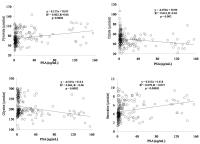
Correlations between cervicovaginal fluid metabolites and
gestational age at delivery
Emmanuel Amabebe1, Steven Reynolds2,
Victoria Stern1, Jennifer Parker3,
Graham Stafford3, Martyn Paley2, and
Dilly Anumba1
1Academic unit of Reproductive and Developmental
Medicine, University of Sheffield, Sheffield, United
Kingdom, 2Academic
unit of Radiology, University of Sheffield, Sheffield,
United Kingdom, 3School
of Dentistry, University of Sheffield, Sheffield, United
Kingdom
Magnetic Resonance Spectroscopy (1H-MRS) can
detect the metabolite profile of the vaginal microniche and
reflects the vaginal bacterial community function. This
study assessed the correlation between 1H-MRS
vaginal fluid metabolites and maternal parameters related to
preterm birth. As expected, vaginal pH, fetal fibronectin,
and cervical length correlated with gestational age at
delivery (GAAD). Vaginal pH also correlated with lactate
and acetate integrals in all study cohorts. Additionally,
lactate and glutamine/glutamate integrals in women studied
at 20-22 gestational weeks; and succinate/lactate ratio in
women studied at 26-28 gestational weeks, correlated
modestly with GAAD. Further correlations between metabolites
were found.
|
|