ISMRM 24th Annual Meeting & Exhibition • 07-13 May 2016 • Singapore

Traditional Poster Session: Neuro

1158 -1176 Normal Brain
1177 -1191 Neuro: Clinical Studies
1192 -1209 Connectomics
1210 -1219 Traumatic Brain Injury
1220 -1254 Neurodegeneration
1255 -1274 Neurodegeneration: Alzheimer's
1275 -1288 Myelin Measurement
1289 -1297 Multiple Sclerosis: Techniques
1298 -1320 Multiple Sclerosis: Studies
1321 -1343 Head & Neck
1344 -1389 Brain Tumours: Pre-Clinical & Clinical Applications
1390 -1399 Psychiatric Disorders: Major Depression
1400 -1415 Pediatrics
1416 -1427 Perfusion in Health & Disease
1428 -1447 Neurovascular Disease & Stroke
1448 -1457 Spine, MRA & Other Clinical Neuro Applications
1458 -1469 Neuroimaging Animal Models


1158.   
Impact of acquisition parameters on cortical thickness and volume derived from Multi-Echo MPRAGE scans
Ross W. Mair1,2, Martin Reuter2,3, and Andre J. van der Kouwe2
1Center for Brain Science, Harvard University, Cambridge, MA, United States, 2A.A. Martinos Center for Biomedical Imaging, Massachusetts General Hospital, Charlestown, MA, United States, 3Department of Mechanical Engineering, Massachusetts Institute of Technology, Cambridge, MA, United States
The multi-echo MPRAGE (MEMPRAGE) sequence was implemented to reduce signal distortion by acquiring at a higher bandwidth and averaging multiple echoes to recover SNR while providing additional T2* information that can enhance cortical segmentation. A rapid 2-minute MEMPRAGE protocol has been implemented for large multi-center studies. Here, we investigate the impact on morphometric results for the cortex by systematically varying all the parameters modified between the rapid 2-minute scan and a conventional 6-minute structural scan. Small but significant differences in cortical thickness and gray matter volume result from a combination of the use of partial fourier acquisition and lowering the spatial resolution to 1.2mm.


1159.   
Quantitative comparison of MP2RAGE skull-stripping strategies
Pavel Falkovskiy1,2,3, Bénédicte Maréchal1,2,3, Shuang Yan4, Zhengyu Jin4, Tianyi Qian5, Kieran O'Brien6,7, Reto Meuli2, Jean-Philippe Thiran2,3, Gunnar Krueger2,3,8, Tobias Kober1,2,3, and Alexis Roche1,2,3
1Advanced Clinical Imaging Technology (HC CMEA SUI DI BM PI), Siemens Healthcare AG, Lausanne, Switzerland, 2Department of Radiology, University Hospital (CHUV), Lausanne, Switzerland, 3LTS5, École Polytechnique Fédérale de Lausanne, Lausanne, Switzerland, 4Department of Radiology, Peking Union Medical College Hospital, Peking Union Medical College, Chinese Academy of Medical Sciences, Beijing, China, People's Republic of, 5MR Collaborations NE Asia, Siemens Helathcare, Beijing, China, People's Republic of, 6Centre for Advanced Imaging, University of Queensland, Brisbane, Australia, 7Siemens Healthcare Pty Ltd., Brisbane, Australia, 8Siemens Medical Solutions USA, Boston, MA, United States
The MP2RAGE pulse sequence exhibits higher grey-matter/white-matter contrast compared to standard MPRAGE acquisitions and provides images with greatly reduced B1 bias. In theory, these qualities of MP2RAGE should lead to more accurate morphometric results. However, a major hindrance to MP2RAGE morphometric processing is the salt-and-pepper noise in the background and cavities. This poses a major problem for the skull-stripping stage of most automated morphometry algorithms. We investigated three skull-stripping strategies using the MorphoBox prototype and FreeSurfer automated-morphometry software packages and compared them to results obtained using the gold-standard MPRAGE contrast.


1160.   
Synthetic Quantitative MRI through Relaxometry Modelling for Improved Brain Segmentation
Martina F Callaghan1, Siawoosh Mohammadi1,2, and Nikolaus Weiskopf1,3
1Wellcome Trust Centre for Neuroimaging, UCL Institute of Neurology, London, United Kingdom, 2Department of Systems Neuroscience, University Medical Center Hamburg-Eppendorf, Hamburg, Germany,3Department of Neurophysics, Max Planck Institute for Human Cognitive and Brain Sciences, Leipzig, Germany
Here we exploit the inter-dependence of quantitative MRI (qMRI) parameters via relaxometry modelling to generate synthetic quantitative maps of magnetisation transfer saturation. The utility of the new concept of synthetic quantitative data is demonstrated by improving image segmentation of deep gray matter structures for neuroimaging applications.


1162.   
DR-TAMAS: Diffeomorphic Registration for Tensor Accurate alignMent of Anatomical Structures
Mustafa Okan Irfanoglu1,2, Amritha Nayak1,2, Jeffrey Jenkins1,2, Elizabeth B Hutchinson1,2, Neda Sadeghi1, Cibu P Thomas1,3, and Carlo Pierpaoli1
1NICHD, NIH, Bethesda, MD, United States, 2Henry Jackson Foundation, Bethesda, MD, United States, 3CNRM, Bethesda, MD, United States
Spatial alignment of diffusion tensor MRI data is of fundamental importance for  voxelwise statistical anaysis and creation of population specific atlases of diffusion MRI metrics. In this work, we propose DR-TAMAS, a novel diffusion tensor imaging registration method which uses a spatially varying metric to achieve accurate alignment not in only in white matter but also in gray matter and CSF filled regions. Our tests indicate that  DR-TAMAS shows excellent overall performance in the entire brain, while being equivalent to the best existing methods in white matter.


1163.   
High resolution anatomical imaging of the human occipital lobe with a large ex-vivo 9.4T RF coil
Shubharthi Sengupta1, Ron Hellenbrand2, René Finger2, Chris Wiggins3, and Alard Roebroeck1
1Dept. of Cognitive Neuroscience, Faculty of Psychology and Neuroscience, Maastricht University, Maastricht, Netherlands, 2Lab Engineering & Instrumentation Department, Maastricht University, Maastricht, Netherlands, 3Scannexus, Maastricht, Netherlands
Small-bore animal scanners or spectroscopy systems have often been used for the investigation of small post-mortem human brain samples. These studies use the high field strengths and strong gradients, but are inherently limited to very small sample sizes. In this abstract, we discuss the acquisition of very high resolution anatomical images (100μm isotropic) of a full occipital sample as large as 80x80x80 mm3, using a customised RF receive coil-array in a large-bore 9.4 T human scanner.


1164.   
Cross-validation of a CSF MRI sequence for calculating brain volume by comparison with brain segmentation methods
Lisa A. van der Kleij1, Jeroen de Bresser1, Esben T. Petersen2, Jeroen Hendrikse1, and Jill B. De Vis1
1Department of Radiology, UMC Utrecht, Utrecht, Netherlands, 2Danish Research Centre for Magnetic Resonance, Centre for Functional and Diagnostic Imaging and Research, Copenhagen University Hospital Hvidovre, Hvidovre, Denmark
We recently introduced a CSF MRI sequence to automatically measure intracranial volume (ICV) and brain parenchymal volume (BPV). This sequence with short imaging time (0:57 min) and fast post processing correlates well with qualitative brain atrophy scores. This study demonstrates that the low resolution and high resolution CSF MRI sequences perform well in the assessment of BPV and ICV, with a precision similar to the conventional brain segmentation methods FSL, Freesurfer and SPM. The CSF MRI sequence showed a good to very good correlation with the conventional segmentation methods for ICV and BPV. 


1165.   
A 7T Human Brain Microstructure Atlas by Minimum Deformation Averaging at 300µm
Andrew L Janke1, Kieran O'Brian2, Steffen Bollmann1, Tobias Kober3, and Markus Barth1
1Centre for Advanced Imaging, University of Queensland, Brisbane, Australia, 2Siemens Healthcare Pty Ltd, Brisbane, Australia, 3Advanced Clinical Imaging Technology, Siemens Healthcare AG, Lausanne, Switzerland
7T provides a method to see detailed image contrast in the human brain; the MP2RAGE sequence allows 500um acquisition resolution.


1166.   
Evaluating the variability of multicenter and longitudinal hippocampal volume measurements.
Stephanie Bogaert1, Michiel de Ruiter2, Sabine Deprez3,4, Ronald Peeters3, Pim Pullens5,6, Frank De Belder6, José Belderbos7, Sanne Schagen2, Dirk De Ruysscher8,9, Stefan Sunaert3,4, and Eric Achten1
1Radiology, Ghent University Hospital, Ghent, Belgium, 2Psychosocial Research and Epidemiology, Netherlands Cancer Institute, Amsterdam, Netherlands, 3Radiology, Leuven University Hospital, Belgium, 4Imaging and Pathology, KU Leuven, Belgium, 5Radiology, University of Antwerp, Belgium, 6Radiology, Antwerp University Hospital, Belgium, 7Radiation Oncology, Netherlands Cancer Institute, Amsterdam, Netherlands,8Radiation Oncology, MAASTRO clinic Maastricht, Netherlands, 9Respiratory Oncology, Maastricht University Medical Center, Netherlands
Longitudinal multicenter MRI studies require stable and comparable measurements. We scanned two subjects in six different scanners (two vendors) at different time points and assessed hippocampal volumes manually and automatically. 

Intrascanner CV was <2.62% for both techniques; Freesurfer is a good alternative for manual delineation for longitudinal studies.

Intervendor variability was sometimes lower than intrascanner variability for the manual technique, which suggests only a modest effect of hardware differences across vendors. Freesurfer results were systematically higher for vendor B compared to A; it is not recommended to compare cross-sectional Freesurfer results between vendors in this multicenter study.


1167.   
Evaluation of 3D T1-weighted imaging at 3 T across scanner vendors and models
Sjoerd B Vos1,2, M Jorge Cardoso1, Marzena Wylezinska-Arridge3, David L Thomas1,3, Enrico De Vita3,4, Marios C Yiannakas5, David Carmichael6, John S Thornton3,4, Olga Ciccarelli5, John S Duncan2,7, and Sebastien Ourselin1
1Translational Imaging Group, University College London, London, United Kingdom, 2MRI Unit, Epilepsy Society, Chalfont St Peter, United Kingdom, 3Neuroradiological Academic Unit, Department of Brain Repair and Rehabilitation, UCL Institute of Neurology, London, United Kingdom, 4Lysholm Department of Neuroradiology, National Hospital for Neurology and Neurosurgery, London, United Kingdom, 5Department of Neuroinflammation, UCL Institute of Neurology, London, United Kingdom, 6Institute of Child Health, University College London, London, United Kingdom, 7Department of Clinical and Experimental Epilepsy, UCL Institute of Neurology, London, United Kingdom
Volumetric analyses of 3D T1-weighted images has become an integral part of the clinical work-up and research studies. Variation between scanners, in both vendors and models, is a major confound in combining imaging-derived biomarkers across sites. In this work, we analyse test-retest data from different days on six 3 T scanners from three vendors to quantify this inter-scanner variability compared to intra-scanner variability. Contrast-to-noise ratios as well as volumetric analyses are performed showing between-scanner variation in total brain volumes – indicating different scanner calibrations – but also tissue-specific differences – possibly arising from different effective contrasts.


1168.   
Basic MR sequence parameters systematically bias automated brain volume estimation
Sven Haller1,2,3,4, Pavel Falkovskiy5,6,7, Reto Meuli6, Jean-Philippe Thiran7, Gunnar Krueger8, Karl-Olof Lovblad1,9, Alexis Roche5,6,7, Tobias Kober5,6,7, and Bénédicte Maréchal5,6,7
1Faculty of Medicine of the University of Geneva, Geneva, Switzerland, 2Affidea Centre de Diagnostique Radiologique de Carouge CDRC, Geneva, Switzerland, 3Department of Surgical Sciences, Radiology, Uppsala University, Uppsala, Sweden, 4Department of Neuroradiology, University Hospital Freiburg, Freiburg, Germany, 5Advanced Clinical Imaging Technology (HC CMEA SUI DI BM PI), Siemens Healthcare AG, Lausanne, Switzerland, 6Department of Radiology, University Hospital (CHUV), Lausanne, Switzerland, 7LTS5, École Polytechnique Fédérale de Lausanne, Lausanne, Switzerland, 8Siemens Medical Solutions USA, Inc., Boston, ME, United States, 9University Hospitals of Geneva, Geneva, Switzerland
Standard MR parameters, notably spatial resolution, contrast and image filtering, systematically bias results of automated brain MRI morphometry by up to 4.8%. This is in the same range as early disease-related brain volume alterations, for example in Alzheimer's disease. Automated brain segmentation software packages should therefore require strict MR parameter selection or include compensatory algorithms to avoid MR-parameter-related bias of brain morphometry results.


1161.   
Evaluation of Pairwise and Groupwise Templates-based Approaches for Automated Segmentation of Structures in Brain MR Images
Subrahmanyam Gorthi1 and Srikrishnan Viswanathan1
1Samsung R&D Institute, Bangalore, India
This work presents a detailed investigation of two multiple-templates based fusion approaches for automated segmentation of structures in the brain MR images: (i) fusion based on direct pairwise registrations between each template and the target image, and (ii) fusion based on an intermediate groupwise template, requiring only a single onsite registration. The key finding from these evaluations is that, if computational time for automated segmentations is a major concern, then groupwise-template based registration followed by fusion is an optimal choice; if time is not a major constraint, then multiple pairwise registrations followed by fusion provides more accurate segmentations.


1169.   
Distortion correction in diffusion weighted imaging of the brain: a quantitative comparison of four correction approaches
Ileana Hancu1, Ek Tsoon Tan1, Luca Marinelli1, Nathan White2, Dominic Holland2, Tim Sprenger3, and Jonathan Sperl3
1GE Global Research Center, Niskayuna, NY, United States, 2University of California San Diego, San Diego, CA, United States, 3GE Global Research Center, Munich, Germany
The performance of four distortion correction algorithms was investigated in a cohort of normal volunteers. While all approaches reduced distortion, it was found that the reversed polarity gradient methods were inherently better than registration or B0-mapping approaches. It was likely that the limited degrees of freedom of the registration approach could not account for localized magnetic field inhomogeneity. The extrapolation of B0 maps in the distorted EPI space introduced errors that decreased the overall performance of the B0-mapping method. 


1170.   
3D FLASH Optimization with Improved Contrast Efficiency and Image Inhomogeneity Correction
Jinghua Wang1, Lili He2, and Zhong-Lin Lu1
1The Ohio State Univeristy, Columbus, OH, United States, 2Center for Perinatal Research, Nationwide Children’s Hospital, Columbus, OH, United States
The 3D FLASH sequence is frequently used in structural imaging of the brain. Tissue contrast inhomogeneity, resulting from inhomogeneous transmit field and receiver sensitivity, significantly affects quantitative structural brain analyses such as classification and quantification of brain tissues in voxel-based morphometry and detection of pathological brain changes in clinical studies.  It is important to optimize the sequence to jointly improve contrast efficiency and image homogeneity.   In this work, we propose optimal imaging parameters and present methods to improve contrast efficiency and reduce or eliminate image inhomogeneity.   


1172.   
Synthetic MRI: an old concept becomes practical
Christina Andica1, Akifumi Hagiwara1,2, Misaki Nakazawa1,3, Masaaki Hori1, Saori Shiota1, Mariko Yoshida1, Kanako Sato1, Yuko Takahashi1, Kanako Kumamaru1, Michimasa Suzuki1, Atsushi Nakanishi1, Kouhei Tsuruta1,3, Ryo Ueda1,3, and Shigeki Aoki1
1Department of Radiology, Juntendo University School of Medicine, Tokyo, Japan, 2Department of Radiology, Graduate School of Medicine, The University of Tokyo, Tokyo, Japan, 3Department of Radiological Sciences, Graduate School of Human Health Sciences, Tokyo Metropolitan University, Tokyo, Japan
Synthetic magnetic resonance imaging (MRI) is a technique which can be used to synthesize contrast-weighted images based on quantification of the longitudinal T1 relaxation, the transverse T2 relaxation, the proton density (PD), and the amplitude of the local radio frequency B1 field. Synthetic MRI images were useful in the evaluation of brain disorders. With Synthetic MRI, echo time (TE), repetition time (TR), and inversion time (TI) of the contrast-weighted image can be freely adjusted retrospectively to optimize image quality. Limitation of synthetic MRI is the partial volume effect.


1171.   
The Neural Basis of Visual Field Asymmetry in Human Visual System by Functional MRI
Caitlin O'Connell1, Leon Ho2,3, Matthew Murphy2, Yolandi van der Merwe1,2, Ian Conner1,2, Gadi Wollstein1,2, Joel Schuman1,2, Rakie Cham1, and Kevin Chan1,2
1Department of Bioengineering, University of Pittsburgh, Pittsburgh, PA, United States, 2UPMC Eye Center, Eye and Ear Institute, Ophthalmology and Visual Science Research Center, Department of Ophthalmology, University of Pittsburgh, Pittsburgh, PA, United States, 3Department of Electrical and Electronic Engineering, The University of Hong Kong, Pokfulam, Hong Kong
Human visual performance has been observed to exhibit superiority in the lower visual field and horizontal meridian compared to the upper visual field and vertical meridian, respectively, in response to many classes of stimuli, but the underlying neural mechanisms remain unclear. This study determines if processing of visual information is dependent on the location of stimuli in the visual field using functional MRI. The results show stronger brain responses and larger activation volumes upon flickering visual stimulation to the lower hemifield compared to upper hemifield, while only the activation size differed between visual presentations to the horizontal and vertical meridians.


1173.   
Towards Higher Spatial Resolution Echo-Planar-Imaging With A Compact Head 3T System
Ek T Tan1, Seung-Kyun Lee1, Paul Weavers2, Matthew Middione3, Matt A Bernstein2, John Huston2, and Thomas KF Foo1
1GE Global Research, Niskayuna, NY, United States, 2Mayo Clinic, Rochester, MN, United States, 3GE Healthcare, Menlo Park, CA, United States
It is challenging to increase spatial resolution (≤1.5 mm) in whole-brain, single-shot echo-planar-imaging (ss-EPI) on conventional whole-body systems due to EPI distortion and limited SNR. A compact head 3T system with an asymmetric head gradient coil capable of high gradient amplitude and 3.5 times the slew rate of whole-body systems can enable ss-EPI acquisition with high spatial resolution and reduced spatial distortion, simultaneously. This work compares spin-echo and gradient-recalled-echo ss-EPI between the compact and whole-body systems, showing substantially reduced distortion and signal dropout, and shorter echo-times. Results with the high performance gradient were also demonstrated in multi-band-accelerated, high b-value diffusion-imaging.


1174.   
Susceptibility Weighted Imaging in Different Regions of Human Brain at 7T
Yeong-Jae Jeon1,2, Sang-Woo Kim1,2, Joo-Yeon Kim1, Young-Seok Park3, and Hyeon-Man Baek1,2
1Bio-Imaging Research Team, Korea Basic Science Institute, Ochang, Korea, Republic of, 2Bio-Analytical Science, University of Science & Technology, Daejeon, Korea, Republic of, 3College of Medicine, Chungbuk National University, Cheongju-si, Korea, Republic of
The purpose of this study was to investigate contrast enhancement difference of SWI from human brain regions at 7T, and to compare contrast enhancement between cortical, anterior septal, and hippocampal veins. Five healthy volunteers (mean±SD, 24.4±1.67 years) participating in this study were scanned on 7T. The observation in this work was the significant difference of contrast enhancement of cortical and other veins, and no significant contrast enhancement difference between anterior septal and hippocampal veins. In conclusion, contrast enhancement of human brain at 7T depends on the regions giving higher cortical vein contrast with respect to anterior septal and hippocampal veins. 


1175.   
The cerebrovascular response to a single session of exercise
Jessica Steventon1, Catherine Foster1, Daniel Helme2, Monica Busse3, and Kevin Murphy1
1CUBRIC, Cardiff University, Cardiff, United Kingdom, 2School of Medicine, Cardiff University, Cardiff, United Kingdom, 3School of Healthcare Sciences, Cardiff University, Cardiff, United Kingdom
Here we examine the acute effects of a single exercise session on cerebrovasculature using a multi-TI arterial spin labelling (ASL) sequence to measure cerebral blood flow (CBF), and a dual-echo ASL sequence with hypercapnia to measure cerebrovascular reactivity (CVR). We show that contrary to previous smaller studies, 20-minutes of aerobic exercise does not affect CBF or CVR in the 60-minute period after exercise. Despite this, changes in CBF after exercise were related to individually-determined systemic physiological changes associated with exercise intensity, informing on moderators of cerebral autoregulation.


1176.   
Vessel-size dependent response of human cerebral arteries to hyperoxia
Esther AH Warnert1, Ian D Driver1, Joseph Whittaker1, and Kevin Murphy1
1Cardiff University Brain Research Imaging Centre, Cardiff University, Cardiff, United Kingdom
Due to the potential of using hyperoxia as a treatment for cerebral ischemic diseases, including stroke, it is important to fully understand the effects of hyperoxia on the cerebrovasculature. Although it is known that breathing of 100% O2 leads to a decrease in cerebral blood flow, it is unclear where along the cerebral arterial tree vasoconstriction occurs. Here we show that, while there is expected constriction of the large arteries, smaller and more distal arteries actually show vasodilation upon hyperoxia.

1177.   
An Investigation of Lateral Geniculate Nucleus(LGN) Volume in Patients with Glaucoma using 7T MRI
Hye Jin Jeong1, Jong Yeon Lee2, Jong Hwan Lee2, Yu Jeong Kim2, Eung Yeop Kim3, Young Yeon Kim4, Zang-Hee Cho1, and Young-Bo Kim1
1Neuroscience Research Institute, Incheon, Korea, Republic of, 2Gil Hospital, Department of Ophthalmology, Incheon, Korea, Republic of, 3Gil Hospital, Department of Radiology, Incheon, Korea, Republic of, 4Korea University College of Medicine, Department of Ophthalmology, Seoul, Korea, Republic of
To investigate lateral geniculate nucleus (LGN) volume of glaucoma patients compared with age-matched normal controls using ultra-high field 7.0-T magnetic resonance imaging (MRI). On high-resolution 7.0-T MRI, LGN volumes in POAG patients are significantly smaller than those of healthy subjects. Furthermore, in patients, LGN volume was found to be significantly correlated with ganglion cell layer and inner plexus layer (GC–IPL) thickness of the contralateral eye.


1178.   
Regional Brain Iron Mapping in Patients with Obstructive Sleep Apnea Syndrome
Sudhakar Tummala1, Daniel W Kang2, Bumhee Park1, Ruchi Vig1, Mary A Woo3, Ronald M Harper4,5, and Rajesh Kumar1,5,6,7
1Anesthesiology, University of California at Los Angeles, Los Angeles, CA, United States, 2Medicine, University of California at Los Angeles, Los Angeles, CA, United States, 3UCLA School of Nursing, Los Angeles, CA, United States, 4Neurobiology, University of California at Los Angeles, Los Angeles, CA, United States, 5Brain Research Institute, University of California at Los Angeles, Los Angeles, CA, United States, 6Radiological Sciences, University of California at Los Angeles, Los Angeles, CA, United States, 7Bioengineering, University of California at Los Angeles, Los Angeles, CA, United States
OSA subjects show brain injury in multiple areas, which may contribute to accumulation of iron in those sites. Deposition of iron in OSA subjects is unclear. We examined regional iron deposition using T2*-relaxometry procedures; R2* values were significantly increased in insular, parietal, cingulate and cingulum bundle, temporal, and cerebellar areas. The increased iron depositions in OSA subjects may result from neural and white matter injury, including myelin and glial dysfunction, with iron potentially accelerating tissue degeneration. These data suggest that interfering with the iron action may reduce the exacerbation of injury in OSA.


1179.   
Measurement of Brain Asymmetry on 3D Magnetic Resonance (MR) Images Obtained for 16 Subjects with Situs Inversus
X. Li1, Neil Roberts1, M. Perrins1, and G. Vingerhoets2
1University of Edinburgh, Edinburgh, United Kingdom, 2Department of Experimental Psychology, Ghent University, Ghent, Belgium
The human brain is structurally asymmetric and typically described as if it has been subject to a rotational moment about the vertical axis of the body, the so-called “Yakovlevian Torque”. In subjects with situs inversus totalis (SI) the internal organs of the body are transposed and it has been obvious to question whether in these subjects brain torque is also reversed? We recruited 16 subjects with SI and 16 age, sex and education matched controls (SS) and applied state of the art image analysis techniques to investigate the extent to which brain asymmetry is reversed on 3D MR images in these subjects. Analysis of the frontal and occipital petalia has confirmed previous reports of significant reversal of the latter but not the former on average in SI, and has also shown that reversed asymmetry is not present in all individuals with SI. 


1180.   
QUANTITATIVE MEASURES OF BRAIN CHANGES IN CHILDREN WHO DO JUDO ON MRI
Tina Seah1, Tang Phua Hwee1, Toh Zhe Han1, Gu Qing Long2, and Wong Weng Hang2
1Diagnostic Imaging, KK women's and children's hospital, singapore, Singapore, 2singapore, Singapore
Quantitative study of the brain changes between young judo athletes and normal children who do not do judo, using diffusion tensor imaging (DTI) and magnetic resonance spectroscopy (MRS). Study shows significant increased fractional anisotropy (FA) of the major white matter tracks (corpus callosum, corticospinal tracks, superior longitudinal fasciculus) with slight increase in N-acetylaspartate to Creatine (NAA/Cr) ratio in the parietal white matter bilaterally.  The increased FA and NAA/Cr ratios support structural changes involving grey matter volumes in the cortical cerebral grey matter described in published literatures on athletes.


1181.   
Visualization of the pituitary gland region’s perforating branch artery before transsphenoidal surgery using a high-spatial-resolution three-dimensional fast spin echo sequence
Keiya Hirata1, Osamu Tachibana2, Chihiro Watari1, Tatsunori Kuroda1, Nanako Miyamoto1, Saeko Tomida1, Masaru Takahashi1, Tomokazu Oku1, Shigeo Miyazaki1, Masahiro Kawashima1, Naoko Tsuchiya3, Ichirou Toyota3, Mariko Doai3, and Hisao Tonami3
1Division of radiology, Kanazawa medical university, Uchinada, Japan, 2Department of neurosurgery, Kanazawa medical university, Uchinada, Japan, 3Department of radiology, Kanazawa medical university, Uchinada, Japan
Transsphenoidal surgery is performed in the surgery of the pituitary region. The perforating branch which performs a nutrient of optic nerve and a mamillary body is present in the cistern around the pituitary gland. We can reduce complications of the surgery if we can identify a perforating branch before surgery.We try to visualize the perforating branch as black blood MRA using the high spatial resolution 3D-FSE sequence. We examined the optimal conditions at phantoms and normal volunteers. The optimal condition was a combination of TR2400msec/2shots, and the imaging time was 20 min and 45 s.


1182.   
Subtractionless MR Angiography of the Neck Using Dixon-based MRI
Ivan E Dimitrov1,2, Qing Yuan3, Sepand Salehian3, Gaurav Khatri3, Marco Pinho3, and Ivan Pedrosa2,3
1Philips Medical Systems, Cleveland, OH, United States, 2Advanced Imaging Research Center, UT Southwestern Medical Center, Dallas, TX, United States, 3Radiology, UT Southwestern Medical Center, Dallas, TX, United States
We investigated the ability of dynamic contrast-enhanced (DCE) dual-echo multi-peak Dixon-based imaging to generate MR angiography of the neck, without the need of subtraction thus eliminating the possibility of errors due to motion. In six patients with multiple sclerosis, DCE MRA based on subtraction of pre-contrast from post-contrast images was compared with MRA generated solely from the post-contrast data where fat suppression was achieved using Dixon-based water imaging. While high levels of vessel-to-background contrast was observed in both methods, the subtractionless DIXON-MRA resulted in higher overall contrast for the aortic arch, the brachiocephalic arteries, and the carotid bifurcation. 


1183.   
Are Negative BOLD Responses Induced by Acupuncture Associated with Neural Inhibitive Effects? : an MRS & BOLD Functional MRI Study
Jiliang Fang1, Yanping Zhao1, Sinyeob Ahn2, Guiyong Liu1, Caixia Fu3, Jin Yang1, Xiaoling Wang1, Bo Hou4, Feng Feng4, and Tianyi Qian5
1Radiology, Guang An Men Hospital, China Academy of Chinese Medical Sciences, Beijing, China, People's Republic of, 2MR Collaboration, Siemens Healthcare, San Francisco, CA, United States, 3APPL, Siemens Shenzhen Magnetic Resonance Ltd., Shenzhen, China, People's Republic of, 4Radiology, Peking Union Medical College Hospital, Beijing, China, People's Republic of, 5MR Collaborations NE Asia, Siemens Healthcare, Beijing, China, People's Republic of
This study investigates the neurotransmitter basis of the negative response in the medial prefrontal cortex induced by acupuncture stimulus. The MEGA-PRESS 1H-MRS sequence was used to detect the gamma-aminobutyric acid (GABA) and Glutamine (Glu) concentration before and after acupuncture stimulation in normal subjects. The result showed that the GABA concentrations were decreased, while the Glu/Gln concentrations were increased. The task-fMRI data acquired during acupuncture stimulation showed deactivation in the same area. These results suggest that the deactivated BOLD response induced by acupuncture might be associated with the neural inhibition effects.


1184.   
MR and Proton MR Spectroscopy Findings of a Pediatric Case with Solitary Intracranial Rosai-Dorfman Disease in the Posterior Fossa
Sehnaz Tezcan1, Muhtesem Agildere1, Taner Sezer2, Ozge Ozturk1, and Aydin Sav3
1Radiology, Baskent University Hospital, Ankara, Turkey, 2Pediatrics, Division of Neurology, Baskent University Hospital, Ankara, Turkey, 3Pathology, Acibadem Maslak Hospital, Istanbul, Turkey
Rosai-Dorfman disease (RDD) is a histioproliferative disorder, rarely affects central nervous system.  A 5-year old boy presented with ptosis, diplopia. MR revealed enhancing mass in the cerebellar pedincle and pons. MR Spectroscopy (MRS) of the lesion showed increased Choline/N-acetyl aspartate ratio and lactate peak.. Histopathology was compatible with RDD. Although intracranial RDD generally presents as dural based lesions and supratentorial in location, intraparencymal lesions may be seen.. In this case report a rare form of RDD, posterior fossa parenchyma involvement presented with particular interest to brain MR, MRS and diffusion findings.


1185.   
Cervical spondyloarthropathy due to the dialysis-related amyloidosis: magnetic resonance imaging findings
Hale Turnaoglu1, Kemal Murat Haberal1, Ozlem Isiksacan Ozen2, and Ahmet Muhtesem Agildere1
1Radiology, Baskent University, Faculty of Medicine, Ankara, Turkey, 2Pathology, Baskent University, Faculty of Medicine, Ankara, Turkey
Dialysis-related amyloidosis that occurs secondarily to the deposition of amyloid fibrils containing beta-2-microglobulin, is a type of amyloidosis affecting patients undergoing long-term hemodialysis. It involves the osteoarticular system predominantly. Destructive spondyloarthropathy, which frequently involves the cervical spine, have been reported only sporadically. CT is the best modality for detecting osseous erosion or small areas of osteolysis in cortical bone. MRI shows the extent and distribution of osseous, articular, spinal cord and soft-tissue involvement and indicates amyloid deposits in the intervertebral disk, synovium of apophyseal joints, and ligaments. The gold standart of the diagnosis is the histological identification of beta-2-microglobulin.


1186.   
Increased Glutamate in Frontal Lobe of HIV Infected Patients with CNS involvement: 3T MRS Study
Virendra Kumar1, Devender Bairwa2, Surabhi Vyas3, Achal Srivastava4, Bimal K Das5, R. M. Pandey6, S. K. Sharma2, Sanjeev Sinha2, and N. R. Jagannathan1
1Department of NMR, All India Institute of Medical Sciences, New Delhi, India, 2Department of Medicine, All India Institute of Medical Sciences, New Delhi, India, 3Department of Radiology, All India Institute of Medical Sciences, New Delhi, India, 4Department of Neurology, All India Institute of Medical Sciences, New Delhi, India, 5Department of Microbiology, All India Institute of Medical Sciences, New Delhi, India,6Department of Biostatistics, All India Institute of Medical Sciences, New Delhi, India
We investigated the effect of HIV infection status on brain metabolites in HIV patients with CNS involvement and asymptomatic HIV patients. 71 subjects were studied including HIV patients with CNS involvement, asymptomatic HIV patients and healthy controls. Single voxel MRS was carried out at 3.0 Tesla MR scanner and metabolite concentrations were determined from three brain regions; left frontal, left basal ganglia and lesion in case of HIV patients with CNS involvement. Glx (Glu+Gln) and creatine were significantly increased in HIV patients in frontal region compared to healthy controls. The concentration of N-acetylaspartate in basal ganglia showed a significant decrease in HIV patients.


1187.   
Regional Brain Myelin Mapping in Patients with Obstructive Sleep Apnea
Sudhakar Tummala1, Bumhee Park1, Ruchi Vig1, Mary A Woo2, Daniel W Kang3, Ronald M Harper4,5, and Rajesh Kumar1,5,6,7
1Anesthesiology, University of California at Los Angeles, Los Angeles, CA, United States, 2UCLA School of Nursing, Los Angeles, CA, United States, 3Medicine, University of California at Los Angeles, Los Angeles, CA, United States, 4Neurobiology, University of California at Los Angeles, Los Angeles, CA, United States, 5Brain Research Institute, University of California at Los Angeles, Los Angeles, CA, United States, 6Radiological Sciences, University of California at Los Angeles, Los Angeles, CA, United States, 7Bioengineering, University of California at Los Angeles, Los Angeles, CA, United States
Obstructive sleep apnea (OSA) patients show gray matter injury in multiple brain areas based on various MRI techniques, which can accompany subcortical and white matter myelin integrity loss in the condition. However, the extent of regional myelin changes in OSA is unclear. We examined regional myelin integrity in newly-diagnosed, treatment-naive OSA patients, and found decreased values, probably resulting from hypoxic/ischemic processes, in critical autonomic, cognitive, respiratory, and mood control sites, functions that are deficient in the condition. These findings show that myelin mapping, based on the ratio of T1- and T2-weighted images, is useful in assessing regional myelin alterations.


1188.   
Investigation of 1H MRS changes in the brain of osteoarthritis patients in relation to perceived pain
Franklyn Arron Howe1, Olakunbi Harrison2, Thomas Richard Barrick1, and Nidhi Sofat2
1Neuroscience Research Centre, St George's, University of London, London, United Kingdom, 2Institute for Infection and Immunity, St George's, University of London, London, United Kingdom
Chronic pain from osteoarthritis (OA) may be aggravated by “central sensitisation”, whereby pain-processing pathways become sensitised by inflammatory and degenerative disease processes. 1H MRS was used to investigate biochemical changes in pain processing brain areas of hand OA patients (n=32) compared to controls (n=14). There were no differences between controls and patients in the anterior cingulate gyrus, nor age related changes. In the insula cortex mI/Glx correlated with the pain score (R2 = 0.52, p = 0.018) after co-varying for age. High mI/Glx in the insula cortex was associated with high pain and may reflect inflammatory effects or neurological changes.


1189.   
Brain bioenergetics as markers of vigilance failure in obstructive sleep apnoea
Caroline D Rae1, Haider Naqvi2, Andrew Vakulin2,3, Angela D'Rozario2, Michael Green1, Hannah Openshaw2, Keith Wong2,4, Jong-Won Kim5, Delwyn J Bartlett6, Doug McEvoy7, and Ronald R Grunstein6
1The University of New South Wales, Randwick, Australia, 2NHMRC Centers of Research Excellence, CIRUS and NeuroSleep, Woolcock Institute of Medical Research, The University of Sydney, Sydney, Australia, 33. Adelaide Institute for Sleep Health: A Flinders Centre of Research Excellence, School of Medicine, Flinders University, Adelaide, Australia, 4Sydney Medical School, The University of Sydney, Sydney, Australia, 5School of Physics, The University of Sydney, Sydney, Australia, 6NeuroSleep and Woolcock Institute of Medical Research, The University of Sydney, Sydney, Australia, 7Adelaide Institute for Sleep Health: A Flinders Centre of Research Excellence, School of Medicine, Flinders University, Adelaide, Australia
Here, we investigated the potential for MRS/MRI markers to differentiate between phenotypes of obstructive sleep apnea patients who are vulnerable, versus resistant to vigilance failure, an indicator of driving impairment and accident risk. Vulnerable patients (N = 15) and resistant patients (N = 30) were differentiated on the basis of left orbito-frontal glutamate and aspartate and also anterior cingulate glutathione levels. There was a trend towards lower orbitofrontal creatine levels in vulnerable OSA subjects, but no group differences in brain volumes.


1190.   
A Diffusional Kurtosis Imaging Study of the White Matter Abnormalities in Type-2 Diabetic Brain
YING XIONG1,2, Shun Zhang1, Qiang Zhang3, and Wenzhen Zhu1
1Radiology Department, Tongji Hospital, Tongji Medical College, Huazhong University of Science and Technology, Wuhan, China, People's Republic of, 2Certer for MR Research, University of Illinois at Chicago, Chicago, IL, United States, 3Neurology Department, Tongji Hospital, Tongji Medical College, Huazhong University of Science and Technology, Wuhan, China, People's Republic of
This study aims at investigating brain microstructural changes in white-matter (WM) of type 2 diabetes mellitus (T2DM) patients using diffusional kurtosis imaging (DKI), and making a comparison with diffusion tensor metrics. Thirty T2DM patients and 28 health controls were recruited and imaged on a 3 Tesla scanner. It was found that in the whole-brain and atlas-based analysis, mean kurtosis (MK) detected more regions with WM alterations than fractional anisotropy (FA), especially in some regions including crossing fibers. DKI can complement conventional DTI and provide more information to characterize and pinpoint brain microstructural changes in WM of T2DM patients.


1191.   
Hematopoietic stem cell transplantation in late-onset Krabbe disease halts demyelination and axonal loss: A 4 year longitudinal case study
Cornelia Laule1,2, Elham Shahinfard1, Burkhard Maedler1, Jing Zhang1, Irene Vavasour1, Ritu Aul3, David K.B. Li1,4, Alex L. MacKay1,5, and Sandra Sirrs6
1Radiology, University of British Columbia, Vancouver, BC, Canada, 2Pathology & Laboratory Medicine, University of British Columbia, Vancouver, BC, Canada, 3Medical Genetics, North York General Hospital, Toronto, ON, Canada, 4Medicine (Neurology), University of British Columbia, Vancouver, BC, Canada, 5Physics & Astronomy, University of British Columbia, Vancouver, BC, Canada, 6Medicine (Endocrinology), University of British Columbia, Vancouver, BC, Canada
Late-onset Krabbe disease is a very rare demyelinating leukodystrophy. We found hematopoietic stem cell transplantation in Krabbe disease halts demyelination and axonal loss up to 4 years post-allograft. Abnormalities far beyond those visible on conventional imaging were detected, suggesting a global pathological process occurs in Krabbe disease with adult onset etiology, with myelin being more affected than axons. However, the degree of Krabbe abnormality did not increase over time for any advanced MR metrics, which supports hematopoietic stem cell transplantation as an effective treatment strategy for stopping progression associated with late-onset Krabbe disease.

1192.   
Phase of quasi-periodic patterns in the brain predicts performance on psychomotor vigilance task in humans
Anzar Abbas1, Waqas Majeed2, Garth Thompson3, and Shella Keilholz4
1Neuroscience Program, Emory University, Atlanta, GA, United States, 2School of Science and Engineering, Lahore University of Management Sciences, Lahore, Pakistan, 3Radiology and Biomedical Imaging, Yale University, New Haven, CT, United States, 4Biomedical Engineering, Emory University and Georgia Institute of Technology, Atlanta, GA, United States
Functional organization of brain networks plays an important role in behavior. Analysis of the dynamics of two functional networks – the default mode (DMN) and task positive (TPN) networks – has shown a dependency of task performance on relative network activation. Fluctuations between these two networks have been seen to occur in humans in a continuous, quasi-periodic fashion. However, the nature of these quasi-periodic patterns (QPPs) and their effect on behavior is not well understood. We show that QPPs do not differ between resting state and task-based scans and that the phase of these QPPs can serve as predictors of performance on the psychomotor vigilance task (PVT).


1193.   
Reduced low frequency band power in resting state activity predicts symptom severity in mild traumatic brain injury (mTBI)
Radhika Madhavan1, Suresh E Joel1, Sumit Niogi2, John A Tsiouris2, Luca Marinelli3, and Teena Shetty2
1GE Global Research, Bangalore, India, 2Hospital for special surgery, New York, NY, United States, 3GE Global Research, Niskayuna, NY, United States
mTBI diagnosis is controversial since although the brain appears normal on CT/MRI scans,  a significant proportion of patients display persistent cognitive impairments up to 6 months post-injury. We recorded rs-fMRI in mTBI patients longitudinally over 3 months, to track functional changes in the brain as patients recovered. Symptom scores were negatively correlated with fractional power in the low-frequency band (0.01-0.1 Hz) of rs-fMRI, and this correlation was most significant in the higher visual, salience and sensorimotor networks. We suggest that low frequency power of rs-fMRI can be used as a biomarker for predicting severity of cognitive impairment in brain injury.


1194.   
Modular changes in functional connectivity associated with clinical symptoms in mild traumatic brain injury (mTBI)
Radhika Madhavan1, Hariharan Ravishankar1, Suresh E Joel1, Rakesh Mullick1, Sumit Niogi2, John A Tsiouris2, Luca Marinelli3, and Teena Shetty4
1GE Global Research, Bangalore, India, 2Weill Cornell Medical College, New York, NY, United States, 3GE Global Research, Niskayuna, NY, United States, 4Hospital for Special Surgery, New York City, NY, United States
Although most mTBI patients recover by 3-6 months, they suffer serious short and long term effects. Additionally, multiple mTBIs may have serious long-term consequences. Here, we correlated brain network-level connectivity features derived from resting state functional magnetic resonance imaging (rs-fMRI) with clinical symptoms, in order to identify neuroimaging biomarkers of mTBI as patients recover over 3 months. We used a machine-learning framework to select connectivity features associated with symptoms and identified functional regions with altered connectivity. These modular network-level features can be used as diagnostic tools for predicting disease severity and recovery profiles.


1195.   
Disruption of the Relationship between Default Mode Network Connectivity and Task-related Deactivation in Patients with Mild Traumatic Brain Injury
David Yen-Ting Chen1,2, Yi-Tien Li1,3, Chien-Yuan Eddy Lin4,5, Chi-Jeng Chen1, and Ying-Chi Tseng1
1Department of Radiology, Shuang-Ho Hospital, Taipei Medical University, New Taipei City, Taiwan, 2Brain and Consciousness Research Center, Taipei Medical University, Taipei City, Taiwan, 3Institute of Biomedical Engineering, National Taiwan University, Taipei City, Taiwan, 4GE Healthcare, Taipei City, Taiwan, 5MR Advanced Application and Research Center, GE Healthcare, Beijing City, China, People's Republic of
Mild traumatic brain injury (MTBI) may cause disruption of default mode network (DMN) in patients. We found differences in both resting state DMN connectivity and task-related deactivation between MTBI patients and healthy controls. Although no significant within-network difference was found in the DMN connectivity between patients and controls, there was increased extra-network connection to the left inferior frontal gyrus in the patients. Significantly more profound task-related deactivation was found in the patients, especially in bilateral IPCs. Increased task-related deactivation may imply the patients need more attention on performing the WM tasks. Furthermore, significant correlation between resting state connectivity and task-related deactivation of DMN was found in healthy controls and this rest-task correlation was disrupted in the patients.


1196.   
mTBI symptom severity is associated with functional connectivity of specific networks
Suresh Emmanuel Joel1, Radhika Madhavan1, Rakesh Mullick1, Sumit Niogi2, John A Tsiouris2, Luca Marinelli3, and Teena Shetty4
1Diagnostic Imaging and Biomedical Technologies, General Electric Global Research, Bangalore, India, 2Weill Cornell Medical College, New York, NY, United States, 3Diagnostic Imaging and Biomedical Technologies, General Electric Global Research, Niskayuna, NY, United States, 4Hospital for Special Surgery, New York, NY, United States
Patients who suffer from mild traumatic brain injury (mTBI) have cognitive and behavioral deficits though MR and CT appear normal. Functional neuroimaging has high promise to provide biomarkers which may enable better prognosis and therapy of mTBI. The work here shows in a large sample (78 mTBI patients and 26 controls in 3 sessions spanning 3 months from injury), significant correlation between functional connectivity in visual, motor and default mode networks and self-reported symptom scores. Given this association, functional connectivity stands to be an important contributor to predict mTBI outcome.


1197.   
Resting-state functional connectivity reveals age-related difference in Valproate-induced rat autism model
Hsin-Yi Lai1, Hui-Ching Lin2,3, Hui-Yu Wang4, Jun-Cheng Weng5, Han-Fang Wu2, and You-Yin Chen4
1Interdisciplinary Institute of Neuroscience and Technology (ZIINT), Zhejiang University, Hangzhou City, China, People's Republic of, 2Department and Institute of Physiology, National Yang-Ming University, Taipei, Taiwan, 3Brain Research Center, National Yang Ming University, Taipei, Taiwan, 4Department of Biomedical Engineering, National Yang-Ming University, Taipei, Taiwan, 5Department of Medical Imaging and Radiological Sciences, Chung Shan Medical University, Taichung, Taiwan
 This study demonstrates changes of functional connectivity in motor related brain areas and it is age-related different in Valproate-induced rat autism model. Our results indicate that the motor cortex and striatum may be crucial areas for treatment and evolution of ASD. rsfMRI has potential to explore functional connectivity in the brain and monitor functional plasticity changes in a specific neuroanatomical pathway in vivo. 


1198.   
The heritability of structural brain network
Xiaopei Xu1, Pek-Lan Khong1, Nichol M. L. Wong2,3, Rainbow T. H. Ho4, C. Mary Schooling5, Pui-sze Yeung6, Tatia M. C. Lee2,3,7,8, and Edward S Hui1
1Department of Diagnostic Radiology, The University of Hong Kong, Hong Kong, Hong Kong, 2Laboratory of Neuropsychology, The University of Hong Kong, Hong Kong, Hong Kong, 3Laboratory of Social Cognitive Affective Neuroscience, The University of Hong Kong, Hong Kong, Hong Kong, 4Department of Social Work and Social Administration, The University of Hong Kong, Hong Kong, Hong Kong, 5School of Public Health, The University of Hong Kong, Hong Kong, Hong Kong, 6Faculty of Education, The University of Hong Kong, Hong Kong, Hong Kong, 7Institute of Clinical Neuropsychology, The University of Hong Kong, Hong Kong, Hong Kong, 8The State Key Laboratory of Brain and Cognitive Science, The University of Hong Kong, Hong Kong, Hong Kong
To better understand the importance of education, genetic, and environmental influences on brain structural connectivity, we used DTI-based tractography and brain network analysis to investigate the thereof in twin pairs.  The correlation between network properties and education was also studied in both twin and non-twin participants. We showed significant correlations between twin pairs for the topology of brain network and the nodal characteristics of brain hubs. Nodal characteristics of hubs were also significantly correlated with education level. These findings suggested that brain topology and cognitive capacity are heritable, and brain network analysis is of potential value in intelligence assessment.


1199.   
Impaired small-world structural brain network in patients with neuropsychiatric systemic lupus erythematosus
Xiaopei Xu1, Henry KF Mak1, MY Mok2, CS Lau2, and Edward S Hui1
1Department of Diagnostic Radiology, The University of Hong Kong, Hong Kong, Hong Kong, 2Department of Medicine, The University of Hong Kong, Hong Kong, Hong Kong
To better understand the underlying mechanisms for various neuropsychiatric symptoms in patients with neuropsychiatric systemic lupus erythematosus (NPSLE), we used DTI-based tractography and graph theory approaches to investigate the change in the global configuration and nodal characteristics of the structural brain network in NPSLE and SLE patients. Our results showed impaired small-world structural network and diminished role of several brain regions as hubs in NPSLE patients, indicating the disruption of brain architecture underlying multiple neuropsychiatric manifestations present in NPSLE. Our results demonstrated that brain network analysis is a reliable method to study systemic disease like NPSLE. 


1200.   
Tractography Study of Brain Asymmetries in a Genetic Mouse Model
Alexandra Petiet1,2, Gonçalo C Vilhais-Neto3,4, Daniel Garcia-Lorenzo1, Stéphane Lehéricy1,2, and Olivier Pourquié3,4,5,6,7
1Center for Neuroimaging Research, Brain and Spine Institute, Paris, France, 2UPMC/Inserm UMRS1127 / CNRS UMR7225, Paris, France, 3Institut de Génétique et de Biologie Moléculaire et Cellulaire, Illkirch, France,4Stowers Institute for Medical Research, Kansas City, MO, United States, 5Howard Hughes Medical Institute, Kansas City, MO, United States, 6Department of Anatomy and Cell Biology, University of Kansas Medical Center, Kansas City, MO, United States, 7Department of Genetics, Harvard Medical School and Department of Pathology, Brigham and Woman’s Hospital, Boston, MA, United States
While humans show clear preference for right hand usage (90%), normal mice show consistent right or left paw usage (50%). We used a Rere-deficient mouse model (Rere+/om) that shows clear right paw usage preference (80%) compared to wild-type (WT) mice (40%) to evaluate structural connectivity changes in the cortico-spinal tract (CST) using diffusion-based tractography. Our results showed significantly reduced and more asymmetric FA along the CST of the dominant hemisphere in the dextral mutant group compared to the WT group. These results show Rere-dependent structural connectivity changes in the brain that could be clinically relevant to human pathologies.


1201.   
Thalamocortical network alteration in Children and Adolescents with Idiopathic Generalized Epilepsy
Tijiang Zhang1, Wuchao Li1, Quanzhong Zhou1, Ganjun Song1, Cong Tian1, Zhen Zeng1, and Xingyu Wang1
1Department of Radiology, Affiliated hospital of Zunyi Medical College, Zunyi, China, People's Republic of
The aim of this study is to investigate FC alterations of thalamocortical network using resting-state fMRI,and correlation FC alterations with Intelligence Quotient (IQ) . 19 patients and 19 healthy volunteers took part in this research. The thalamocortical FC seeding at the left thalamus in the IGE patients showed a significant increase in left inferior temporal gyrus and right supramarginal gyrus, but decrease in left anterior cingulate, bilateral posterior cingutate, bilateral dorsolateral frontal gyrus, whereas, seeding the right thalamus as seed showed increase in right cerebellum, right supramarginal gyrus, but decrease in bilateral dorsolateral frontal gyrus, bilateral PCC and right supramarginal gyrus. Correlation analysis revealed that IQ positively correlated with FC strength between thalamus and left anterior cingulate, left dorsolateral superior frontal gyrus. The alteration of  FC may reflect the progress of long-term destruction of functional architecture, and may be served as a potential biomarker to examine subtle brain abnormalities in children and adolescence with IGE.


1202.   
Network centrality insights into the effects of Dexamethasone on brain function in healthy subjects
Fatima Nasrallah1,2,3, Bernice OH4, Trina Kok2, Mary Stephenson2, Tony Chin-Ian Tay4, Edwin Kean-Hui Chiew4, Jiesen Wang5, Alexandre Schaefer5, Adriana Benzoic5, Johnson Fam4, and Allen Eng-Juh Yeoh4
1Clinical Imaging Research Centre, NUS/A*STAR, St Lucia, Australia, 2Clinical Imaging Research Centre, NUS/A*STAR, Singapore, Singapore, 3Queensland Brain Institute, Queensland, Australia, 4National University Hospital, Singapore, Singapore, 5Clinical Imaging Research Centre, NUS, Singapore, Singapore
Dexamethasone is a glucocorticoid which has demonstrated clinical improvement in acute lymphoblastic leukaemia patients but has been associated with diminished memory and executive function. Because it is normally administered as a cocktail of drugs during the treatment regimen, understanding its main mechanism of action has been hindered. We investigate the effect of dexamethasone on brain function in healthy volunteers using resting state fMRI connectivity


1203.   
Altered structural network connectivity in non-neuropsychiatric systemic lupus erythematosus: a graph theoretical analysis
Man Xu1, Xiangliang Tan2, Patrick Peng GAO3, Ed.X. Wu3,4, Yingjie Mei1,5, Xixi Zhao2, Yikai Xu2, and Yanqiu Feng1,3,4
1School of Biomedical Engineering and Guangdong Provincial Key Laboratory of Medical Image Processing, Southern Medical University, Guangzhou, China, People's Republic of, 2Department of Medical Imaging Center, Nanfang Hospital, Southern Medical University, Guangzhou, China, People's Republic of, 3Laboratory of Biomedical Imaging and Signal Processing, The University of Hong Kong, Hong Kong SAR, China, People's Republic of, 4Department of Electrical and Electronic Engineering, The University of Hong Kong, Hong Kong SAR, China, People's Republic of, 5Philips Healthcare, Guangzhou, China, People's Republic of
The character of the brain structural connectivity in patients with non-neuropsychiatric systemic lupus erythematosus (non-NPSLE) has not been well studied. The aim of the study was to investigate the alterations of the topological metrics in non-NPSLE networks and to identify the regions in which the metrics were significantly different. A structural connectivity matrix was constructed for each subject using PANDA toolbox. Then graph theoretical analysis was applied to investigate the alteration of the metrics. The results revealed that the non-NPSLE group exhibited a trend of decreased global network properties and changed betweenness and degree in several brain regions.


1204.   
White matter parcellation on the basis of probabilistic fiber pathway reconstructions
Patrick Schiffler1 and Jan-Gerd Tenberge1
1University of Münster, Münster, Germany
We present an approach that permits a fiber association based definition of white matter regions of interest, which offers region specific analysis of the white matter.


1205.   
Quality Control measures for Constrained Spherical Deconvolution MR diffusion tractography in clinical use.
Donald W McRobbie1,2 and Marc Agzarian1
1Medical Imaging, Flinders Medical Centre, Adelaide, Australia, 2Surgery, Imperial College, London, United Kingdom
Quality Control (QC) methods for clinical MR tractography using whole-brain Constrained Spherical Deconvolution (CSD) in individual patients are used to assess the quality of the acquired data. Clinical scoring of the resulting tractograms demonstrates robust depiction of anatomically realistic tracts over a range of MR scanners, acquisitions, and with varying raw image quality. Whole brain CSD shows potential for clinical use subject to suitable QC measures.


1206.   
Gray matter networks in the mouse brain
Marco Pagani1,2, Angelo Bifone1, and Alessandro Gozzi1
1Istituto Italiano di Tecnologia, Rovereto, Italy, 2Center for Mind and Brain Sciences, Rovereto, Italy
Structural covariance MRI (scMRI) has highlighted robust gray matter networks encompassing known neuroanatomical systems of the human brain. The application of scMRI in the mouse can provide insights on the elusive neurobiological determinants underlying the emergence of this phenomenon. We show that the mouse brain contains robust inter-hemispheric anatomical covariance networks recapitulating anatomical features observed in humans. Our findings pave the way to the use of mouse genetics to investigate the biological underpinnings of scMRI networks and their aberration in brain disorders.


1207.   
Altered Default Mode Network in Developmental Stages of ADHD Rats
Sheng-Min Huang1, Kun-I Chao1, Kung-Chu Ho2, and Fu-Nien Wang1
1Department of Biomedical Engineering and Environmental Sciences, National Tsing Hua University, Hsinchu, Taiwan, 2Division of Nuclear Medicine, Chang Gung Memorial Hospital, Taoyuan, Taiwan
We investigated the DMN in ADHD rats of different ages. A major difference of DMN between SHR and WKY rats was found in caudate putamen area. As age increasing, the striatal activation presented in the DMN of 6-week SHR started to decrease at 8-week and tend to fade out at 10-week. Since the volume difference of striatal region between SHR and WKY rats has been reported, our result may suggest that the structural development is followed by persisted functional network alteration. The correlation of development of striatal volume and striatal resting state activity both suggest that the timing is important. 


1208.   
Changes in brain Connectivity and Its Correlation with idiopathic complex partial seizures epilepsy Patients: Evidence from Resting-State fMRI
Peng-fei Qiao1, Guang-ming Niu1, Yang Gao1, and Ai-shi Liu1
1Affiliated Hospital of Inner Mongolia Medical University, HOHHOT, China, People's Republic of
     In order to detect the resting state fMRI (rfMRI) change of the complex partial seizures(CPS) epilepsy patients by employing the regional homogeneity(ReHo)?the amplitude of low frequency fluctuation (ALFF) and the functional connectivity(FC) techniques.And we found there were important values to study epilepsy using 3 above techniques at the resting state.


1209.   
Graph-Theoretical Analysis of BOLD-fMRI Using Nociceptive Stimuli Unravels Characteristics of Pain Chronification
Isabel Wank1, Silke Kreitz1, and Andreas Hess1
1Institute of Pharmacology and Toxicology, University of Erlangen-Nuremberg, Erlangen, Germany
Pain is a warning sign and a highly potent modulator of behavior. This naturally very useful mechanism evolves into a central healthcare problem, when pain becomes chronic and highly impacting the patient's daily life. By means of fMRI and modern graph theoretical analyses, we surveyed dynamic changes of functional connectivity within the mouse brain evoked within 7 sessions of noxious thermal stimulation of the hind paw. With no evidence of peripheral hyperalgesia, we found noticeable alterations of connectivity especially within cognitive and associative-evaluative brain structures. We hypothesize that these findings reflect profound changes that central sensitization impresses on the brain.

1210.   
Quantitative sodium MRI in traumatic brain injury (TBI): Pilot study
Guillaume Madelin1, Jonathan M Silver2, Tamara Bushnik3, and Ivan I Kirov1
1Department of Radiology, New York University Langone Medical Center, New York, NY, United States, 2Department of Psychiatry, New York University Langone Medical Center, New York, NY, United States,3Department of Rehabilitation Medicine, New York University Langone Medical Center, New York, NY, United States
In this pilot quantitative sodium MRI study, 4 patients in the chronic stage after traumatic brain injury (TBI) and 6 controls were scanned at 3 T. Intracellular sodium concentration (C1) and extracellular volume fraction (α2) were calculated in lesions, as well as in whole grey and white matters. Global C1 skewness and kurtosis showed significant differences between patients and controls, and regional measurements in lesions presented large increases of C1 and α2 compared to normal tissue. The results indicate that quantitative sodium MRI shows promise as an imaging biomarker of cell death in chronic TBI.


1211.   
GlucoCEST matches 18F-FDG PET on a pulse focus ultrasound induced traumatic brain injury
Tsang-Wei Tu1, Zsofia I. Kovacs1, George Z. Papadakis1, Neekita Jikaria1, William Reid1, Dima Hammoud 1, and Joseph A. Frank1
1Radiology and Imaging Sciences, National Institute of Health, Bethesda, MD, United States
18F-FDG positron emission tomography (PET) is  used to non-invasively measure the glucose metabolism in the brain. However PET imaging is also limited on the longitudinal monitoring of glucose due low spatial and anatomical resolution. This study compares the glucoCEST and 18F-FDG PET in detecting the glucose concentration in a new traumatic brain injury model using MRI guided pulsed focus ultrasound. Our data show that the glucoCEST could deliver comparable results with the 18F-FDG PET results in detecting the event of hypo-metabolism in the traumatized brain with greater higher image resolution as compared to PET scans.


1212.   
Therapeutic potential of mesenchymal stem cells after transplantation in traumatic brain injury mice: an in vivo 1H MRS and behavioural study
Sushanta Kumar Mishra1,2, Subash Khushu1, and Gurudutta Gangenahalli2
1NMR Research Centre, Institute of Nuclear Medicine and Allied Sciences, Delhi, India, 2Division of Stem Cell and Gene Therapy Research, Institute of Nuclear Medicine and Allied Sciences, Delhi, India
Mesenchymal stem cells have been shown to be effective against neuronal degeneration through mechanisms that include both the recovery of neurometabolites and behavioural activity. This study demonstrates that intravenous administration of MSCs in traumatic brain injury mice alter the neurometabolic concentration at lesion site and improve the behavioural functional outcome. The concentrations of metabolites like phosphocholine and inositol were increased, while other metabolites like NAA, GABA, Cr+PCr, Glu+Gln and taurine were decreased at injury site after MSCs transplantation and become its normal concentrations. The functional activities like stress level, grip strength and depression index were improved in transplanted TBI mice. 


1213.   
Effect of football position played on brain metabolite concentrations in retired NFL players
Alexander Lin1, Jeffery K Cooper1, Molly Charney1, Huijun Liao1, Benjamin C Rowland1, Martha E Shenton2, and Robert A Stern3
1Radiology, Brigham and Women's Hospital / Harvard Medical School, Boston, MA, United States, 2Psychology, Brigham and Women's Hospital / Harvard Medical School, Boston, MA, United States, 3Chronic Traumatic Encephalopathy Center, Boston University School of Medicine, Boston, MA, United States
Repetitive brain trauma (RBT) from playing American football places athletes at risk for chronic traumatic encephalopathy (CTE).  While all confirmed cases of CTE have had exposure to RBT, not all those exposed develop the disease, suggesting the importance of factors such as impact severity in its development.  In this study we utilize magnetic resonance spectroscopy to measure brain chemistry levels in retired NFL players and compare differences in neurochemistry of different player positions and their related concussion burden.  Results show significant changes in glutamate and creatine that provide a potential mode for understanding excitoxic changes as a result of RBT.


1214.   
Characterization of white matter changes in a mouse model of mild blast traumatic brain injury
Sujith Sajja1, Jiangyang Zhang1, Jeff W.M. Bulte1, Robert Stevens2, Joseph Long3, Piotr Walczak1,4, and Miroslaw Janowski1,5
1The Russell H. Morgan Department of Radiology and Radiological Science, Johns Hopkins University, Baltimore, MD, United States, 2Departments of Anesthesiology/Critical Care Medicine, Neurology, Neurosurgery, and Radiology, Johns Hopkins University, Baltimore, MD, United States, 3Walter Reed Army Institute of Research, Silver Spring, MD, United States, 4Department of Radiology, University of Warmia and Mazury, Olsztyn, Poland, 5NeuroRepair Department, Mossakowski Medical Research Centre PAS, Warsaw, Poland
White matter abnormalities in veterans with behavioral symptoms following blast exposure have been detected with diffusion tensor imaging (DTI) without changes in T1/T2-weighted anatomical MRI. Our aim was to reproduce the battlefield scenario in a mouse model. We observed no focal anatomical changes, while diffuse white matter abnormalities were observed with DTI, and CEST MRI. They coincided with behavioral abnormalities and post-mortem neuropathological changes. The use of MRI may facilitate non-invasive and longitudinal monitoring of blast injury, and aid in developing therapeutics aimed to minimize further damage progression.


1215.   
Volumetric analysis of structural brain changes in acute and sub-acute mild traumatic brain injury
Tianhao Zhang1, Sumit Niogi2, John A. Tsiouris2, Luca Marinelli3, and Teena Shetty4
1GE Healthcare, Waukesha, WI, United States, 2Weill Cornell Medical Center, New York, NY, United States, 3GE Global Research, Niskayuna, NY, United States, 4Hospital for Special Surgery, New York, NY, United States
Mild traumatic brain injury (mTBI) is a heterogeneous disease with a variety of symptoms associated with brain function alterations after the trauma. There is still limited understanding of the relationship between physiological and structural changes and recovery rate. In this work, we aim to identify structural brain changes in a mTBI population at 4 time points. The analysis is in two folds: 1) correlation analysis between brain volumes and clinical scores; and 2) longitudinal analysis across different encounters.  The results revealed significant brain volume changes over time, and at 3 months post-injury, volumes demonstrated significant negative correlations with clinical scores.


1216.   
Distribution of brain sodium after mild traumatic brain injury
Yvonne W Lui1, Yongxian Qian1, Karthik Lakshmanan1, Jacqueline Smith1, Graham Wiggins1, Steven Flanagan2, and Fernando E Boada1
1Radiology, New York University, New York, NY, United States, 2Rehabilitation Medicine, New York University, New York, NY, United States
Mild traumatic brain injury (mTBI) is a growing public health problem with more than 1.5 million cases a year in the United States. The pathophysiological processes underlying mTBI are complex, including biomechanical injury induced stretching of the axons and depolarization of the normal resting voltage across the cell membrane. Sodium handling by the brain is critical to restore ionic homeostasis after injury and disordered handling is implicated in the long-term pathophysiology of concussion. With state-of-the-art sodium (23Na) MR imaging, one can obtain high quality sodium images in a clinical setting at 3T. Here we seek to observe patterns of total sodium distribution in brain in individuals with mTBI.


1217.   
Assessment of mild traumatic brain injury due to blast overpressure in breachers: A 31P MRS Study.
Mary C Stephenson1,2, Trina Kok1, Fatima A Nasrallah1, Pamela Boon Li Pun3, Melissa Ai Ling Teo3, Julie Su Li Yeo3, Lu Jia3, Benjamin A Thomas1, Mary K Enci3, and John J Totman1
1Clinical Imaging Research Centre, A*STAR-NUS, Singapore, Singapore, 2Department of Medicine, NUS, Singapore, Singapore, 3Defense Science Organization, Singapore, Singapore
Traumatic Brain Injury (TBI) is identified as the signature injury of soldiers involved in the Iraq and Afghanistan wars. Mild TBI (mTBI) often goes undetected, meaning vital opportunities for early treatment are missed. In this study we use 31P MRS to investigate whether changes in 31P metabolites can be identified in soldiers at risk of mTBI due to blast overpressure. Measurements of brain volumes and 31P MRS are made at baseline and 1, 3, 7 and 28 days following training with low level explosives. We show a tendency for decreases in Pi/PCr ratio which reach significance 28 days after training.


1218.   
Quantitative Tissue Specific R2* Measurements detect mTBI related damage in brain areas without evident anatomical changes
Jie Wen1, Serguei V. Astafiev2, Kristina L. Zinn2, Anne H. Cross2, Dmitriy A. Yablonskiy1, and Maurizio Corbetta2
1Radiology, Washington University, Saint Louis, MO, United States, 2Neurology, Washington University, Saint Louis, MO, United States
In this study we used quantitative tissue specific R2* measurements to detect brain abnormalities in chronic subjects with mild traumatic brain injury (mTBI). mTBI patients demonstrated decreased R2* values in frontal pole and hippocampus. Reduced R2* values in the white matter of the hippocampus were strongly related to the reported memory problems typical for mTBI. Importantly, this R2* value reduction was not accompanied by decreased volume of white matter and grey matter inside those regions, suggesting that R2* values may detect mTBI related abnormalities before detectable anatomical changes appear.


1219.   
Single-subject level inference for volumetry features in mild traumatic brain injury using machine learning methods
Venkata Veerendranadh Chebrolu1, Tianhao Zhang2, Hariharan Ravishankar1, Sumit Niogi3, John A Tsiouris3, and Luca Marinelli4
1GE Global Research, Bangalore, India, 2GE Healthcare, Waukesha, WI, United States, 3Weill Cornell Medical Center, New York, NY, United States, 4GE Global Research, Niskayuna, NY, United States
The purpose of this work is to derive single-subject level inferences for volumetry features in mild traumatic brain injury (mTBI) at multiple time points after initial trauma using machine learning methods. 78 uncomplicated mTBI subjects were scanned three days (32 subjects), seven days (61 subjects), one month (56) and three months (42 subjects) post injury to derive volumetery features. 23 controls were also scanned. Logistic-regression models were used to identify important volumetry features that jointly describe the mTBI effects at single-subject level. Pallidus, supratentorial and whole-brain volumetry features together provide single-subject level signature for mTBI at multiple time-points after injury.

1220.   
Periventricular Longitudinal Neural Tracts Are Implicated in Postural Instability Gait Disorder
Shawn Tan1, Nicole Keong2, Ady Thien2, HuiHua Li1, Helmut Rumpel1, EK Tan2, and Ling Chan1
1Singapore General Hospital, Singapore, Singapore, 2National Neuroscience Institute, Singapore, Singapore
Postural instability gait disorder (PIGD) is associated with predominant gait dysfunction compared to typical tremor dominant Parkinson’s disease (PD). We evaluated the periventricular longitudinal neural tracts in PIGD using DTI compared to PD and controls, and examined their clinical correlates. We showed for the first time that these neural tracts are more affected in PIGD than PD or HC, and their DTI measures correlate with clinical gait severity. It has been postulated that disconnection of motor networks served by these tracts linking brain regions involved in executive function and visuoperception with those involved in gait control leads to gait decline.  


1221.   
1H NMR-based Metabolomics study of saliva samples in Patients with Parkinson’s disease
Sadhana Kumari1, Senthil S Kumaran1, Vinay Goyal2, Madhuri Behari2, S N Dwivedi3, Achal Srivastava2, and Naranamangalam R Jagannathan1
1Department of NMR and MRI Facility, All India Institute of Medical Sciences, New Delhi, India, 2Department of Neurology, All India Institute of Medical Sciences, New Delhi, India, 3Department of Biostatistics, All India Institute of Medical Sciences, New Delhi, India
NMR techniques play a major role in understanding the metabolic changes associated with neurological disorders.  We used 1H NMR spectra at 700 MHz for identification of biomarkers in PD from saliva samples. The data were processed using MestReNova software (version 10.0) and PLS-DA multivariate analysis using MetaboAnalyst (version 3.0) software. We observed significantly elevated level of butyrate, glycine, phenyl alanine, tyrosine and decreased level of lactate, which may be attributed to poor intestinal absorption in PD patients.


1222.   
Quantitative Susceptibility Mapping (QSM) detects iron deposition and demyelination in mouse model of Huntington Disease
Xuan Vinh To1, Hongjiang Wei2, Reshmi Rajendran1, Marta Garcia-Miralles3, Ling Yun Yeow1, Chunlei Liu2, Hong Xin1, Mahmoud A. Pouladi3, and Kai-Hsiang Chuang1
1Singapore Bioimaging Consortium, Agency for Science, Technology and Research (A*STAR), Singapore, Singapore, 2Brain Imaging and Analysis Center, School of Medicine, Duke University, Durham, NC, United States,3Translational Laboratory in Genetic Medicine, Agency for Science, Technology and Research (A*STAR), Singapore, Singapore
This study looked at the potential for Quantitative Susceptibility Mapping (QSM) for longitudinal detection of demyelination and iron accumulation in YAC128 mouse model of Huntington disease. Control and YAC128 mice were scanned at 9, 12, and 15 months of age; with a number of mice sacrificed after each timepoint for histological validation and correlation (ongoing). Current results shows the potential for QSM in detecting demyelination is several white matter regions and iron accumulation in grey matter.


1223.   
Tract-based Spatial Statistics of DTI Metrics in Parkinson's Disease
Yong Zhang1, Hailong Luo2, Changzheng Shi2, and Li Guo3
1GE Healthcare MR Research China, Beijing, China, People's Republic of, 2Medical Imaging Center, the First Affiliated Hospital of Jinan University, Guangzhou, China, People's Republic of, 3Neurology, the First Affiliated Hospital of Jinan University, Guangzhou, China, People's Republic of
Tract-based spatial statistics of average and directional DTI-derived metrics were analyzed between the Parkinson's patients and age-matched healthy controls. It was found that MD increased and FA decreased across WM in accordance with previous studies. The more widespread change of MD compared with FA suggests higher sensitivity of MD to WM degenerations. Besides, it was observed that the change of perpendicular diffusivity was more profound compared with that of axial diffusivity, suggesting the existence of demyelination in PD patients


1224.   
Identifying Brain Connectomic Alterations Specific to Mild Cognitive Impairment and Depression Co-morbid with Parkinson’s Disease
Sinan Zhao1, Peipeng Liang2,3,4, and Gopikrishna Deshpande1,5,6
1AU MRI Research Center, Department of Electrical and Computer Engineering, Auburn University, Auburn, AL, United States, 2Department of Radiology, Xuanwu Hospital, Capital Medical University, Beijing, China, People's Republic of, 3Beijing Key Lab of MRI and Brain Informatics, Beijing, China, People's Republic of, 4Key Laboratory for Neurodegenerative Diseases, Ministry of Education, Beijing, China, People's Republic of,5Department of Psychology, Auburn University, Auburn, AL, United States, 6Alabama Advanced Imaging Consortium, Auburn University and University of Alabama Birmingham, Auburn, AL, United States
Resting state fMRI has been used to investigate connectomic alterations in Parkinson’s disease (PD). These studies used conventional connectivity analysis where in connectivity is assumed to be stationary over time. However, recent work suggests that temporal variability of connectivity is sensitive to human behavior in health and disease. Therefore, we estimated static functional connectivity (SFC), dynamic FC (DFC) from: PD, PD subjects with mild-cognitive-impairment (PDMCI), Depressed PD subjects with MCI (DPDMCI) and Normal Controls (NC). We hypothesized that increased disease burden would lead to reduced strength of SFC and the variability of DFC. We provide evidence to support this hypothesis.


1225.   
Cerebral diffusion-weighted spectroscopy in Duchenne muscular dystrophy patients shows higher diffusion in all intra-cellular metabolites compared to controls
Nathalie Doorenweerd1,2, Ece Ercan1, Melissa T Hooijmans1, Jedrek Burakiewicz1, Andrew Webb1, Jos G.M. Hendriksen3,4, Jan J.G.M. Verschuuren2, Erik H Niks2, Hermien E. Kan1, and Itamar Ronen1
1C.J. Gorter Center for High Field MRI, Leiden University Medical Center, Leiden, Netherlands, 2Neurology, Leiden University Medical Center, Leiden, Netherlands, 3Neurological Learning Disabilities, Kempenhaeghe Epilepsy Center, Heeze, Netherlands, 4Neurology, Maastricht University Medical Centre, Maastricht, Netherlands
Patients with Duchenne muscular dystrophy (DMD) suffer from behavioural or neurocognitive problems in addition to muscle weakness. Using DTI, we previously showed reduced white matter FA and increased ADC, especially radial diffusivity, in DMD patients indicating microstructural alterations. We now apply diffusion weighted spectroscopy in temporo-parietal white matter to study if these alterations are likely intracellular or extracellular.  N-acetylaspartate, creatine and choline ADCs were higher in patients compared to controls. These results show higher diffusion both within cells and across membranes, irrespective of cell-type.


1226.   
Distinct atrophy of subcortical structures demonstrates gender-specific changes in ALS
Qiuli Zhang1, Ming Zhang1, Jingxia Dang2, Jiaoting Jin2, Fang Hu2, Haining Li1, Dandan Zheng3, and Yuchen Zhang4
1Medical Imaging, the First Affiliated Hospital of Xi'an Jiaotong University, Xi'an, China, People's Republic of, 2Department of Neurology, the First Affiliated Hospital of Xi'an Jiaotong University, Xi'an, China, People's Republic of, 3GE Healthcare, MR Research China, Beijing, China, People's Republic of, 4Zonglian College, Xi'an Jiaotong University, Xi'an, China, People's Republic of
Clinical heterogeneity is a feature in ALS. Here we analyzed subcortical structure volume and executive function between male and female ALS patients, compared with corresponding normal controls. Our results showed that male and female patients exhibited distinct subcortical structure atrophy. The linear regression results also indicated that compared with male patients, whose cognitive status was mostly related with age and education level, the executive dusfunction in female patients may be deteriorated by emotional disorder.


1227.   
The Association Between Structural Brain Connectivity With Plasma APO-A1 Levels In Parkinson Disease: Connectometry Approach
Farzaneh Rahmani1 and MohammadHadi Aarabi1
1Students'Scientific Research Center, Tehran University of Medical Sciences, Tehran, Iran
 The basis of Parkinson disease (PD) pathology is accumulation of α-synuclein particles (Lewy bodies) in the presynaptic terminal and perikaria of neocortex, cerebellum, thalamus and SN.

Features of the lipid profile specially cholesterol levels are association with PD risk. However no such data exists on the association of these plasma markers with structural brain changes in PD. The primary site of PD pathology is the nigrostriatal tract which then progresses to the cingulium. The nigrostriatal tract is extensively damaged prior to PD onset. Lower plasma levels of apoA-I is associated with earlier onset of PD and greater putaminal DAT deficit and a more rapid motor decline in PD . However apoA-I levels have never been investigated regarding changes in structural brain connectivity.  The our results show that apoA-I levels in drug_naďve patients are associated with structural changes in the even prior to pathologic involvement of cingulium.


1228.   
Direct visualization human pedunculopontine nucleus: validation and new coordinate establishing
Fei Cong1, Jiawei Wang2, Zhangyan Yang1, Bo Wang1, Yuqing Zhang2, and Yan Zhuo1
1Institute of Biophysics, Chinese Academy of Science, Beijing, China, People's Republic of, 2Functional neurosurgery, Xuanwu Hospital, Capital Medical University, Beijing, China, People's Republic of
The pedunculopontine nucleus (PPN), as a potential Deep brain stimulation (DBS) target for the patients to improve gait and posture. Until now, only a few results of the location of PPN has been published. In this study, 7T ultra-high field MR system and high resolution MP2RAGE sequence were used to locate the PPN by a direct view, and a new coordinate designed for PPN location was introduced and test. The boundary of PPN was display and a more consistency coordinate used for localizing PPN was presented.


1229.   
Comparison of Thalamic GABA and Glx Levels in Patients with Essential Tremor and Parkinson's Disease
Ruoyun Ma1,2, Johnathan P Dyke3, Shalmali Dharmadhikari4, Nora Hernandez5, Elizabath Zauber6, Elan Louis5,7,8, and Ulrike Dydak1,2
1School of Health Sciences, Purdue University, West Lafayette, IN, United States, 2Department of Radiology and Imaging Sciences, Indiana University School of Medicine, Indianapolis, IN, United States, 3Department of Radiology, Weill Cornell Medical College, New York, NY, United States, 4Radiology and Imaging Sciences, Emory University, Atlanta, GA, United States, 5Department of Neurology, Yale School of Medicine, New Heaven, CT, United States, 6Department of Neurology, Indianapolis University School of Medicine, Indianapolis, IN, United States, 7Department of Chronic Disease Epidemiology, Yale School of Public Health, New Heaven, CT, United States, 8Center for Neuroepidemiology and Clinical Neurological Research, Yale School of Medicine, New Heaven, CT, United States
Essential tremor (ET) and Parkinson’s disease (PD) are two most prevalent movement disorders. It was suggested that tremors in both diseases, though of different types, may be modulated by neuropathways involving the thalamus. We found a significant trend of elevated thalamic GABA levels from controls to ET patients to PD patients, which may be related to the increased risk of ET patients to develop PD, and thus suggesting thalamic GABA as imaging marker of preclinical parkinsonism. However, thalamic GABA is not associated with tremor of either type.


1230.   
Associations between Brain Microstructural and Motor Severity of Parkinsonian Symptoms in Elderly Parkinson Diseases
MohammadHadi Aarabi1, Farzaneh Rahmani1, Ahmad Shojaei2, and Hamidreza Safabakhsh2
1Students'Scientific Research Center, Tehran University of Medical Sciences, Tehran, Iran, 2Basir Eye Health Research Center, Tehran, Iran., Tehran, Iran
Parkinson's Disease (PD) is a progressive neurodegenerative disorder assumed to involve different areas of CNS and PNS. Thus, Diffusion Tensor Imaging (DTI) is used to examine the areas engaged in PD neurodegeneration. We studied the relationship between local connectome alterations obtained by connectometry approach and motor severity of elderly PD as measured with Unified Parkinson's disease 3. Our findings demonstrate the fornix and cingulium fibers in limbic system have association with motor severity in elderly PD patients in onset.


1231.   
?-aminobutyric acid spectroscopy of the thalamus in diabetic neuropathy
Iain D Wilkinson1, Pillai Shillo1, Marni Greig1, Solomon Tesfaye1, Richard A Edden2, and Dinesh Selvarajah1
1Academic Radiology, University of Sheffield, Sheffield, United Kingdom, 2Kennedy Krieger Institute, Baltimore, MA, United States
The sensory system is affected in diabetic neuropathy (DN), a common ailment associated with diabetes mellitus. The thalamus is part of the brain’s sensory pathway. This study applies MEGA-PRESS to assess thalamic GABA in-vivo in patients with and without DN. Differences in GABA/H20 ratios were identified between those with and without DN, demonstrating potential differences in the neuronal inhibitory status of the thalamus.


1232.   
Brain structural changes in type 2 diabetes mellitus: a DTI and VBM study
Qian Sun1, YuChuan Hu1, LinFeng Yan1, Ying Yu 1, Xin-tao Hu2, Yu Han1, DanDan Zheng3, Xu-Feng Liu 4, Wen Wang1, and GuangBin Cui1
1Department of Radiology, Tangdu Hospital, Fourth Military Medical University, Xi’an, China, People's Republic of, 2Northwestern Polytechnical University, Xi’an, China, People's Republic of, 3MR Research China, GE Healthcare China, Beijing, China, People's Republic of, 4Department of Endocrinology, Tangdu Hospital, Fourth Military Medical University, Xi’an, China, People's Republic of
More than 20.4% of the elderly population have diabetes in china, among which Type 2 Diabetes Mellitus (T2DM) accounts for 90%. T2DM is a major risk factor for cardiovascular disease and has been consistently associated with an increased risk of incident dementia, as well as with cognitive deficits and increased brain atrophy. T2DM related cognitive decline may be partly due to neuroanatomical alterations revealed by structural MR. Diffusion tensor imaging (DTI) has been used to quantify microstructural alterations in white matter that may also impact cognition. Fractional anisotropy (FA) and average diffusion coefficient( DCavg ) value derived from DTI reflect verall white matter health, maturation, and organization6.Voxel-based morphometry (VBM), which reflects brain volume, can be used in early detecting brain structural abnormalities in T2DM patients. Our research aims to detect brain microstructure changes in T2DM patients both in white matter (WM) and grey matter (GM) based on global DTI and VBM.


1233.   
Regeneration of olfactory performance after sinus surgery correlates with white matter changes in cingulum bundle
Daniel Güllmar1, Tabea Witting2, Thomas Bitter2, Orlando Guntinas-Lichius2, and Jürgen R Reichenbach1
1Medical Physics Group / IDIR, Jena University Hospital - Friedrich Schiller University Jena, Jena, Germany, 2Department of Otolaryngology and the Institute of Phoniatry and Pedaudiology, Jena University Hospital - Friedrich Schiller University Jena, Jena, Germany
In this study we have investigated neuronal changes in a longitudinal study using anatomical and structural MRI before and after pansinus surgery. Neuronal changes measured by means of DTI before and after surgery were compared to changes in olfactory performance. The analysis was carried out using a tract specific analysis involving probabilistic tractography and subsequent alignment of control points along the cingulum bundles. Patients, which showed an improvement >10 in olfactory performance measure, showed also a significant increase in radial diffusivity in the middle segment of the left cingulum bundle.


1234.   
Increased slow diffusion in cortical gray matter is related with cognitive decline in severe white matter hyperintensity
Yerfan Jiaerken1, Xinfeng Yu1, and Minming Zhang1
1Radiology, The second affiliated hospital of Zhejiang university school of medicine, Hangzhou, China, People's Republic of
We used MRI IVIM technique to investigate how is microstructure in cortical gray matter (CGM) affected by white matter hyperintensity (WMH), and how does it affect cognitive function. We found that diffusion in WMH is correlated with diffusion in CGM. And diffusion in CGM is connected with cognitive state, while diffusion in WMH isn’t. This may suggest that CGM damage is secondary to microstructural damage in WMH. And CGM damage may lead to cognitive dysfunction, while WMH can only affect cognitive state by damaging gray matter.


1235.   
Regional specificity of obesity-induced neurochemical modifications measured in vivo by proton MRS at 14.1 T
Joăo M.N. Duarte1, Blanca Lizarbe1, Rolf Gruetter1,2,3, and Ana Francisca Soares1
1LIFMET, EPFL, Lausanne, Switzerland, 2UNIL, Lausanne, Switzerland, 3UNIGE, Geneva, Switzerland
Insulin resistance has deleterious effects on memory performance, brain morphology and the neurochemical profile of the cortex and hippocampus. We now investigated the neurochemical modifications in the hippocampus, cortex and hypothalamus of mice exposed to high-fat diet, a model of obesity-associated insulin resistance. In long-term high-fat diet-exposed mice, obesity-associated insulin resistance affects the neurochemical profiles of the hippocampus, cortex and hypothalamus in a region-specific manner.


1236.   
?ADC in Idiopathic Normal Pressure Hydrocephalus After Shunt Surgery
Ryoko Yamamori1, Tosiaki Miyati1, Naoki Ohno1, Mitsuhito Mase2, Tomoshi Osawa2, Shota Ishida1, Hiroto Kan3, Nobuyuki Arai3, Harumasa Kasai3, and Yuta Shibamoto3
1Division of Health Sciences, Graduate School of Medical Sciences, Kanazawa University, Kanazawa, Japan, 2Department of Neurosurgery, Nagoya City University Graduate School of Medical Sciences, Nagoya, Japan,3Department of Radiology, Nagoya City University Hospital, Nagoya, Japan
Apparent diffusion coefficient (ADC) in brain significantly changed during the cardiac cycle, and this change (ΔADC) in patients with idiopathic normal pressure hydrocephalus (iNPH) characterized by low intracranial compliance was significantly higher than those in control subjects. Shunt surgery is the most common treatment for iNPH. In this study, we determined and compared ΔADC values of the white matter in iNPH before and after shunt surgery. ΔADC in the frontal white matter decreases with the shunt surgery. ΔADC analysis makes it possible to noninvasively provide detailed information on change in the intracranial condition due to the shunt surgery.


1237.   
Magnetic susceptibility in primary motor cortex correlates with iron concentration and upper motor neuron impairment in amyotrophic lateral sclerosis
Mauro Costagli1, Graziella Donatelli2, Laura Biagi3, Elena Caldarazzo Ienco2, Gabriele Siciliano2, Michela Tosetti3, and Mirco Cosottini2
1Imago7, Pisa, Italy, 2University of Pisa, Pisa, Italy, 3IRCCS Stella Maris, Pisa, Italy
3D gradient-recalled multi-echo sequences were used on a 7 Tesla MR system for Quantitative Susceptibility Mapping (QSM) targeting M1 at high spatial resolution in patients with Amyotrophic Lateral Sclerosis (ALS) and Healthy Controls (HC). The magnetic susceptibility of the deep cortical layers of patients’ M1 subregions corresponding to Penfield's areas of the hand and foot significantly correlated with the clinical scores of UMN impairment. QSM might therefore prove useful in measuring M1 iron concentration, as a possible in vivo biomarker of UMN burden and neuroinflammation in ALS patients.


1238.   
7T MR spectroscopy reflects disease severity in a large animal model of neurologic disease and the effects of gene therapy
Heather Gray-Edwards1, Nouha Salibi2, Anne Maguire1, Taylor Voss1, Lauren Ellis1, Ashley Randle1, Ronald Beyers1, Miguel Sena-Esteves3, Thomas Denney1, and Douglas Martin1
1Auburn University, Auburn, AL, United States, 2Siemens Healthcare, Malvern, PA, United States, 3University of Massachusetts, Worcester, MA, United States
GM1 gangliosidosis is a fatal neurodegenerative disorder of children and currently only palliative care is available to patients.  Preclinical gene therapy experiments in the GM1 cat resulted in >5 fold increased lifespan, prompting human clinical trials, however objective markers are lacking.   7T MR spectroscopy reliably predicts feline GM1 neurodegeneration with several alterations occurring presymptomatically and worsening with disease progression.  Gene therapy results in partial correction of several parameters and changes correlate with clinical assessment scores.  Post-mortem analyses included assessment of microgliosis and demyelination, and these findings also correlated with MRS.


1239.   
White matter connectome in patients with genetic dystonia
Silvia Basaia1, Federica Agosta1, Alexandra Tomic2, Elisabetta Sarasso1, Nikola Kresojevic2, Sebastiano Galantucci1, Marina Svetel2, Vladimir S. Kostic2, and Massimo Filippi1,3
1Neuroimaging Research Unit, San Raffaele Scientific Institute, Vita-Salute San Raffaele University, Milan, Italy, 2Clinic of Neurology, Faculty of Medicine, University of Belgrade, Belgrade, Yugoslavia, 3Department of Neurology, San Raffaele Scientific Institute, Vita-Salute San Raffaele University, Milan, Italy
We investigated structural neural pathways in asymptomatic and symptomatic mutation carriers with several dystonia (DYT) genotypes using a network approach. Both symptomatic and asymptomatic mutation carriers showed an alteration of structural connectivity compared to controls, beyond the basal ganglia/sensorimotor cortex regions. No differences were found between symptomatic and asymptomatic DYT subjects. The structural connectome offered the possibility of identifying genotype-related trait characteristics, even in the preclinical phase of the disease, providing new insights into understanding DYT generation.


1240.   
MEMRI Detects Neuronal Loss in MPTP-Intoxicated Mice.
Aditya N Bade1, Katherine Olson1, Charles Schutt1, Jingdong Dong2, R Lee Mosley1, Howard E Gendelman1, Michael D Boska1,3, and Yutong Liu1,3
1Department of Pharmacology and Experimental Neuroscience, University of Nebraska Medical Center, Omaha, Omaha, NE, United States, 2Second Affiliated Hospital, Dalian Medical University, Dalian, China, People's Republic of, 3Department of Radiology, University of Nebraska Medical Center, Omaha, Omaha, NE, United States
This study showed that neuronal loss in 1-methyl-4-phenyl-1,2,3,6-tetrahydropyridine (MPTP) injected mice as confirmed by immunohistology caused signal change in manganese-enhanced MRI (MEMRI). Both gliosis and neuronal loss occur in the progress of neurodegenerative diseases. Previous studies have shown that MEMRI signal is associated with he gliosis in rodents. With the findings in this study, it is demonstrated the combined pathologic effects of neuronal damage and gliosis determine MEMRI results. The study suggested that MEMRI is an in vivo imaging tool to study the progress of neurodegenerative disease in rodents.


1241.   
Inline Morphometric Analysis of Temporal-Lobe Epilepsy Patients
Tianyi Qian1, Yi Shan2, Peipei Wang2, Bénédicte Maréchal3,4,5, Jie Lu2, and Kuncheng Li2
1MR Collaborations NE Asia, Siemens Healthcare, Beijing, China, People's Republic of, 2Radiology, Xuanwu Hospital, Capital Medical University, beijing, China, People's Republic of, 3Advanced Clinical Imaging Technology (HC CMEA SUI DI BM PI), Siemens Healthcare, Lausanne, Switzerland, 4Radiology, University Hospital (CHUV), Lausanne, Switzerland, 5LTS5, École Polytechnique Fédérale de Lausanne, Lausanne, Switzerland
Quantitative measurement of hippocampal volume using high-resolution MRI provides morphological and clinically relevant information in medial temporal lobe epilepsy with hippocampal sclerosis. In this study we applied an inline morphometry package in temporal-lobe HS epilepsy patients to investigate the degenerative patterns of this patient group. The volume computed by the inline segmentation tool could provide accurate information about the brain volume changes of temporal-lobe HS epilepsy patients. The tool also provided whole-brain structure volumetric information which was valuable for surgical or treatment planning.


1242.   
Clinical imaging at 7T: Initial results in epilepsy patients
Se-Hong Oh1, Irene Wang2, Stephen E. Jones1, and Mark J. Lowe1
1Imaging Institute, Cleveland Clinic Foundation, Cleveland, OH, United States, 2Epilepsy Center, Cleveland Clinic Foundation, Cleveland, OH, United States
This is an initial study of epilepsy patient scan using 7T. This study serves as a starting point toward 7T clinical scanning. In addition, it gives some insight as to current challenges and future work.


1243.   
Relationship between hippocampal volume, white matter and cognition in temporal lobe epilepsy
Amanda K.W. Buck1,2, Lauren M. Severence2, Benjamin N. Conrad1, Bennett A. Landman1,3, Adam W. Anderson1,2, Bassel Abou-Khalil4, Monica L. Jacobs5, and Victoria L. Morgan1,2
1Institute of Imaging Science, Vanderbilt University, Nashville, TN, United States, 2Biomedical Engineering, Vanderbilt University, Nashville, TN, United States, 3Electrical Engineering and Computer Science, Vanderbilt University, Nashville, TN, United States, 4Neurology, Vanderbilt University, Nashville, TN, United States, 5Psychiatry, Vanderbilt University, Nashville, TN, United States
Temporal lobe epilepsy (TLE) is associated with changes in regional brain structure, function, and cognition. This study demonstrates an indirect link between right hippocampal volume reductions and extratemporal cognition in right TLE. As hippocampal volume decreases, the right uncinated fasciculus (RUF) axial diffusivity (AD) increases.  This increase is correlated with verbal comprehension index (VCI) score decrease. Considering that VCI deficits are related to inferior frontal cortex lesions, these results imply that the RUF, which structurally connects the hippocampus to the frontal lobe, is the mediator of impairment between the seizure focus in the hippocampus and VCI deficits in right TLE.


1244.   
Arterial Spin Labelling perfusion measurements in Prion Disease: relation with restricted diffusion
Enrico De Vita1,2, Andrew Melbourne3, Marie-Claire Porter4,5, David L Thomas6, Sebastien Ourselin3, Tarek Yousry1,2, Xavier Golay2, Rolf Jager1,2, Simon Mead4,5, and John S Thornton1,2
1Lysholm Department of Neuroradiology, National Hospital for Neurology and Neurosurgery, London, United Kingdom, 2Academic Neuroradiological Unit. Department of Brain Repair and Rehabilitation, UCL Institute of Neurology, London, United Kingdom, 3Medical Physics and Biomedical Engineering, University College London, London, United Kingdom, 4National Prion Clinic, National Hospital for Neurology and Neurosurgery, London, United Kingdom, 5MRC Prion Unit, Department of Neurodegenerative Diseases, UCL Institute of Neurology, London, United Kingdom, 6Dementia Research Centre, UCL Institute of Neurology, London, United Kingdom
Perfusion in Prion disease has only been explored with SPECT, except in 2 single-case studies.

We aimed to evaluate perfusion abnormalities with ASL-MRI in prion patients and compare the findings with clinically diagnostic high b-value diffusion weighted MRI. We observed high correlation between diffusion abnormalities and hypoperfusion. ASL-MRI could help to shed light non-invasively on the neurovascular aspects of prion disease


1245.   
Superior sensitivity to focal cortical dysplasia obtained by a multivariate analysis of MRI and PET image features
Hosung Kim1, Yee-Leng Sung Tan2, Tarik Tihan3, Anthony James Barkovich1, Duan Xu1, and Robert C Knowlton2
1Radiology & Biomedical Imaging, University california San francisco, San Francisco, CA, United States, 2Neurology, University california San francisco, SAN FRANCISCO, CA, United States, 3Pathology, University california San francisco, SAN FRANCISCO, CA, United States
Focal cortical dysplasia (FCD) is an epileptogenic developmental malformation. Identification of this lesion can lead to a successful surgery.  We propose to analyze a combined feature-set extracted from MRI and PET. Studying 29 FCD patients and 23 controls, classification using the combined MRI and PET features demonstrated superior performance to the analysis of MRI as it resulted in a lower false positive (FP) rate in controls (1.3% lower) and a higher sensitivity in FCD (7% higher). Analysis of the combined MRI and PET revealed a larger FP rate in FCD compared to MRI-only, suggesting the presence of extralesional pathology.


1246.   
MR-based brain Morphometry Improves Localization of Focal Cortical Dysplasia at Individual Level
Xin Chen1, Tianyi Qian2, Bénédicte Maréchal3,4,5, Nan Chen1, and Kuncheng Li1
1Radiology, Xuanwu Hospital, Capital Medical University, Beijing, China, People's Republic of, 2MR Collaborations NE Asia, Siemens Healthcare, Beijing, China, People's Republic of, 3Advanced Clinical Imaging Technology (HC CMEA SUI DI BM PI), Siemens Healthcare AG, Lausanne, Switzerland, 4LTS5, Ecole Polytechnique Fédérale de Lausanne, Lausanne, Swaziland, 5Radiology, University Hospital (CHUV), Lausanne, Switzerland
       In order to explore the potential of volume-based morphometry for computer-aided diagnosis on an individual level, we evaluated a volume-based morphometric MRI analysis prototype for detection of cortical abnormalities in individual focal cortical dysplasia (FCD) patients for whom no lesion was reported after routine MR exam. Using intracranial EEG as the gold standard, the results of a performed ROC analysis show good detection performance with AUC=0.882, sensitivity =93.88%, and specificity 79.57% at the optimal cut-off point. These results suggest that such automated methods provide additional value for MR-based diagnostics.


1247.   
High-quality MRS detects metabolic changes in mice at different stages of prion disease
Eleni Demetriou1, Mohamed Tachrount2, Karin Shmueli3, Mark Farrow4, and Xavier Golay1
1Brain Repair and Rehabilitation, Institute of Neurology, London, United Kingdom, 2Brain repair and rehabilitation, Institute of Neurology, London, United Kingdom, 3Medical Physics and Biomedical Engineering, University College of London, London, United Kingdom, 4MRC prion unit, University College of London, London, United Kingdom
The neurochemical profile of prion disease in mice at different disease stages was evaluated using high-quality MR spectra obtained in thalamus. Seven metabolites were measured in vivo and longitudinally providing substantial metabolic information.  Metabolic changes were obtained throughout the disease course, however only glutamate and myo-inositol were significantly different at all stages of the disease. We conclude that MR spectroscopy provides additional information over previous histological studies [1].


1248.   
Alterations Of Functional Connectivity in Resting-State Networks Following Medial Temporal Lobectomy in Patients With Unilateral Hippocampal Sclerosis
Arzu Ceylan Has1, Irsel Tezer2, Burcak Bilginer3, Serap Saygi2, and Kader Karli Oguz4
1National Magnetic Resonance Research Center (UMRAM), Bilkent University, Ankara, Turkey, 2Department of Neurology, Hacettepe University, Ankara, Turkey, 3Department of Neurosurgery, Hacettepe University, Ankara, Turkey, 4Department of Radiology, Hacettepe University, Ankara, Turkey
Temporal lobe epilepsy with unilateral hippocampal sclerosis patients benefit from the medial temporal lobectomy. Since the hippocampus is involved in many cognitive tasks, we hypothesized that resting-state(rs) network alterations would occur in these patients following temporal lobectomy. All patients had pre- and post-operative neurocognitive tests, rs-fMRI and structural T1-weighted imaging . Post-operative studies were performed at 1-year-follow-up. Following temporal lobectomy, left- and right-HS patients showed significantly decreased and increased activations in default-mode-network and fronto-parietal-network. A pre-operative extent of tissue damage or dominancy of the epileptic hemisphere may be responsible for the different patterns of adaptation/change of brain networks after lobectomy.


1249.   
Investigation of the Healthy Nigrosome-1 for the Diagnosis of Parkinson’s Disease using Multiple Susceptibility based MRI Techniques
Kyung Mi Lee1 and Hyug-Gi Kim2
1Radiology, Kyung Hee University Hospital, Seoul, Korea, Republic of, 2Biomedical Engineering, College of Electronic Information Engineering, Kyung Hee University, Gyeonggi-do, Korea, Republic of
Nigrosome-1 region that is affected to the loss of dopaminergic neurons is one of the important characteristics of Parkinson’s disease (PD). To evaluate the early stage of PD and investigate the main mechanisms of nigrosome degeneration using MR images, the susceptibility based MRI techniques were performed: R2* (=1/T2*) map, SWI and QSM map in seven elderly healthy subjects that are reference subjects for PD.


1250.   
Structural and Functional Reorganization of the Rat Brain in the 6-OHDA Model of Parkinson’s Disease
Vincent Perlbarg1,2, Benjamin Butler3, Justine Lambert3, Romain Valabrčgue3, Anne-Laure Privat3, Chantal François3, Stéphane Lehéricy4,5, and Alexandra Petiet4,5
1Bioinformatics and Biostatistics Platform, Brain and Spine Institute, Paris, France, 2UPMC/Inserm UMRS1146 / CNRS UMR7371, Paris, France, 3Brain and Spine Institute, Paris, France, 4Center for Neuroimaging Research, Brain and Spine Institute, Paris, France, 5UPMC/Inserm UMRS1127 / CNRS UMR7225, Paris, France
Parkinson’s disease (PD) is characterized by neurodegeneration of the dopaminergic neurons in the substantia nigra pars compacta (SNc), which can be recapitulated in the 6-hydroxydopamine (6-OHDA) rat model. To evaluate structural and functional cerebral reorganization after induction of the lesion, we performed a longitudinal study up to 6 weeks using diffusion and resting-state functional MRI. Our results showed increased fractional anisotropy in the striatum ipsilateral to the lesion and increased bilateral functional connectivity between the striatum, the globus pallidus and the sensorimotor cortex in the 6-OHDA group. These results will help improve our understanding of cerebral alterations and reorganization in PD pathology.


1252.   
Neuropsychological Measures of Parietal Lobe Thickness
Christopher Bird1, Sarah Banks1, and Dietmar Cordes1
1Cleveland Clinic Lou Ruvo Center For Brain Health, Las Vegas, NV, United States
We report relationships between cortical thickness as assessed with Freesurfer, and performance on three tests: Judgment of Line Orientation (JOLO), Brief Visuospatial Memory Test Copy Trial (BVMT-C), and Block Design (BD) in 122 consecutive memory clinic patients. Geometric construction tests (BD) was more sensitive to right sided thickness, while judgment of angles and simple construction of shapes was more sensitive to the left parietal lobe.


1253.   
Conformity between Brain structures and Neuropsychological tests in Methamphetamine Abusers
Artit Rodkong1, Nuttawadee Intachai1, Suwit Saekho1,2, Apinun Aramrattana3, Kanok Uttawichai4, Mekkla Thomson5, Bangorn Sirirojn6, Daralak Thavornprasit6, Sineenart Taejaroenkul6, Kamolrawee Sintupat6, Victor Valcour7, Robert Paul8, and Napapon Sailasuta9
1Radiological Technology, Chiang Mai University, Chiang Mai, Thailand, 2Biomedical Engineering Center, Chiang Mai University, Chiang Mai, Thailand, 3Family Medicine, Chiang Mai University, Chiang Mai, Thailand,4Thanyarak Hospital, Chiang Mai, Thailand, 5Westat, Rockville, MD, United States, 6Research Institute for Health Sciences, Chiang Mai, Thailand, 7Neurology, University of California, San Francisco, San Francisco, CA, United States, 8Psychology, University of Missouri, St. Louis, MO, United States, 9Huntington Medical Research Institute, Pasadena, CA, United States
Magnetic resonance imaging (MRI) studies show evidence of brain alteration in Methamphetamine (MA) users. We compare brain structures including gray matter (GM), white matter (WM) and cortical thickness between MA abusers and Healthy control (HC) group, and explore relationship between brain structures and neuropsychological performance (NP) in MA compared with HC. The results demonstrated that MA group revealed poorer cognitive function and reduced volumetrics in critical brain regions that underlie cognitive performance compared to that of the HC group.


1254.   
Modifications of gray matter volume in migraine patients over four years: a tensor-based morphometry study
Elisabetta Pagani1, Maria Assunta Rocca1,2, Roberta Messina1, Bruno Colombo2, Giancarlo Comi2, Andrea Falini3, and Massimo Filippi1,2
1Neuroimaging Research Unit, San Raffaele Scientific Institute, Vita-Salute San Raffaele University, Milan, Italy, 2Department of Neurology, San Raffaele Scientific Institute, Vita-Salute San Raffaele University, Milan, Italy, 3Department of Neuroradiology, San Raffaele Scientific Institute, Vita-Salute San Raffaele University, Milan, Italy
Aim of the study was to explore longitudinal gray matter (GM) changes over a four-year follow up in migraine patients and their association with patients’ clinical characteristics and disease activity. Brain dual-echo and 3D T1-weighted scans were acquired from 25 patients with migraine and 25 healthy controls at baseline and after 4 years. At follow up, compared to controls, migraine patients had an increased volume of fronto-parietal regions, whereas they developed atrophy of the right thalamus and occipital areas. The migraine brain changes dynamically over time. Various pathophysiological mechanism might affect different brain regions in migraineurs after 4 years.


1251.   
Towards Quantitation of Nerve Trauma Using SHINKEI Based MR Neurography of Brachial Plexus at 1.5T
Prashant Nair1, Lalit Gupta2, Rajagopal K.V.1, Praveen Mathew1, Rolla Narayana Krishna2, and Indrajit Saha3
1Radiodiagnosis and Imaging, KMCH, Manipal University, Manipal, India, 2Philips Healthcare, Bangalore, India, Bangalore, India, 3, Philips India Ltd., Gurgaon, India, Gurgaon, India
The purpose of this study was to make an image processing marker using 3D SHINKEI based MR Neurography images of brachial plexus to identify nerve conditions and establish the condition for normalcy and abnormality by extracting the contrast property from the second order gray level Co-occurrence Matrix. The images from fourteen healthy volunteers and three patients were studied. The contrast in perpendicular direction of nerve anatomy was twice as high as other contrasts among normal subjects, while in patients, there was no such difference. Our ongoing work has the potential to classify the severity of the detected nerve lesion.

1255.   
3D Texture Analyses of Quantitative Susceptibility Maps to Differentiate Alzheimer’s Disease from Cognitive Normal and Mild Cognitive Impairment
Eo-Jin Hwang1, Hyug-Gi Kim1, Chanhee Lee1, Hak Young Rhee2, Chang-Woo Ryu1, Dal-Mo Yang1, Tian Liu3, Yi Wang3, and Geon-Ho Jahng1
1Department of Radiology, Kyung Hee University Hospital at Gangdong, Kyung Hee University, Seoul, Korea, Republic of, 2Department of Neurology, Kyung Hee University Hospital at Gangdong, Kyung Hee University, Seoul, Korea, Republic of, 3Biomedical Engineering and Radiology, Cornell University, New York, NY, United States
To investigate QSM textures in the subjects with cognitively normal (CN), mild cognitive impairment (MCI) and Alzheimer’s disease (AD) and to compare the QSM texture results with those of 3D T1W images, 18 elderly CN, 18 MCI, and 18 AD subjects were scanned both 3D multi-echo gradient-echo and 3D T1-weighted sequences. The 1st and 2nd ordered texture parameters of the QSMs and 3DT1W images were calculated and compared among the three subject groups to differentiate the subject groups. Our results suggest that the demyelination effect could be more dominant than the metal accumulations in AD progression.


1256.   
Longitudinal DTI detects ApoE isoforms dependent change in white matter
Ling Yun Yeow1, Xuan Vinh To1, Xin Hong1, Boon Seng Wong2, and Kai-Hsiang Chuang1,2
1Neuro Imaging Group, Singapore Bioimaging Consortium, A*STAR, Singapore, Singapore, 2Department of Physiology, National University of Singapore, Singapore, Singapore
To understand the genetic influence of ApoE isoforms on the brain aging, we conducted longitudinal diffusion tensor imaging (DTI) on transgenic mice expressing human ApoE3 (hApoE3) or hApoE4 gene. Mean FA showed a general trend of hApoE4 » WT > hApoE3 from 12 to 18 months of age. There was age-dependent reduction of FA in all the animals, which was due to increased radial diffusivity. The hApoE4 mice also showed larger increase of parallel diffusivity. These indicate ApoE isoform dependent axonal change with aging.  


1257.   
Assessment of metabolism, perfusion and diffusion changes in the hippocampal subfields of MCI and AD using simultaneous PET-MR
Maged Goubran1, Audrey Peiwan Fan1, Praveen Gulaka1, David Douglas1, Steven Chao2, Andrew 5 Graduates of Quon1, Greg Zaharchuk1, Minal Vasanawala1, and Michael Zeineh1
1Department of Radiology, Stanford University, Stanford, CA, United States, 2Department of Neurology, Stanford University, Stanford, CA, United States
Hippocampal subfields are selectively affected in AD, however the hippocampus is assessed as a whole in PET studies. In this work we investigate the metabolic, perfusion and diffusion changes within the subfields of patients with MCI and AD using a simultaneous PET-MR scanner. Our preliminary results demonstrate significant reduction in metabolism and perfusion that are appreciated on the subfield level but not when assessing the hippocampus as a whole. This work suggests that subfield assessment is potentially more sensitive to pathological changes in AD than whole hippocampus analysis, and highlights the utility of simultaneous PET-MR as a tool for discovering novel biomarkers in neurodegenerative diseases. 


1258.   
Brain iron accumulation in Alzheimer’s disease evaluated using susceptibility-weighted imaging
Amir Fazlollahi1,2, Pierrick Bourgeat1, Ashley I. Bush3,4, Fabrice Meriaudeau 2, David Ames4, Colin L. Masters3,4, Christopher C. Rowe4,5, Victor L. Villemagne3,4,5, and Olivier Salvado1
1Australian e-Health Research centre, CSIRO, Brisbane, Australia, 2University of Burgundy, Le Creusot, France, 3Florey Institute of Neuroscience & Mental Health, Melbourne, Australia, 4The University of Melbourne, Melbourne, Australia, 5Austin Health, Melbourne, Australia
MRI susceptibility weighted imaging (SWI) has shown a promising sensitivity in visualizing iron deposits, while less effort is made to establish a pseudo-quantitative estimate of iron. In this study, an image processing framework was employed to normalize the uncalibrated intensity of SWI with respect to the corresponding value of cerebrospinal fluid. After excluding large detectable veins, the resulting pseudo-quantitative image along with a standard brain atlas, were used to compute regional concentrations of iron in a cohort of Alzheimer’s disease. A group-wise analysis was then showed a stepwise increment in SWI-iron along the progression of the disease.


1259.   
Cerebral Blood Flow Measured by Arterial Spin Labeled MRI Predicts Longitudinal Hippocampal Atrophy in Mild Cognitive Impairment
Long Xie1,2, Sandhitsu R. Das1,3, Arun Pilania3,4, Molly Daffner3,4, Grace E. Stockbower3,4, Sudipto Dolui3,5,6, John A. Detre3,5,6, and David A. Wolk3,4
1Penn Image Computing and Science Laboratory (PICSL), Department of Radiology, University of Pennsylvania, Philadelphia, PA, United States, 2Department of Bioengineering, University of Pennsylvania, Philadelphia, PA, United States, 3Department of Neurology, University of Pennsylvania, Philadelphia, PA, United States, 4Penn Memory Center, University of Pennsylvania, Philadelphia, PA, United States, 5Center for Functional Neuroimaging, Department of Radiology, University of Pennsylvania, Philadelphia, PA, United States, 6Department of Radiology, University of Pennsylvania, Philadelphia, PA, United States
In this study, we compared regional cerebral blood flow (CBF) measured by arterial spin labeled perfusion MRI (ASL-MRI) with baseline hippocampal volume from structural MRI in predicting likely Alzheimer’s disease (AD) progression measured by longitudinal hippocampal atrophy. Stepwise linear regression analyses demonstrated that CBF measurements were significantly associated with longitudinal hippocampal atrophy in entire cohort, as well as just within the MCI patients, while baseline hippocampal volume does not provide complementary information. Our results indicate ASL-MRI could potentially have important utility in identifying candidates for AD related therapeutic intervention studies and clinical trials.


1260.   
Nested support vector machine applied to structural and diffusion MR features for Alzheimer's disease prediction
Giovanni Giulietti1, Mara Cercignani2, and Marco Bozzali1
1Neuroimaging Laboratory, Santa Lucia Foundation, Rome, Italy, 2Clinical Imaging Sciences Centre, Brighton and Sussex Medical School, University of Sussex, Brighton, United Kingdom
The current study is an application of nested support vector machine (SVM) to distinguish healthy subjects and patients with Alzheimer’s disease using very few features coming from structural (T1) and diffusion (DWI) MR. After having segmented the T1 images in GM, WM and CSF, mean values of fractional_anisotropy, mean_diffusivity, radial_diffusivity and axial_diffusivity were computed in GM and WM; volume of GM and WM as percentage of total_intracranial_volume were also assessed. Therefore we computed 1023 different SVMs, one for each possible combination of the 10 features. Surprisingly, the WM diffusion measuresresulted to be the most specific of dementia status.


1261.   
Quantitative Susceptibility Mapping in Patients with Alzheimer's Disease and Mild Cognitive Impairment
Hyug-Gi Kim1, Chan-Hee Lee1, Chang-Woo Ryu2, Soonchan Park2, Hak Young Rhee3, Kyung Mi Lee4, Wook Jin2, Dal-Mo Yang2, Soo Yeol Lee1, Tian Liu5, Yi Wang5, and Geon-Ho Jahng2
1Biomedical Engineering, Kyung Hee University, Gyeonggi-do, Korea, Republic of, 2Radiology, Kyung Hee University Hospital at Gangdong, Seoul, Korea, Republic of, 3Neurology, Kyung Hee University Hospital at Gangdong, Seoul, Korea, Republic of, 4Radiology, Kyung Hee University Hospital, Seoul, Korea, Republic of, 5Biomedical Engineering and Radiology, Cornell University, New York, NY, United States
One of the important characteristics of Alzheimer’s disease (AD) is the iron accumulations in the brain. To estimate the quantitative susceptibility effects in AD brain, the susceptibility changes were investigated in subjects with 19 cognitive normal (CN), 19 mild cognitive impairment (MCI) and 19 AD. Seven-echo 3D gradient-echo images were obtained to map quantitative susceptibility mapping (QSM) and 3D T1-weighted images using the MPRAGE sequence were also obtained to map gray matter volume (GMV). Both voxel-based and ROI-based analyses were performed to evaluate the group differences. The result showed that QSM can be useful to evaluate the AD brain.


1262.   
Multilevel classification of Alzheimer’s and Mild Cognitive Impairment patients by using Diffusion Tensor Imaging data
Ranganatha Sitaram1, Josué Luiz Dalboni da Rocha2,3, Ivanei Bramati4, Gabriel Coutinho4, and Fernanda Tovar Moll4
1Institute for Medical and Biological Engineering and Department of Psychiatry, Pontificia Universidad Católica de Chile, Santiago, Chile, 2Biomedical Engineering, University of Florida, Gainesville, FL, United States,3University of Florida, Gainesville, FL, United States, 4Instituto D'Or de Pesquisa e Ensino, Rio de Janeiro, Brazil, Rio de Janeiro, Brazil
The proposed novel approach is based on three levels of analyses of DTI data: 1) voxel level analysis of Fractional Anisotropy, 2) connection level based on fiber tracks between brain regions, and 3) network level based connections among multiple brain regions. This novel approach was applied to differentiate between AD, MCI and controls. We achieved accuracy of 93% between AD and controls, 90% between AD and MCI. Main discriminative areas were Hippocampal Cingulum and Parahippocampal Gyrus. The results suggest that our multilevel DTI analysis not only informs difference between brain conditions, but also shows strong potential as diagnostic tool.


1266.   
The effect of the medical food Souvenaid on brain phospholipid metabolism in patients with mild Alzheimer’s disease: a randomised controlled 31P-MRS study
Anne Rijpma1,2, Marinette van der Graaf3,4, Olga Meulenbroek1,2, Marieke Lansbergen5, John Sijben5, Marcel Olde Rikkert1,2, and Arend Heerschap3
1Geriatric Medicine, Radboud university medical center, Nijmegen, Netherlands, 2Radboud Alzheimer Centre, Donders Institute for Brain, Cognition and Behaviour, Radboud university medical center, Nijmegen, Netherlands, 3Radiology and Nuclear Medicine, Radboud university medical center, Nijmegen, Netherlands, 4Paediatrics, Radboud university medical center, Nijmegen, Netherlands, 5Nutricia Research, Nutricia Advanced Medical Nutrition, Utrecht, Netherlands
Loss of neuronal membranes and synaptic integrity are major factors that contribute to the development of cognitive impairment in individuals with Alzheimer’s disease (AD). Membrane phospholipid metabolism can be investigated non-invasively using phosphorus Magnetic Resonance Spectroscopy (31P-MRS). Here we report on the results of a double-blind randomised controlled study investigating the effects of the medical food Souvenaid on brain phospholipid metabolism in patients with mild AD. 3D 31P-MRS imaging was performed at baseline and after 4 weeks intervention. 


1267.   
The Influence of Cerebrovascular Disease on Structural Covariance Networks in Prodromal and Early Stages of Alzheimer’s Disease
Joanna Su Xian Chong1, Yng Miin Loke1, Saima Hilal2,3, Mohammad Kamran Ikram3,4, Xin Xu2,3, Boon Yeow Tan5, Narayanaswamy Venketasubramanian6, Christopher Li-Hsian Chen2,3, and Juan Zhou1,7
1Centre for Cognitive Neuroscience, Neuroscience and Behavioural Disorders Programme, Duke-National University of Singapore Graduate Medical School, Singapore, Singapore, 2Department of Pharmacology, National University Health System, Clinical Research Centre, Singapore, Singapore, 3Memory Ageing & Cognition Centre, National University Health System, Singapore, Singapore, 4Duke-National University of Singapore Graduate Medical School, Singapore, Singapore, 5St. Luke's Hospital, Singapore, Singapore, 6Raffles Neuroscience Centre, Raffles Hospital, Singapore, Singapore, 7Clinical Imaging Research Centre, The Agency for Science, Technology and Research and National University of Singapore, Singapore, Singapore
Cerebrovascular disease (CVD) frequently co-occurs with Alzheimer’s disease (AD), however its effects on the organization of brain networks in AD patients remain unknown. This study aimed to examine the influence of CVD on grey matter (GM) structural covariance (SC) networks in prodromal and early AD patients. Divergent changes in GM volumes and SC of higher-order networks were found between CVD and non-CVD subtypes. Specifically, the default mode network showed changes in non-CVD subtypes but was spared in CVD subtypes. These findings highlight the different pathophysiology underlying AD patients with CVD and those without CVD.


1268.   
The joint effects of APOE genotype and age on functional network in non-demented old adults
Liang Gong1, Hao Su1, Cancan He1, Qing Ye1, Feng Bai1, Chunming Xie1, and Zhijun Zhang1
1Department of Neurology, Affiliated ZhongDa Hospital, School of Medicine, Southeast University, Nanjing, China, People's Republic of
A cross-sectional resting-state functional magnetic resonance imaging study was conducted with 84 aMCI subjects (including 9 APOE ε2, 45 ε3, 28 ε4 carriers) and well-matched 124 cognitively normal (CN) healthy elders (including 35 APOE ε2, 43 ε3, 46 ε4 carriers). The finding revealed that the ε2 carriers and ε4 carriers showed convergent effects on right AFC but divergent effects on left AFC network when CN compared to aMCI patients. Interactive effects of APOE genotypes and age on AFC network further revealed neural basis of ten years earlier on the age of onset in aMCI patients. Further, mediation analysis suggested that connectivity strength mediated the effects of APOE genotypes and age on the cognitive function in aMCI patients.


1269.   
Intrinsic Functional Connectivities of “Where” Visual Network in Patients with Mild Cognitive Impairment
Yanjia Deng1, Lin Shi2,3, Defeng Wang1,4,5, and ADNI Alzheimer’s Disease Neuroimaging Initiative6
1Imaging and Interventional Radiology, The Chinese University of Hong Kong, Hong Kong, China, People's Republic of, 2Department of Medicine and Therapeutics, The Chinese University of Hong Kong, Hong Kong, China, People's Republic of, 3Chow Yuk Ho Technology Centre for Innovative Medicine, The Chinese University of Hong Kong, Hong Kong, China, People's Republic of, 4Shenzhen Research Institute, The Chinese University of Hong Kong, Shenzhen, China, People's Republic of, 5Research Center for Medical Image Computing, Department of Imaging and Interventional Radiology, The Chinese University of Hong Kong, Hong Kong, China, People's Republic of, 6Los Angeles, CA, United States
In order to extend the knowledge on the impaired pattern of “where” visual perception in mild cognitive impairment (MCI) patients, we investigated the connectivity of the “where” visual networks in terms of intrinsic interaction in early and late MCI patients. Resting-state functional MRI data of late MCI, early MCI and matched healthy controls from Alzheimer’s Disease Neuroimaging Initiative dataset were analyzed to investigate the alterations of interregional connections of “where” visual networks. Significant increased interregional connectivties in late MCI patients were found, which may extend the current knowledge on the pattern of visual perceptual impairment in MCI patients.


1270.   
Estimation of Water Exchange across the Blood Brain Barrier using Contrast-enhanced ASL
Helen Beaumont1, Aimee Pearson2, Matthias J van Osch3, and Laura M Parkes1
1Centre for Imaging Sciences, University of Manchester, Manchester, United Kingdom, 2Dept of Physics, University of Manchester, Manchester, United Kingdom, 3C.J. Gorter Center for High Field MRI, Dept of Radiology, Leiden University Medical Centre, Leiden, Netherlands
This study investigates the possibility of estimating water exchange across the blood-brain barrier by manipulating the T1 of blood using a gadolinium-based contrast agent, together with pre- and post-contrast Arterial Spin Labelling measurements. Gadolinium lowers the T1 of blood, but not of tissue, allowing the proportions of label in intra- and extra-vascular tissue to be estimated. A Look-Locker readout was used to measure the temporal evolution of the ASL signal at four doses of contrast agent. Even with T1 of approximately 500ms, an ASL subtraction signal was still detected at an inversion time of 2s, indicating that labelled blood water has exchanged with tissue water.


1271.   
Profiling patterns of white matter injury in normal pressure hydrocephalus pre- and post-intervention using diffusion tensor imaging
Nicole Chwee Har Keong1,2, Alonso Pena3, Stephen J Price4, Marek Czosnyka4, Zofia Czosnyka4, Elise DeVito5, Charlotte Housden6, Jonathan H Gillard7, Barbara Sahakian6, and John D Pickard4
1Neurosurgery, National Neuroscience Institute, Singapore, Singapore, 2Neurosurgery, University of Cambridge, Cambridge, United Kingdom, 3SDA Bocconi School of Management, Milan, Italy, 4Neurosurgical Division, Dept of Clinical Neurosciences, University of Cambridge Hospitals NHS Foundation Trust, Cambridge, United Kingdom, 5Dept of Psychiatry, Yale University School of Medicine, New Haven, CT, United States,6Department of Psychiatry and MRC/Wellcome Trust Behavioural and Clinical Neuroscience Institute, University of Cambridge, Cambridge, United Kingdom, 7Department of Radiology, University of Cambridge, Cambridge, United Kingdom
Normal pressure hydrocephalus (NPH) is a confounding condition of gait disturbance, cognitive decline and urinary incontinence remediable with surgical intervention.  We have used diffusion tensor imaging (DTI) to demonstrate patterns of white matter injury pre- and post-surgical intervention


1272.   
Quantifying the effects of age and the apolipoprotein E e4 allele on Alzheimer’s disease progression
Hao Shu1, Guangyu Chen1, Gang Chen1, B. Douglas Ward1, Piero G. Antuono2, and Shi-Jiang Li1
1Department of Biophysics, Medical College of Wisconsin, Milwaukee, WI, United States, 2Department of Neurology, Medical College of Wisconsin, Milwaukee, WI, United States
Aging and the apolipoprotein E (APOE) ε4 allele are two established factors advancing Alzheimer’s disease (AD) progression; however, the extent to which these factors effect AD remains unclear. In this study, we employed the event-based probabilistic model to develop an index for characterizing Alzheimer’s disease risk event (CARE); we then used the CARE index to quantify the effects of age and the APOE ε4 allele on AD progression. This study demonstrated an aging-related increase in CARE index scores and its exacerbation by the APOE ε4 allele, thus providing a surrogate to quantitatively assess aging and the APOE ε4 allele modulations on AD progression.


1273.   
Dissecting the Role of Gender in Alzheimer's Disease: A 1H-[13C]-NMR Study in APP-PS1 Mice
Anant Bahadur Patel1, Niharika Rajnala1, and Kamal Saba1
1NMR Microimaging and Spectroscopy, CSIR-Centre for Cellular and Molecular Biology, Hyderabad, India
The population of Alzheimer's disease (AD) is increasing due to increased longevity in human. The dementing condition associated with AD is reported to be more in female than male. In this study, we explored the neurotransmitter metabolism in APP-PS1 female mice, and compared with age matched males, using 1H-[13C]-NMR spectroscopy following an administration of [1,6-13C2]glucose. The cerebral metabolic rates of glucose oxidation by glutamatergic and GABAergic neurons was found to be reduced in the cerebral cortex, striatum and hippocampus of the transgenic male mice. In contrary, transgenic female mice did not show change in metabolic rates when compared with wild type controls.


1274.   
Analysis of Functional Connectivity between Hippocampus Subfields and Perirhnial / Parahippocampal in patient with Mild Cognitive Impairment and Alzheimer’s Disease
Yafei Wang1, Yu Sun1, Lingyi Xu1, Yue Zhang1, Jiaming Lu2, Bing Liu3, Bing Zhang2, and Suiren Wan1
1The Laboratory for Medical Electronics, School of Biological Sciences and Medical Engineering, Southeast University, Nanjing, China, People's Republic of, 2Department of Radiology, The affiliated Drum Tower hospital of Nanjing University Medical School, Nanjing, China, People's Republic of, 3National Laboratory of Pattern Recognition, Institute of Automation, Chinese Academy of Science, Beijing, China, People's Republic of
The functional connectivity between hippocampus subfields and perirhnial cortices (PRC)/parahippocampal cortices (PHC) among normal cognition controls (NC), mild cognitive impairment (MCI) and Alzheimer’s disease (AD) was investigated in this study. The result shows the significant differences of functional connectivity in 3 pairs of ROIs among NC, AD and MCI. It may reveal that the difference of functional connectivity can be the marker to diagnosis AD and MCI.


1265.   
Regional CBF and Cognition in Longitudinal ADNI Disease Groups
Sudipto Dolui1,2, Long Xie3,4, David A. Wolk2, and John A. Detre1,2
1Department of Radiology, University of Pennsylvania, Philadelphia, PA, United States, 2Department of Neurology, University of Pennsylvania, Philadelphia, PA, United States, 3Penn Image Computing and Science Laboratory (PICSL), Department of Radiology, University of Pennsylvania, Philadelphia, PA, United States, 4Department of Bioengineering, University of Pennsylvania, Philadelphia, PA, United States
We evaluated longitudinal changes in regional cerebral blood flow (CBF) for patients at different stages of Alzheimer’s disease and correlated CBF with cognition assessed by the clinical dementia rating scale sum of boxes (CDR-SB). Mean CBF in precuneus, posterior cingulate cortex (PCC) and hippocampus were statistically significantly correlated with CDR-SB. However, longitudinal changes in CDR-SB only correlated with CBF change in PCC. There was a statistically significant group difference in baseline PCC-CBF between incipient Alzheimer’s patients whose cognitive function deteriorated versus those who didn’t, demonstrating that CBF can be used as a predictor of disease progression.


1264.   
Impact of image acquisition systems on Alzheimer's disease-related atrophy detection
Pavel Falkovskiy1,2,3, Bénédicte Maréchal1,2,3, Tobias Kober1,2,3, Philippe Maeder1, Reto Meuli1, Jean-Philippe Thiran3, and Alexis Roche1,2,3
1Department of Radiology, University Hospital (CHUV), Lausanne, Switzerland, 2Advanced Clinical Imaging Technology (HC CMEA SUI DI BM PI), Siemens Healthcare AG, Lausanne, Switzerland, 3LTS5, École Polytechnique Fédérale de Lausanne, Lausanne, Switzerland
We investigate the potentially confounding effect of using different image acquisition systems (field strength, manufacturers) on automated Alzheimer's disease detection using standardized Alzheimer's Disease Neuroimaging Initiative (ADNI) data. Disease classifiers based on brain volumetric markers computed by FreeSurfer and the MorphoBox prototype were evaluated with and without correcting for variations in acquisition systems. While the correction overall had limited impact on Alzheimer's disease detection, it enabled significant error reduction for the classification of mildly cognitively impaired patients versus both healthy controls and Alzheimer's patients.


1263.   
Correlation of 2-year longitudinal structural changes with basal CSF Alzheimer's Disease biomarkers in elderly cognitive healthy subjects
Carles Falcon1,2, Alan Tucholka1, Juan Domingo Gispert1,2, Gemma Cristina Monte-Rubio3, Lorena Rami3,4, and Jose Luis Molinuevo3,4
1BarcelonaBeta Brain Research Center. Pasqual Maragall Foundation, Barcelona, Spain, 2CIBER-BBN, Barcelona, Spain, 3Institut d'Investigacions Biomediques August Pi i Sunyer (IDIBAPS), Barcelona, Spain,4Neurology, Hospital Clinic of Barcelona, Barcelona, Spain
We report the correlation of two-year gray matter (GM) changes with basal levels of Aβ42, p-tau and p-tau/Aβ42 in CSF on a sample of 62 cognitively normal subjects (18 Aβ42 positive and 26 p-tau positive), aged 60-80. GM volume decrease was correlated with Aβ42 in medial and orbital frontal, precuneus, cingulate, medial temporal regions and cerebellum. Correlations with p-tau were located in left hippocampus, parahippocampus and striatal nuclei and with p-tau/Aβ42 in ventral and medial temporal areas. We conclude that diverse pathological mechanisms in the preclinical stage could underpin atrophy rates in different regions known to be altered in AD

1279.   
Clinical Feasibility of Myelin Water Fraction (MWF) Imaging Based on 3D Non-selective GRASE Sequence
Dushyant Kumar1,2, Patrick Borchert1, Jens Fiehler1, Susanne Siemonsen1,2, and Jan Sedlacik1
1Dept. of Diagnostic and Interventional Radiology, Universitätsklinikum Hamburg-Eppendorf, Hamburg, Germany, 2Institute of Neuroimmunology and Multiple Sclerosis, Universitätsklinikum Hamburg-Eppendorf, Hamburg, Germany
Problem: The clinical utility of myelin imaging based on “gold standard” multi echo spin echo (MESE) T2 relaxometry is currently impeded due to requirement of high SNR and need to account for contributions from stimulated pathways. We compare faster GRASE based myelin quantification against those from MESE. Methods: 3D non-selective GRASE and MESE were optimized. Implemented post processing method combines T2-decay model based extended phase graph with spatial regularization framework to improve on noise robustness and accurately account for B1-error. Results & Conclusions: Results demonstrate good consistency between MWF-maps from both sequences, except in left part of frontal brain.


1275.   
Analyzing Myelin Water Fraction using mcRISE
Fang Liu1, Andrew Alexander2, and Alexey Samsonov1
1Department of Radiology, University of Wisconsin-Madison, Madison, WI, United States, 2Department of Medical Physics, University of Wisconsin-Madison, Madison, WI, United States
Myelin, a thin layer of sheath-like cell, provides an important role in protecting nerve axon and accelerating neural impulse transmission. Myelin water fraction (MWF) mapping has been recently proposed for assessing myelin content in-vivo. One quantitative MR method called mcDESPOT has shown promising results for assessing myelin content. However, this method is lack of consideration of magnetization transfer (MT) effect leading to the complication of interpretation for MWF values. In this study, we proposed a method called mcRISE to account for MT effect and investigate the feasibility of assessing myelin content with MT-insensitive MWF as well as additional MT parameters.


1276.   
Quantification of Myelin by Solid-State MRI of the Lipid Matrix Protons
Cheng Li1, Alan C. Seifert2, Suzanne L. Wehrli3, and Felix W. Wehrli1
1Radiology, University of Pennsylvania, Philadelphia, PA, United States, 2Translational and Molecular Imaging Institute, Icahn School of Medicine at Mount Sinai, New York, NY, United States, 3NMR Core Facility, Children’s Hospital of Philadelphia, Philadelphia, PA, United States
Myelin is a lamellar liquid crystal consisting of a variety of phospholipids and cholesterol, water and proteins. So far quantitative information on myelin density has been obtained primarily indirectly via myelin water quantification or quantitative magnetization transfer. Here, we examined 3D UTE and ZTE methods at 400 MHz demonstrating the feasibility of MRI quantification of reconstituted myelin suspended in D2O as well as of myelin in lamb spinal cord in situ. Results show the magnitude signal amplitude to be linearly correlated with actual myelin content, allowing estimation to be made of myelin fraction in neural tissues. 


1278.   
A Quantitative Evaluation of Normal Appearing White Matter in Multiple Sclerosis: ViSTa MWI and SE MWI.
Joon Yul Choi1, In Hye Jeong2, Se-Hong Oh3, Chang-Hyun Oh4,5, Ho Jin Kim2, and Jongho Lee1
1Laboratory for Imaging Science and Technology, Department of Electrical and Computer Engineering, Seoul National University, Seoul, Korea, Republic of, 2Department of Neurology, Research Institute and Hospital of National Cancer Center, Goyang, Korea, Republic of, 3Imaging Institute, Cleveland Clinic Foundation, Cleveland, OH, United States, 4Department of Electronics and Information Engineering, Korea University, Seoul, Korea, Republic of, 5ICT Convergence Technology Team for Health&Safety, Korea University, Seoul, Korea, Republic of
This study investigated the applicability of ViSTa-MWI for the detection of myelin damage in MS. The results show ViSTa-MWI sensitively detects normal appearing white matter damage with better reliability than SE-MWI. Additionally, ViSTa-MWI can discriminate T1 isointense lesions from T1 hypointense lesions.


1283.   
Direct phase imaging of myelin: a validation study using ultrashort echo time (UTE) sequence and myelin phantoms
Qun He1,2, Vipul Sheth1, Hongda Shao1, Jun Chen1, Graeme Bydder1, and Jiang Du1
1University of California, San Diego, San Diego, CA, United States, 2Ningbo Jansen NMR Technology Co., Ltd., Cixi, Zhejiang, China, People's Republic of
Phase images have been a valuable source of contrast in applications such as venography and for depicting gray-white matter differences with high contrast in high field MR imaging. Phase differences evolve during typical TEs of 10 - 30ms in brain studies. It has been uncertain whether it would be possible to detect signal and obtain phase maps from ultrashort non-water protons in myelin which have typical mean T2s of 0.2 - 0.5 ms. In this study single and bicomponent T2* were measured in bovine myelin lipid, brain extract, and myelin basic proton and synthetic myelin and high quality phase maps were produced in each case.


1284.   
Direct IR-UTE imaging of myelin in healthy volunteers: the effect of T1 variation
Rong Luo1, Soorena Azam ZAnganeh1, Hongda Shao1, Jun Chen1, Graeme Bydder1, and Jiang Du1
1Radiology, University of California, San Diego, San Diego, CA, United States
MS is a disease that relatively specifically affects myelin which is invisible with conventional sequences. Adiabatic inversion recovery prepared ultrashort echo time (IR-UTE) sequences have been proposed to directly image myelin protons and suppress the long T2 water signal by adiabatic inversion and signal nulling. However, water signal contamination is a major challenge. T1 variation in long T2 white matter, and thus imperfect choice of TI is a potential source of error in direct myelin imaging. We aimed to investigate the T1 variation in long T2 white matter in volunteers, and the effects of this on IR-UTE imaging of myelin.


1285.   
RAFF4 MRI in detection of demyelinating lesions induced by lysophosphatidyl choline injections in rat
Lauri J Lehto1,2, Alejandra Sierra1, Aloma Albors1, Shalom Michaeli2, Antti Nurmi3, Laura Tolppanen3, Lynn E Eberly4, Silvia Mangia2, and Olli Gröhn1,2
1A. I. Virtanen Institute for Molecular Sciences, University of Eastern Finland, Kuopio, Finland, 2Center for Magnetic Resonance Research, University of Minnesota, Minneapolis, MN, United States, 3Charles River Laboratories, Kuopio, Finland, 4Division of Biostatistics, University of Minnesota, Minneapolis, MN, United States
An MRI contrast sensitive to demyelination would be invaluable in assessing a multitude of neurodegenerative diseases. Here, we demonstrate the benefits of a novel rotating frame method entitled Relaxation Along a Fictitious Field in the rotating frame of rank n (RAFFn) in detection of demyelinating lesions induced by lysophosphatidyl choline (LPC) injections in the rat corpus callosum (CC) and dorsal tegmental tract (DTG). RAFFn performed better than magnetization transfer in CC and DTG, and clearly outperformed diffusion tensor imaging in DTG, an area with heterogeneous fiber orientation distribution. Our results demonstrate high potential of RAFFn for imaging demyelinating lesions.


1281.   
Using T1 and Quantitative Magnetization Transfer to Monitor Tissue Myelin Content in the Lysolecithin Model of Multiple Sclerosis
Raveena Dhaliwal1,2,3, Daniel J. Korchinski1,2,3, Samuel K. Jensen1,2, V. Wee Yong1,2, and Jeff F. Dunn1,2,3
1Neuroscience, University of Calgary, Calgary, AB, Canada, 2Hotchkiss Brain Institute, Calgary, AB, Canada, 3Radiology, University of Calgary, Calgary, AB, Canada
Multiple Sclerosis requires treatments that stimulate remyelination and reduce demyelination. Currently, both T1 and the quantitative magnetization transfer parameter bound pool fraction (f) have been found to correlate strongly with myelin content but little is known about the sensitivity of these techniques at different signal to noise ratios. This work demonstrates that T1 is highly sensitive to changes in myelin content but f can miss significant differences in tissue myelin content at a standard signal to noise. MS treatments should be developed using a multi-modal approach that combines techniques with high sensitivity (T1) and those that have high specificity (f).


1280.   
Water content changes in new multiple sclerosis lesions have minimal effect on myelin water fraction
Irene Vavasour1, Kimberley Chang2, Anna Combes3, Sandra Meyers4, Shannon Kolind2, Alexander Rauscher5, David Li1, Anthony Traboulsee2, Alex MacKay1,4, and Cornelia Laule1,6
1Radiology, University of British Columbia, Vancouver, BC, Canada, 2Medicine, University of British Columbia, Vancouver, BC, Canada, 3Neuroimaging, King's College London, London, United Kingdom, 4Physics and Astronomy, University of British Columbia, Vancouver, BC, Canada, 5Pediatrics, University of British Columbia, Vancouver, BC, Canada, 6Pathology and Laboratory Medicine, University of British Columbia, Vancouver, BC, Canada
Myelin water fraction (MWF) is a useful technique for measuring myelination changes in vivo. However, since MWF is the fraction of myelin water over the total water, changes in water content (WC) can influence this measurement. This is particularly relevant in new multiple sclerosis (MS) lesions which may have demyelination but also show significant increases in WC at first appearance that resolve at later times. We compared MWF and myelin water content (MWC=MWF×WC) in new MS lesions. Similar patterns of change were seen with both MWF and MWC indicating that changes in WC had minimal effect on the MWF.


1286.   
Using diffusion MRI to study demyelination in cortex and deep gray matter in animal model of multiple sclerosis
Tina Pavlin1,2, Vanja Flatberg3, Renate Gruner2,4, Erlend Hodneland5,6, and Stig Wergeland7,8
1Molecular Imaging Center, Department of Biomedicine, University of Bergen, Bergen, Norway, 2Department of Radiology, Haukeland University Hospital, Bergen, Norway, 3Department of Physics, University of Bergen, Bergen, Norway, 4Department of Physics and Technology, University of Bergen, Bergen, Norway, 5Christian Michelsen Research, Bergen, Norway, 6MedViz Research Cluster, Bergen, Norway, 7KG Jebsen Centre for MS-Research, Department of Clinical Medicine, University of Bergen, Bergen, Norway, 8The Norwegian Multiple Sclerosis Competence Centre, Department of Neurology, Haukeland University Hospital, Bergen, Norway
We have applied a biophysical model of diffusion to study dendrite density and diffusion in cortex and deep gray matter in an animal model of MS. We have performed DTI on mice brains ex-vivo at baseline, after 3 and 5 weeks of cuprizone exposure, and 4 weeks after termination of exposure. We observed a significant drop in neurite density and an increase in intra-axonal diffusion at 3 and 5 weeks of exposure, and a recovery to baseline values after remyelination. Our study shows the potential of DTI to detect subtle changes in myelin content in gray matter, thereby improving out understanding of the disease.  


1282.   
Longitudinal Observation of Individual Multiple Sclerosis White Matter Lesions Using Quantitative Myelin Imaging
Hagen H Kitzler1, Köhler Caroline1, Wahl Hannes1, Eisele C Judith2, Sean C Deoni3, Brian K Rutt4, Tjalf Ziemssen2, and Jennifer Linn1
1Neuroradiology, Technische Universität Dresden, Dresden, Germany, 2Neurology, Technische Universität Dresden, Dresden, Germany, 3Children's Hospital, Colorado, University of Colorado Medical School, Denver, CO, United States, 4Radiology, Stanford University, Stanford, CA, United States
Myelin Imaging is a potential tool to study demyelination and remyelination in inflammatory central nervous system diseases. This work presents a specific approach of tracking the individual myelination in single Multiple Sclerosis lesions and their pattern in Clinically Isolated Syndrome and early MS. Within n = 137 lesions of n = 15 patients we found 25% constant myelin loss, 14% permanent myelin regain, 56% fluctuating myelin content, and, 5% stable myelin reduction. These findings demonstrate an in vivo measurable highly dynamic individual lesion myelination status in inflammatory early disease. This method may facilitate to observe damage and reparative mechanism distribution in individual patients.


1287.   
The reproducibility and statistical power of brain T2 mapping at 7 tesla in naďve rats in vivo
Serguei Liachenko1
1National Center for Toxicological Research, US FDA, Jefferson, AR, United States
The baseline behavior of T2 relaxation at 7 tesla in different parts of the rat brain was studied to provide the foundation for possible biomarker performance evaluation.


1288.   
High-resolution myelin water imaging using Direct Visualization of Short Transverse Relaxation Time Component (ViSTa) at 7T
Se-Hong Oh1 and Mark J. Lowe1
1Imaging Institute, Cleveland Clinic Foundation, Cleveland, OH, United States
Recently, a new high quality myelin water imaging method, direct visualization of short transverse relaxation time component (ViSTa) has been developed. The ViSTa signal is primarily from short T2* in the range of myelin water. In this study, we assessed 3D ViSTa sequence for 7T MR imaging which can cover the whole brain. Taking advantage of higher SNR at 7T, ViSTa using 7T MRI provided high-resolution and high-quality myelin water images generating a whole brain volume in clinically reasonable time. And the method successfully detected demyelinated MS lesions.


1277.   
Quantitative Estimates of Myelin Volume Fraction from T2 and Magnetization Transfer
Kathryn L West1,2, Nathaniel D Kelm1,2, Daniel F Gochberg2,3, Robert P Carson4, Kevin C Ess4, and Mark D Does1,2
1Biomedical Engineering, Vanderbilt University, Nashville, TN, United States, 2Vanderbilt University Institute of Imaging Science, Vanderbilt University, Nashville, TN, United States, 3Radiology and Radiological Sciences, Vanderbilt University, Nashville, TN, United States, 4Neurology, Vanderbilt University, Nashville, TN, United States
Multiexponential T2 and quantitative magnetization transfer experiments provide quantitative measures of myelin water fraction (MWF) and bound pool fraction (BPF), respectively. These measures are known to correlate with myelin content in white matter; however discrepancies between the two have been shown. We display that by correcting for all proton pools contributing to MWF and BPF in white matter, we are able to show similar absolute measures of myelin content from MWF and BPF that are nearly equal to each other and close to myelin content measured by quantitative histology.

1289.   
Longitudinal automated detection of white-matter and cortical lesions in relapsing-remitting multiple sclerosis
Mário Joăo Fartaria1,2, Guillaume Bonnier1,2, Tobias Kober1,2,3, Alexis Roche1,2,3, Bénédicte Maréchal1,2,3, David Rotzinger2, Myriam Schluep4, Renaud Du Pasquier4, Jean-Philippe Thiran2,3, Gunnar Krueger2,3,5, Reto Meuli2, Meritxell Bach Cuadra2,3,6, and Cristina Granziera1,4,7
1Advanced Clinical Imaging Technology (HC CMEA SUI DI BM PI), Siemens Healthcare AG, Lausanne, Switzerland, 2Department of Radiology, Centre Hospitalier Universitaire Vaudois (CHUV) and University of Lausanne (UNIL), Lausanne, Switzerland, 3Signal Processing Laboratory (LTS 5), Ecole Polytechnique Fédérale de Lausanne (EPFL), Lausanne, Switzerland, 4Neuroimmunology Unit, Neurology, Department of Clinical Neurosciences, Centre Hospitalier Universitaire Vaudois (CHUV) and University of Lausanne (UNIL), Lausanne, Switzerland, 5Siemens Medical Solutions USA, Inc., Boston, MA, United States, 6Signal Processing Core, Centre d'Imagerie BioMédicale (CIBM), Lausanne, Switzerland, 7Martinos Center for Biomedical Imaging, Massachusetts General Hospital and Harvard Medical School, Boston, MA, United States
Magnetic Resonance Imaging(MRI) plays an important role for lesion assessment in early stages of Multiple Sclerosis(MS). This work aims at evaluating the performance of an automated tool for MS lesion detection, segmentation and tracking in longitudinal data, only for use in this research study. The method was tested with images acquired using both a "clinical" and an "advanced" imaging protocol for comparison. The validation was conducted in a cohort of thirty-two early MS patients through a ground truth obtained from manual segmentations by a neurologist and a radiologist. The use of the "advanced protocol" significantly improves lesion detection and classification in longitudinal analyses.


1290.   
New insight in perivenular lesion formation in multiple sclerosis on weekly susceptibility weighted images
Simon Mure1, Charles Guttmann2, Thomas Grenier1, Hugues Benoit-Cattin1, and François Cotton3
1CREATIS, Villeurbanne cedex, France, 2Center for Neurological Imaging, Brigham and Women's Hospital, Boston, MA, United States, 3CREATIS - HCL, Villeurbanne cedex, France
In this paper, we take advantage of a unique longitudinal MRI dataset acquired at weekly intervals on untreated multiple sclerosis patients. We study the signal dynamics of relapsing-remitting multiple sclerosis lesions on SWI MRI and show, thanks to an unsupervised spatiotemporal clustering algorithm, that specific signal intensity behaviors exist between the veins and the lesions that are synchronous with contrast enhancement on gadolinium-enhanced T1-weighted MRI. Our study shows that vein narrowing depicted on SWI is an early event that appears to precede blood-brain barrier disruption signified by contrast-enhancement.


1291.   
Lobule-wise quantitative T1 and T2* analysis of cerebellar grey matter in multiple sclerosis patients at 7T MRI
Yohan Boillat1, Kieran O'Brien2,3, Mário Joăo Fartaria de Oliveira4,5, Guillaume Bonnier1,5,6, Gunnar Krueger5,7, Wietske van der Zwaag1,8, and Cristina Granziera1,5,6,9
1Ecole Polytechnique Fédérale de Lausanne, Lausanne, Switzerland, 2Siemens Healthcare Pty Ltd., Brisbane, Australia, 3University of Queensland, St-Lucia, Australia, 4University of Lausanne, Lausanne, Switzerland,5Advanced Clinical Imaging Technology Group, Siemens, Lausanne, Switzerland, 6Centre Hospitalier Universitaire Vaudois, Lausanne, Switzerland, 7Healthcare Sector IM&WS S, Siemens Schweiz AG, Lausanne, Switzerland, 8Spinoza Centre for Neuroimaging, Amsterdam, Switzerland, 9Massachusetts General Hospital and Harvard Medical School, Charlestown, MA, United States
We compared ultra-high field, high resolution quantitative T1 and T2* measurements in the cerebellum of MS patients to that of healthy controls. A correlation between the multiple sclerosis functional scale scores and local T2* values was found for several motor and cognitive related lobules. No significant differences between groups were found.


1292.   
Edema-Correction is Essential for Monitoring Brain Atrophy with BPF
Marcel Warntjes1,2, Anders Tisell1,3, Irene Hĺkansson4, and Peter Lundberg1
1Center for Medical Imaging Science and Visualization, Linköping, Sweden, 2SyntheticMR AB, Linköping, Sweden, 3Department of Medical and Health Sciences, Radiation Physics, Linköping, Sweden, 4Department of Clinical and Experimental Medicine, Neurology, Linköping, Sweden
The rate of brain atrophy in neuro-degenerative diseases is monitored using the brain parenchymal fraction (BPF, the ratio of brain volume and intracranial volume). The true atrophy, however, may be obscured by the simultaneous brain swelling due to inflammatory processes, disease activity and medication. Measurement of the average relaxation rates and proton density of the brain allows correction for the presence of edemic water. The edema-corrected BPF showed a higher rate of atrophy, 0.495%/year (p = 0.003), in comparison to the uncorrected BPF, 0.175%/year (p = 0.12), in a group of early-onset Multiple Sclerosis patients.


1293.   
T1? MRI Demonstrates Increased Contrast-to-Noise-Ratio in MS Lesions Compared to T2
Jay V Gonyea1, Richard Watts1, Angela Applebee2, Trevor Andrews1,3, Scott Hipko1, Joshua P Nickerson1, Lindsay Thornton4, and Christopher G Filippi5
1Radiology-MRI Center for Biomedical Imaging, University of Vermont College of Medicine, Burlington, VT, United States, 2Neurological Sciences, University of Vermont College of Medicine, Burlington, VT, United States, 3Philips Health Tech, Cleveland, OH, United States, 4Radiology, University of Florida, Gainesville, FL, United States, 5Radiology, North Shore University Hospital-Long Island Jewish, New York, NY, United States
Quantitative MRI measures such as T2 are limited in their ability of staging MS progression. T may be sensitive to low-frequency chemical exchange between proteins and extracellular water. A 3D TSE with whole-brain coverage, spin-lock times of 0, 20, 40, 60, 80, and 100 ms spin-lock times was acquired at 500 Hz. We found that T provides better contrast-to-noise-ratio (CNR) than T2.


1294.   
Assessment of whole brain blood flow changes in multiple sclerosis: phase contrast MRI versus ASL
Yulin Ge1, Olga Marshall1, Ilya Kister1, Jean-Christophe Brisset1, Louise Pape1, Jacqueline Smith1, and Robert I Grossman1
1Radiology, New York University School of Medicine, New York City, NY, United States
Cerebral blood flow (CBF) is an important characteristic of the brain since it reflects the availability of blood to enable healthy neuronal function. Previous studies in multiple sclerosis (MS) have shown regional hemodynamic changes indicating a state of both increased or decreased perfusion, which may reflect underlying neuroinflammatory activity and impaired vascular perfusion of the disease, respectively. However, it is still unclear how the whole brain blood supply or blood flow changes in MS. This study was to investigate whether global CBF levels are effected in MS compared to controls, while evaluating with two different imaging techniques to confirm the findings.


1295.   
Focal cerebellar pathology in early relapsing-remitting multiple sclerosis patients: a MP2RAGE study at 3T and 7T MRI
Mário Joăo Fartaria1,2, Guillaume Bonnier1,2, Tobias Kober1,2,3, Kieran O'Brien4,5, Alexis Roche1,2,3, Bénédicte Maréchal1,2,3, Reto Meuli2, Jean-Philippe Thiran2,3, Gunnar Krueger6, Meritxell Bach Cuadra2,3,7, and Cristina Granziera1,7,8,9
1Advanced Clinical Imaging Technology (HC CMEA SUI DI BM PI), Siemens Healthcare AG, Lausanne, Switzerland, 2Department of Radiology, Centre Hospitalier Universitaire Vaudois (CHUV) and University of Lausanne (UNIL), Lausanne, Switzerland, 3Signal Processing Laboratory (LTS 5), Ecole Polytechnique Fédérale de Lausanne (EPFL), Lausanne, Switzerland, 4Centre for Advanced Imaging, University of Queensland, Brisbane, Queensland, Australia, 5Siemens Healthcare Pty Ltd., Brisbane, Queensland, Australia, 6Siemens Medical Solutions USA, Inc., Boston, Switzerland, 7Signal Processing Core, Centre d'Imagerie BioMédicale (CIBM), Lausanne, Switzerland, 8Martinos Center for Biomedical Imaging, Massachusetts General Hospital and Harvard Medical School, Boston, MA, United States, 9Neuroimmunology Unit, Neurology, Department of Clinical Neurosciences, Centre Hospitalier Universitaire Vaudois (CHUV) and University of Lausanne (UNIL), Lausanne, Switzerland
In this work, we assessed the sensitivity of MP2RAGE at 7T MRI to detect focal cerebellar pathology, both in grey and white matter. To do this, we compared cerebellar lesion count in 7T and 3T MP2RAGE images in a cohort of MS patients. Lesion detection rate at 7T MRI was higher than the one at 3T, yet the total lesion volume was comparable at different field strengths. Lesion volumes calculated on 7T MP2RAGE images showed higher correlations with clinical scores than the ones at 3T, pointing at a clinical value of 7T MRI for complex regions such as cerebellum.


1296.   
Phosphorus MR Spectroscopy as a biomarker of improved tissue metabolism after aerobic exercise in Multiple Sclerosis at 7T
Manoj K Sammi1, Rebecca Spain2, Bharti Garg3, Kerry Kuehl3, and William D Rooney1
1Advanced Imaging Research Center, Oregon Health & Science University, Portland, OR, United States, 2Department of Neurology, Oregon Health & Science University, Portland, OR, United States, 3Department of Medicine, Oregon Health & Science University, Portland, OR, United States
Moderate exercise has been shown to benefit several aspects of brain health.  We investigate the feasibility of aerobic exercise in subjects with Multiple Sclerosis and use of 31P MR spectroscopic imaging as a biomarker.


1297.   
Exploring Sodium MRI Contrast Beyond Concentration in Multiple Sclerosis Lesions
Robert Stobbe1, Penny Smyth2, Roxane Billey2, Leah White2, Fabrizio Giuliani2, Derek Emery3, and Christian Beaulieu1
1Biomedical Engineering, University of Alberta, Edmonton, AB, Canada, 2Neurology, University of Alberta, Edmonton, AB, Canada, 3Radiology, University of Alberta, Edmonton, AB, Canada
Two different relaxation-weighted 23Na sequences were compared to density-weighted 23Na imaging in the context of multiple sclerosis (MS) and the lesion contrast produced by each sequence was significantly different, thus identifying the presence of substantial 23Na relaxation change. Given that macromolecular density and structure directly influence the electric field gradients driving orientation of the nuclear electric quadrupole moment and 23Na relaxation, exploration of 23Na relaxation change may help in the assessment of MS including axonal loss and demyelination. The use of relaxation-weighted sequences and their relative combination to eliminate sodium concentration dependence is a starting point for 23Na relaxation exploration.

1298.   
MRI detects the effects of demyelination and remyelination on hippocampal structure and function
Harsha Battapady1, Jacqueline Chen1, and Bruce D Trapp1
1Neurosciences, Cleveland Clinic, Cleveland, OH, United States
Multiple sclerosis (MS) features demyelination of the brain and spinal cord, resulting in impaired and eventual loss of neuronal function. Approximately 65% of MS patients experience cognitive impairment and memory dysfunction. Postmortem analyses reveal hippocampal demyelination and glutamate receptor loss in MS patients, suggesting impaired synaptic function in this brain region critical for memory and learning. Using a mouse model of reversible demyelination, we demonstrate that MRI can detect the loss and restoration of myelin and neuronal function in the hippocampus. Our results suggest that MRI is a powerful pre-clinical tool for testing neuroprotective and reparative therapies targeting the hippocampus.


1299.   
Cerebellar contribution to motor and cognitive impairment in multiple sclerosis patients: a sub-regional structural MRI analysis
Elisabetta Pagani1, Maria Assunta Rocca1,2, Alessandro D'Ambrosio1,3, Gianna Carla Riccitelli1, Bruno Colombo2, Mariaemma Rodegher2, Andrea Falini4, Giancarlo Comi2, and Massimo Filippi1,2
1Neuroimaging Research Unit, San Raffaele Scientific Institute, Vita-Salute San Raffaele University, Milan, Italy, 2Department of Neurology, San Raffaele Scientific Institute, Vita-Salute San Raffaele University, Milan, Italy, 3I Division of Neurology, Department of Medical, Surgical, Neurological, Metabolic and Aging Sciences, Second University of Naples, Naples, Italy, 4Department of Neuroradiology, San Raffaele Scientific Institute, Vita-Salute San Raffaele University, Milan, Italy
Aim of the study was to assess the role of cerebellar global and sub-regional involvement on motor and cognitive impairment in multiple sclerosis (MS) patients. Cerebellar segmentation and lobular parcellation was performed on T1 weighted images from 95 MS patients and 32 healthy controls using SUIT tool. The Nine Hole Peg Test was obtained as a measure of motor performance; patients also underwent cognitive evaluation. Cerebellar posterior-inferior volume accounted for variance in cognitive measures in MS patients, whereas anterior cerebellar volume accounted for variance in motor performance, supporting a critical contribution of regional cerebellar damage to clinical manifestations of MS.


1300.   
Cross-modal plasticity among sensory networks in neuromyelitis optica spectrum disorders
Paola Valsasina1, Maria Assunta Rocca1, Filippo Savoldi1, Marta Radaelli2, Paolo Preziosa1, Giancarlo Comi2, Andrea Falini3, and Massimo Filippi1
1Neuroimaging Research Unit, San Raffaele Scientific Institute, Vita-Salute San Raffaele University, Milan, Italy, 2Department of Neurology, San Raffaele Scientific Institute, Vita-Salute San Raffaele University, Milan, Italy, 3Department of Neuroradiology, San Raffaele Scientific Institute, Vita-Salute San Raffaele University, Milan, Italy
This study gives a comprehensive description of sensory and motor resting state functional connectivity abnormalities in patients with neuromyelitis optica spectrum disorders (NMOSD) fulfilling the new 2015 diagnostic criteria. Functional connectivity abnormalities found in these patients were compared with isolated optic neuritis and myelitis. Our results suggest different mechanisms of brain reorganization in NMOSD vs isolated optic neuritis and myelitis, with a more evident cross-modal plasticity between sensory systems in NMOSD patients. This result might help to better characterize the different pathophysiological mechanisms occurring in these conditions.


1301.   
Correlation of transcallosal motor network resting state connectivity with motor performance after 12 months of Fingolimod treatment
Pallab K Bhattacharyya1, Robert Fox2, Jian Lin1, Ken Sakaie1, and Mark Lowe1
1Imaging Institute, Cleveland Clnic, Cleveland, OH, United States, 2Neurological Institure, Cleveland Clnic, Cleveland, OH, United States
Resting state functional connectivity (fcMRI) between left and right primary motor cortices in  MS patients on Fingolimod treatment was studied at baseline (just before the start of the treatment), 6 months and 12 months after start of treatment. Since such fcMRI metric has been previously reported to be reduced, changes in fcMRI over every 6 months  interval were correlated with changes in clinical score as measured by 9 hole peg test duing the treatment course. A significant difference in the correlation was observed between dominant and non-dominant hand performance in between 6 and 12 months. 


1302.   
Contribution of cortical lesion volume detected with 7T MRI to cortical thinning, thalamic and callosal atrophy in multiple sclerosis
Tobias Granberg1,2,3,4, Russell Ouellette1,2, Constantina Andrada Treaba1,2, Celine Louapre1,2, Sindhuja T Govindarajan1,2, Costanza Gianně1,2, Elena Herranz1,2, Revere P Kinkel5, and Caterina Mainero1,2
1Athinoula A. Martinos Center for Biomedical Imaging, Department of Radiology, Massachusetts General Hospital, Charlestown, MA, United States, 2Harvard Medical School, Boston, MA, United States, 3Department of Clinical Science, Intervention and Technology, Karolinska Institutet, Stockholm, Sweden, 4Department of Radiology, Karolinska University Hospital, Stockholm, Sweden, 5Department of Neurosciences, University of California, San Diego, CA, United States
Grey matter pathology contributes to disability in multiple sclerosis (MS), but in vivo sensitivity for cortical lesions is low with conventional MRI. The role of cortical pathology in the dynamic atrophy processes in MS is, therefore, uncertain. Using 7T MRI and longitudinal 3T imaging (mean follow-up 1.9 years), we showed, in a small MS cohort, that cortical lesion volumes at follow-up correlated with cortical thinning in areas known to be predilection sites for cortical demyelination in MS, while thalamic atrophy was more strongly associated with white matter lesions. No effect of cortical lesions was found on corpus callosal atrophy. 


1303.   
Regional analysis of diffusion MRI (DKI/DTI) in patients with multiple sclerosis: Correlation with cognitive function and clinical measures
Phil Lee1,2, Peter Adany1, Douglas R. Denney3, Abbey J. Hughes3, Sharon G. Lynch4, and In-Young Choi1,2,4
1Hoglund Brain Imaging Center, University of Kansas Medical Center, Kansas City, KS, United States, 2Molecular & Integrative Physiology, University of Kansas Medical Center, Kansas City, KS, United States,3Psychology, University of Kansas, Lawrence, KS, United States, 4Neurology, University of Kansas Medical Center, Kansas City, KS, United States
Diffusion kurtosis imaging (DKI) and diffusion tensor imaging (DTI) techniques were used to evaluate microstructure changes multiple brain regions as well as gray and white matter in patients with multiple sclerosis at various disease stages and types. DKI/DTI parameters in various brain regions were able to distinguish MS subtypes, and to discriminate patients from controls. Microstructure alterations measured by DKI/DTI were region-specific and correlated with cognitive function and clinical status of patients, providing promising metrics in clinical applications to assess disease status and progression. 


1304.   
Investigating Cerebrovascular Reactivity in MS with BOLD, ASL and EEG
Mark J Lowe1, Wanyong Shin1, Balu Krishnan2, Lael Stone2, and Andreas Alexopoulos2
1Imaging Institute, Cleveland Clinic, Cleveland, OH, United States, 2Neurlogic Institute, Cleveland Clinic, Cleveland, OH, United States
Recent reports indicate that cerebrovascular reactivity (CR) may be impaired in multiple sclerosis (MS). Here we report initial studies to use simultaneous measurements of electroencephalography (EEG), regional cerebral blood flow, and BOLD during performance of a motor task. We show that it is possible to produce EEG estimators of a healthy control subject that correlate very highly with BOLD measurements, while the same measurements in an age and gender matched MS patient have a much lower correspondence. Although inconclusive due to the small sample, the methodology shows promise for helping to understand possible CR issues in MS.


1305.   
Blood brain barrier alterations precede ventriculomegaly in experimental autoimmune encephalomyelitis
Sonia Waiczies1, Laura Boehmert1, Jason M. Millward2, Stefanie Kox1, Joao dos Santos Perquito1, Till Huelnhagen1, Carmen Infante-Duarte2, Andreas Pohlmann1, and Thoralf Niendorf1,3
1Berlin Ultrahigh Field Facility (B.U.F.F.), Max Delbrück Center for Molecular Medicine in the Helmholtz Association, Berlin, Germany, 2Institute for Medical Immunology, Charité - Universitätsmedizin Berlin, Berlin, Germany, 3Experimental and Clinical Research Center (ECRC), a joint cooperation between the Charité Medical Faculty and the Max Delbrück Center for Molecular Medicine in the Helmholtz Association, Berlin, Germany
Previously, we observed an enlargement of cerebral ventricles, prior to clinical disease manifestation, in experimental autoimmune encephalomyelitis (EAE). In this study we investigated the kinetics of blood brain barrier (BBB) leakage in relation to changes in ventricle size during EAE progression using pre- and post-contrast T1-weighted imaging and T1-mapping. We show that BBB integrity is compromised even earlier than ventriculomegaly, which already occurs prior to the occurrence of neurological symptoms. Furthermore, a partial renormalization and reappearance of BBB disruptions was observed throughout the disease course and these changes appear to occur prior to the normalization and re-expansion of ventricle size. 


1306.   
Investigating the Correlation between Cognitive Fatigue and Brain Iron Deposition in Basal Ganglia in Multiple Sclerosis
Sarah Wood1, Emilyrose Havrilla1,2, Ekaterina Dobryakova3, Zhiguo Jiang4, and Bing Yao1,5
1Rocco Ortenzio Neuroimaging Center, Kessler Founadation, West Orange, NJ, United States, 2Department of Psychology, Montclair State University, Montclair, NJ, United States, 3Traumatic Brain Injury Laboratory, Kessler Founadation, West Orange, NJ, United States, 4Human Performance Engineering Laboratory, Kessler Founadation, West Orange, NJ, United States, 5Department of Physical Medicine & Rehabilitation, Rutgers, the State University of New Jersey, Newark, NJ, United States
Basal ganglia play important roles in cognitive fatigue, which is one of the most common symptoms in multiple sclerosis. This study examined the correlation between brain iron concentration measured by MR susceptibility contrast imaging in the basal ganglia and the severity of fatigue in the individuals with multiple sclerosis. 


1307.   
Disruption of functional connectivity of M1 and cerebellum in Multiple sclerosis: a long-range functional dysconnection?
Adnan A.S. Alahmadi1,2, Carmen Tur1, Matteo Pardini1,3, Peter Zeidman4, Rebecca S. Samson1, Egidio D'Angelo5,6, Ahmed T. Toosy1,7, Karl J. Friston4, and Claudia Angela Michela Gandini Wheeler-Kingshott1,6
1NMR Research Unit, Queen Square MS Centre, Department of Neuroinflammation, UCL Institute of Neurology, London, United Kingdom, 2Department of Diagnostic Radiology, Faculty of Applied Medical Science, KAU, Jeddah, Saudi Arabia, 3Department of Neurosciences, Ophthalmology and Genetics, University of Genoa, Genoa, Italy, 4Wellcome Centre for Imaging Neuroscience, UCL, Institute of Neurology, London, United Kingdom, 5Department of Brain and Behavioral Sciences, University of Pavia, Pavia, Italy, 6Brain Connectivity Center, C.Mondino National Neurological Institute, Pavia, Italy, Pavia, Italy, 7NMR Research Unit, Department of Brain Repair and Rehabilitation, Queen Square MS Centre, UCL Institute of Neurology, London, United Kingdom
This study investigated changes in functional and effective connectivity with M1 and anterior cerebellum using psychophysiological interaction (PPI) and resting-state fMRI (rsfMRI), applied to a motor task fMRI dataset in healthy subjects and multiple sclerosis (MS) patients.  Results show that M1 in MS patients has reduced long-range connectivity to the contra-lateral hemisphere and the cerebellum and vice versa. Furthermore, MS patients lose visuo-motor integration with parietal areas. This is in contrast to rsfMRI functional connectivity, where connectivity of M1 to areas identified by the PPI network is increased. Results indicate a task-specific disconnection reflecting increased disability, associated also with low frequency maladaptive increased rsfMRI connectivity. 


1308.   
Functional response to a complex visuo-motor task supports local compensatory mechanisms in Multiple Sclerosis
Adnan A.S. Alahmadi1,2, Matteo Pardini1,3, Rosa Cortese1, Niamh Cawley1, Rebecca S. Samson1, Egidio D'Angelo4,5, Karl J. Friston6, Ahmed T. Toosy1,7, and Claudia Angela Michela Gandini Wheeler-Kingshott1,5
1NMR Research Unit, Queen Square MS Centre, Department of Neuroinflammation, UCL Institute of Neurology, London, United Kingdom, 2Department of Diagnostic Radiology, Faculty of Applied Medical Science, KAU, Jeddah, Saudi Arabia, 3Department of Neurosciences, Ophthalmology and Genetics, University of Genoa, Genoa, Italy, 4Department of Brain and Behavioral Sciences, University of Pavia, Pavia, Italy, 5Brain Connectivity Center, C.Mondino National Neurological Institute, Pavia, Italy, Pavia, Italy, 6Wellcome Centre for Imaging Neuroscience, UCL, Institute of Neurology, London, United Kingdom, 7NMR Research Unit, Department of Brain Repair and Rehabilitation, Queen Square MS Centre, UCL Institute of Neurology, London, United Kingdom
We investigated simple and complex (non-linear) relationships between BOLD signals and different applied grip forces in multiple sclerosis (MS) patients and healthy volunteers (HV). Using a power grip event-related paradigm and modelling BOLD responses with a polynomial expansion of force, we show profound and distributed functional network reorganizations in sensorimotor, associative and cerebellar areas, probably indicating compensatory mechanisms in MS.


1309.   
Patterns of Regional Gray Matter and White Matter Atrophy Progression Contributing to Clinical Deterioration in MS: A 5-Year Tensor-Based Morphometry Study
Elisabetta Pagani1, Maria Assunta Rocca1,2, Paolo Preziosa1, Sarlota Mesaros3, Jelena Drulovic3, and Massimo Filippi1,2
1Neuroimaging Research Unit, San Raffaele Scientific Institute, Vita-Salute San Raffaele University, Milan, Italy, 2Department of Neurology, San Raffaele Scientific Institute, Vita-Salute San Raffaele University, Milan, Italy, 3Clinic of Neurology, Faculty of Medicine, University of Belgrade, Belgrade, Yugoslavia
In this study we investigated the regional patterns of atrophy progression over a five year follow-up in multiple sclerosis (MS) patients and their association with clinical and cognitive deterioration. Clinical (EDSS and phenotype changes), neuropsychological (Rao’s battery) and brain MRI assessment were performed at baseline and after 5 years from 66 MS patients. Compared to stable MS patients, those with clinical and cognitive worsening showed a left-lateralized pattern of atrophy. A different vulnerability of the two brain hemispheres to irreversible structural damage may be among the factors contributing to clinical and cognitive worsening in these patients.


1310.   
Age at disease onset influences gray matter and white matter damage in adult multiple sclerosis patients
Elisabetta Pagani1, Maria Assunta Rocca1,2, Laura Vacchi1, Bruno Colombo2, Mariaemma Rodegher2, Lucia Moiola2, Angelo Ghezzi3, Giancarlo Comi2, Andrea Falini4, and Massimo Filippi1,2
1Neuroimaging Research Unit, San Raffaele Scientific Institute, Vita-Salute San Raffaele University, Milan, Italy, 2Department of Neurology, San Raffaele Scientific Institute, Vita-Salute San Raffaele University, Milan, Italy, 3Multiple Sclerosis Study Center, Hospital of Gallarate, Gallarate, Italy, 4Department of Neuroradiology, San Raffaele Scientific Institute, Vita-Salute San Raffaele University, Milan, Italy
Aim of the study was to explore the extent and distribution of brain gray matter (GM) atrophy and white matter (WM) microstructural abnormalities in adult multiple sclerosis (MS) patients according to their age at disease onset. High-resolution T1-weighted and diffusion tensor MRI scans were acquired from 58 pediatric-onset MS patients, 58 age-matched and 58 disease duration-matched adult-onset MS patients, and 58 healthy controls. The distribution of atrophy and microstructural WM damage were assessed using voxel-wise approaches. Neurodegenerative and inflammatory-demyelinating processes seemed less pronounced in pediatric-onset MS patients. However, with increasing disease duration, an accelerated normal appearing WM damage occurred.


1311.   
Quantitative T2 and atrophy in multiple sclerosis: A retrospective 7-year study using standard clinical brain images
Md. Nasir Uddin1, Kelly C. McPhee2, Gregg Blevins3, and Alan H. Wilman1
1Department of Biomedical Engineering, University of Alberta, Edmonton, AB, Canada, 2Department of Physics, University of Alberta, Edmonton, AB, Canada, 3Division of Neurology, Department of Medicine, University of Alberta, Edmonton, AB, Canada
Proton density and T2-weighted images are frequently used in clinical MS exams.  These two images can be used to obtain accurate T2 by fitting them with prior knowledge of RF pulse shapes and refocusing flip angles in order to compensate for indirect and stimulated echo contributions.  After demonstrating feasibility in healthy controls, we investigate 7-year changes in T2 in subcortical grey matter in 14 relapsing remitting MS patients and related these with disease severity and brain atrophy.


1312.   
MRI of Cuprizone Induced Demyelination in Rat Brain
Wendy Oakden1, Nicholas A Bock2, Alia Al-Ebraheem3, Michael J Farquharson3, and Greg J Stanisz1,4,5
1Physical Sciences, Sunnybrook Research Institute, Toronto, ON, Canada, 2Psychology, Neuroscience and Behavior, McMaster University, Hamilton, ON, Canada, 3Medical Physics and Applied Radiation Sciences, McMaster University, Hamilton, ON, Canada, 4Medical Biophysics, University of Toronto, Toronto, ON, Canada, 5Neurosurgery and Pediatric Neurosurgery, Medical University of Lublin, Lublin, Poland
The cuprizone mouse model of demyelination is widely used. While initial histological studies in rats reported only spongiform encephalopathy, more recent work has also demonstrated demyelination. In this study we use a high-resolution myelin-contrast optimized MRI protocol to identify regions of altered myelin content in the rat brain. Wistar rats were imaged after 2, 4, and 6 weeks on a cuprizone diet. Luxol fast blue was used to assess demyelination, and X-Ray fluorescence for quantification of transition metals which also affect MRI contrast.  This study demonstrates that cuprizone-induced demyelination in the rat brain can be observed in vivo using MRI.


1313.   
In vivo white matter development of Fmr1 knockout mice
Da Shi1, Jiachen Zhuo1, Su Xu1, Mary C. McKenna2, and Rao P. Gullapalli1
1Diagnostic Radiology, University of Maryland Baltimore, Baltimore, MD, United States, 2Department of Pediatrics, University of Maryland Baltimore, Baltimore, MD, United States
Fragile X syndrome is the most common genetic cause of autism and is modeled with the Fmr1 knockout mouse. To investigate recent report of myelination delay in Fragile X, this study used translational imaging techniques including T2 mapping and magnetization transfer imaging to determine myelination changes in the developing Fmr1 knockout mouse. Age-related trajectory changes in regional white matter development were observed between the genotypes and may provide insights into the pathophysiology of Fragile X.


1314.   
Induction of Experimental Autoimmune Encephalomyelitis (EAE) in cynomolgus monkey: a valuable model of auto-immune demyelinating diseases
Julien Flament1,2, Claire-Maëlle Fovet1,3, Lev Stimmer1,3, Philippe Hantraye1,2,4, and Ché Serguera1,2
1CEA/DSV/I2BM/MIRCen, Fontenay-aux-Roses, France, 2INSERM UMS 27, Fontenay-aux-Roses, France, 3INSERM UMR 1169, Fontenay-aux-Roses, France, 4CNRS Université Paris-Saclay UMR 9199, Fontenay-aux-Roses, France
Acquired demyelinating diseases are a major cause of neurological disabilities. If Experimental Autoimmune Encephalomyelitis (EAE) model has been widely used in rodents, it does not recapitulate disease variability observed in humans. We propose for the first time a primate model of EAE without immunomodulatory treatment in Macaca fascicularis which exhibited a more developed immune system than rodents. All monkeys developed MRI visible lesions that were significantly correlated to clinical signs onset. Our longitudinal follow up allows a precise monitoring of lesions and may offer the opportunity to better understand biological and physiological processes underlying the pathology of demyelinating diseases. 


1315.   
Spatial and temporal characterization of blood brain barrier permeability with disease progression in the NOD-EAE mouse model using MRI and histology
Mohammed Salman Shazeeb1, Nellwyn Hagan2, Xiaoyou Ying1, and Andrea Edling2
1DSAR Bioimaging, Sanofi, Framingham, MA, United States, 2Neuroimmunology, Sanofi, Framingham, MA, United States
Blood brain barrier (BBB) dysregulation is one of the earliest signs of multiple sclerosis (MS) and the mechanism underlying BBB breakdown in not completely understood. The non-obese diabetic experimental allergic encephalomyelitis (NOD-EAE) mouse model of secondary progressive MS offers a preclinical tool to understand BBB breakdown and explore potential therapeutics. MRI is capable of quantifying BBB permeability using gadolinium contrast agent. In this study we quantified the spatial and temporal characterization of BBB permeability in NOD-EAE mice with progressing disease using MRI. These quantifying parameters can potentially be used to test the effect of therapeutic agents on BBB breakdown.


1316.   
Fluctuations in Ventricle Size during the Progression of Experimental Autoimmune Encephalomyelitis
Laura Boehmert1, Henning Reimann1, Stefanie Kox1, Andreas Pohlmann1, Thoralf Niendorf1,2, and Sonia Waiczies1
1Berlin Ultrahigh Field Facility (B.U.F.F.), Max Delbrück Center for Molecular Medicine in the Helmholtz Association, Berlin, Germany, Berlin, Germany, 2Experimental and Clinical Research Center (ECRC), a joint cooperation between the Charité Medical Faculty and the Max Delbrück Center for Molecular Medicine in the Helmholtz Association, Berlin, Germany, Berlin, Germany
Multiple sclerosis is an autoimmune condition that involves immune cell infiltration through the blood brain barrier, during the initial stages of disease. In the experimental autoimmune encephalomyelitis animal model, we previously observed an increase in ventricle size prior to neurological manifestation. In this study we extended these findings by showing a dynamic fluctuation in ventricle size, with successive re-normalization and re-expansion. Fluctuations in ventricle size commonly ran ahead of clinical relapses and remissions during disease progression. We could identify these findings by following ventricle size for a long period of time (64 days) during the progression of encephalomyelitis.


1317.   
Whole-Brain Ex-Vivo Imaging of Demyelination in the Cuprizone Mouse with mcDESPOT and DTI
Tobias C Wood1, Camilla Simmons1, Joel Torres1, Flavio Dell' Acqua1, Anthony Vernon2, Samuel A Hurley3, Steve CR Williams1, and Diana Cash1
1Neuroimaging, IoPPN, King's College London, London, United Kingdom, 2Basic and Clinical Neuroscience, IoPPN, King's College London, London, United Kingdom, 3FMRIB Centre, University of Oxford, Oxford, United Kingdom
We demonstrate the feasibility of full-brain high-resolution ex-vivo imaging and analysis of demyelination in the Cuprizone mouse model using multi-component DESPOT and DTI. We found evidence of demyelination in the Cerebellum as well as the Corpus Callosum.


1318.   
Longitudinal characterization of the Theiler's Murine Encephalomyelitis Virus (TMEV) mouse model using a cryogenic brain coil at 9.4T
Nicola Bertolino1, Claire M Modica1,2, Michael G Dwyer1, Paul Polak1, Trina Ruda1, Marilena Preda1,3, Jacqueline C Krawiecki1,4, John M Barbieri1,5, Michelle L Sudyn1,2, Danielle M Siebert1,6, Robert Zivadinov1,3, and Ferdinand Schweser1,3
1Buffalo Neuroimaging Analysis Center, Department of Neurology, Jacobs School of Medicine and Biomedical Sciences, The State University of New York at Buffalo, Buffalo, NY, United States, 2Neuroscience Program, Jacobs School of Medicine and Biomedical Sciences, The State University of New York at Buffalo, Buffalo, NY, United States, 3MRI Molecular and Translational Research Center, Jacobs School of Medicine and Biomedical Sciences, The State University of New York at Buffalo, Buffalo, NY, United States, 4Department of Geology, The State University of New York at Buffalo, Buffalo, NY, United States, 5Department of Biological Sciences, The State University of New York at Buffalo, Buffalo, NY, United States, 6Department of Exercise and Nutritional Sciences, School of Public Health and Health Professions, The State University of New York at Buffalo, Buffalo, NY, United States
Theiler's Murine Encephalomyelitis Virus (TMEV) infection is a mouse model of multiple sclerosis (MS) with a similar disease course to human MS. In susceptible breeds TMEV infections gives way to a progressive demyelinating course and a chronic, immune-mediated, demyelinating, neurodenegerative condition that persists for the remainder of the natural life of the animal.  

While post mortem tissue and motor disability are well-characterized in TMEV, structural and metabolite tissue damage associations are not thoroughly understood. In this work, we studied the TMEV model over 2 months after the infection using advanced MRI with a cryogenic brain coil at 9.4 Tesla. 


1319.   
Lesion Distribution Probability in Japanese Macaque Encephalomyelitis: A Comparison to Human Demyelinating Diseases
Ian Tagge1, Steven Kohama2, Dennis Bourdette3, Randy Woltjer3, Scott Wong2, and William Rooney1
1Advanced Imaging Research Center, Oregon Health & Science University, Portland, OR, United States, 2Oregon National Primate Research Center, Oregon Health & Science University, Beaverton, OR, United States,3Neurology, Oregon Health & Science University, Portland, OR, United States
Japanese Macaque Encephalomyelitis bears marked clinical and pathological similarities to multiple sclerosis (MS), acute disseminated encephalomyelitis (ADEM), and neuromyelitis optica. Here, we describe lesion topography typical of JME. This represents an important step in not only understanding this disease, but also in making meaningful comparisons to human demyelinating diseases. Animals most commonly presented with lesions in the cerebellum, followed by the brainstem, internal capsule, and upper cervical spinal cord, most similarly to pediatric MS or ADEM. JME is a novel and exciting non-human primate model of MS-like disease that may help elucidate pathomechanisms of human disease.


1320.   
Volume differences on quantitative susceptibility map and T2-weighted image of multiple sclerosis lesions at different disease stages and indication for iron activity
Yan Zhang1,2, Dong Zhou2, Ajay Gupta2, Susan A. Gauthier3, and Yi Wang2
1Radiology, Tongji hospital, Tongji Medical College, Huazhong University of Science & Technology, Wuhan, China, People's Republic of, 2Radiology, Weill Cornell Medical College, New York, NY, United States,3Neurology, Weill Cornell Medical College, New York, NY, United States
 In this study, we examed different multiple sclerosis (MS) lesion patterns using quantitative susceptibility map (QSM) and compared lesion volumes on T2-weighted (T2w) and QSM images at different lesion stages. The relative lesion QSM/T2w volume increases over 1 year of the lesion onset indicates the participation of iron in chronic active lesions.

1321.   
Non-invasive Assessments of Biomechanical and Biochemical Properties in Animal and Human Eyes using Multi-modal MRI
Leon C. Ho1,2, Ian A. Sigal1,3, Ning-juan Jan1,3, Chan Hong Moon4, Xiaoling Yang1, Yolandi van der Merwe1,3, Tao Jin4, Ed X. Wu2, Seong-Gi Kim4,5, Gadi Wollstein1,3, Joel S. Schuman1,3, and Kevin C. Chan1,3
1UPMC Eye Center, Eye and Ear Institute, Ophthalmology and Visual Science Research Center, Department of Ophthalmology, University of Pittsburgh, Pittsburgh, PA, United States, 2Department of Electrical and Electronic Engineering, The University of Hong Kong, Pokfulam, Hong Kong, 3Department of Bioengineering, Swanson School of Engineering, University of Pittsburgh, Pittsburgh, PA, United States, 4Department of Radiology, School of Medicine, University of Pittsburgh, Pittsburgh, PA, United States, 5Center for Neuroscience Imaging Research, Institute for Basic Science, Suwon, Korea, Republic of
The microstructural organization and compositions of the corneoscleral shell are central to ocular biomechanics, and are important in diseases such as glaucoma and myopia. In this study, we showed that T2-weighted MRI, diffusion tensor MRI and magnetization transfer MRI can be used to detect and differentiate microstructural and macromolecular changes in freshly prepared ovine eyes under different abnormal conditions including intraocular pressure loading, cross-linking and glycosaminoglycans depletion. We also demonstrated the feasibility of assessing the human sclera with in vivo MRI. Multi-modal MRI may be useful for evaluating the biomechanical and pathophysiological mechanisms in the corneoscleral shell non-invasively and quantitatively. 


1322.   
SHINKEI Quant: Simultaneous Acquisition of MR Neurography and T2 Mapping for Quantitative Evaluation of Chronic Inflammatory Demyelinating Polyneuropathy
Masami Yoneyama1, Osamu Togao2, Akio Hiwatashi2, Yuriko Ozawa3, Makoto Obara1, Tomoyuki Okuaki4, and Marc Van Cauteren4
1Philips Electronics Japan, Tokyo, Japan, 2Department of Clinical Radiology, Graduate School of Medical Sciences, Kyushu University, Fukuoka, Japan, 3Yaesu Clinic, Tokyo, Japan, 4Philips Healthcare Asia Pacific, Tokyo, Japan
MR neurography achieves selective depiction of peripheral nerves and detects pathological changes related to neuropathies as a signal abnormality. Recently, we proposed a novel MR neurography sequence (SHINKEI) that provides high-quality MR neurography in the brachial plexus and the lumbosacral plexus. However, SHINKEI could not quantitatively assess the nerve pathology. In this study, we developed a new sequence (SHINKEI-Quant) to simultaneously acquire MR neurography and T2 mapping by further optimizing the iMSDE preparation. SHINKEI-Quant could simultaneously provide both MR neurography and T2 maps without prolongation of acquisition time compared with the conventional SHINKEI sequence. This quantitative sequence may be helpful to quantitatively assess the nerve pathology such as chronic inflammatory demyelinating polyneuropathy.


1323.   
Development of a dedicated solenoidal ring-based RF Coil for MRI of the Larynx
Christoph Leussler1, Christian Findeklee1, Peter Mazurkewitz1, Jürgen Gieseke2, and Peter Börnert1
1Philips GmbH Innovative Technologies, Hamburg, Germany, 2Philips Deutschland GmbH, Hamburg, Germany
A dedicated solenoidal RF coil for imaging of the larynx was developed for cylindrical MRI systems using a static magnetic field in axial direction. The coil consists of two flexible solenoidal windings entangling the cervix anatomy. Numerical and experimental evaluation demonstrates higher SNR for the region of the larynx compared with conventional neck coil designs.


1324.   
Non-Gaussian diffusion weighted imaging in the head and neck; how we can improve the clinical diagnostic accuracy beyond ADC
Mami Iima1,2, Akira Yamamoto1, Shigeru Hirano3, Ichiro Tateya3, Morimasa Kitamura3, and Kaori Togashi1
1Department of Diagnostic Imaging and Nuclear Medicine, Graduate Schoolof Medicine, Kyoto University, Kyoto, Japan, 2The Hakubi Center for Advancer Research, Kyoto University, Kyoto, Japan, 3Department of Otolaryngology, Graduate Schoolof Medicine, Kyoto University, Kyoto, Japan
The usefulness of non-Gaussian diffusion parameters for the clinical diagnostic ability in the head and neck was evaluated. 135 (62 malignant/63 benign/10 inflammation) patients were prospectively recruited, and non-Gaussian DWI and IVIM parameters as well as synthetic ADC, which comprises both Gaussian and non-Gaussian effect, were estimated from the DWI datasets with multiple b values. Significant difference in each parameter was observed between malignant and benign lesions. There was a significant difference between inflammation or lymphatic vascular malformation and tumors in K or fIVIM values, providing the potential to improve the DWI diagnostic accuracy by complementing their diagnostic abilities.


1325.   
Improving Visualization of Superficial Temporal Artery Using Segmented TOF MR Angiography at 7T
Zihao Zhang1,2, Ning Wei1,2, Xiaofeng Deng3, Dehe Weng4, Jing An4, Yan Zhuo1, Xiaohong Joe Zhou5, and Rong Xue1,6
1State Key Laboratory of Brain and Cognitive Science, Beijing MR Center for Brain Research, Institute of Biophysics, Chinese Academy of Sciences, Beijing, China, People's Republic of, 2Graduate School, University of Chinese Academy of Sciences, Beijing, China, People's Republic of, 3Department of Neurosurgery, Beijing Tiantan Hospital, Capital Medical University, Beijing, China, People's Republic of, 4Siemens Shenzhen Magnetic Resonance Ltd., Shenzhen, China, People's Republic of, 5Center for MR Research and Department of Radiology, University of Illinois at Chicago, Chicago, IL, United States, 6Beijing Institute for Brain Disorders, Beijing, China, People's Republic of
    Time-of-Flight MR Angiography (TOF-MRA) can benefit from better contrast and higher spatial resolution using ultra-high field 7T MRI. In a segmented TOF technique at 7T, the Specific Absorption Rate (SAR) of saturation pulses was reduced to enable the suppression of venous blood signal. In this study, the TOF technique was successfully used to discriminate between the superficial temporal artery (STA) and vein (STV), and depict blood flow in tiny vessels, facilitating future applications in pre-operative assessment for STA-MCA bypass surgery.


1326.   
Multiparametric MR neurographic orthopantomogram of the mandibular bone and nerve using ultra-short echo-time imaging, simultaneous multi-slice readout-segmented echo planar imaging and 3D reversed fast imaging with steady state free procession
Andrei Manoliu1, Michael Ho1, Daniel Nanz1, Marco Piccirelli2, Evelyn Dappa1, Lukas Filli1, Andreas Boss1, Gustav Andreisek1, and Felix Pierre Kuhn1
1Institute for Diagnostic and Interventional Radiology, University Hospital Zurich, University of Zurich, Zurich, Switzerland, 2Department of Neuroradiology, University Hospital Zurich, University of Zurich, Zurich, Switzerland
We propose a new technique for ‘MR neurographic orthopantomograms' using ultra-short echo-time imaging of bone and teeth with morphological and functional neurography. Ten healthy volunteers were scanned at 3.0T. Bone images were acquired using a ultra-short TE sequence. Morphological neurography was performed using dedicated PSIF and SPACE STIR sequences. Functional neurography was accomplished using readout-segmented EPI with simultaneous multi-slice excitation. Image acquisition and post-processing were feasible in all volunteers. All mandibular bones and nerves were assessable and considered normal. Fiber tractography yielded physiological diffusion properties. The presented technique allowed robust assessment of osseous and neuronal structures in a single examination. 


1327.   
One-Second 3-D-Imaging of the Vocal Tract to Measure Dynamic Articulator Modifications
Michael Burdumy1,2, Matthias Echternach2, Jan Gerrit Korvink3, Bernhard Richter2, Jürgen Hennig1, and Maxim Zaitsev1
1Medical Physics, University Medical Center Freiburg, Freiburg, Germany, 2Institute of Musicians' Medicine, University Medical Center Freiburg, Freiburg, Germany, 3Institute of Microstructure Technology, Karlsruhe Institute of Technology, Karlsruhe, Germany
To accelerate dynamic 3-D imaging of the vocal tract during articulation, a stack-of-stars sequence with golden angle rotation and iterative reconstruction was implemented. Phase correction, peripheral under-sampling, temporal and spatial regularization were applied to reach an acquisition time of 1.3 seconds. The vocal tract modifications of one subject could be successfully analyzed at discrete time steps during phonation of a long note.


1328.   
The pointwise encoding time reduction with radial acquisition (PETRA) sequence: visualization of intracranial arteries and facial nerve canals
Sachi Okuchi1, Yasutaka Fushimi1, Tomohisa Okada1,2, Akira Yamamoto1, Tsutomu Okada1, Takuya Hinoda1, Yutaka Natsuaki3, and Kaori Togashi1
1Diagnostic Imaging and Nuclear Medicine, Graduate School of Medicine, Kyoto University, Kyoto, Japan, 2Human Brain Research Center, Kyoto University Graduate School of Medicine., Kyoto, Japan, 3Siemens Medical Solutions USA, Inc., Huntington Beach, CA, United States
The PETRA sequence provided good image quality. We compared the visualization of the intracranial arteries between TOF-MRA and PETRA-MRA, and evaluated the visualization of the facial nerve canal among PETRA and other 3D sequences (MPRAGE and SPACE). PETRA-MRA was less visualized at the peripheral artery, but PETA-MRA was as well as TOF-MRA at the main trunk. In the visualization of the facial nerve canal, PETRA was better than MPRAGE at all segments and best at labyrinthine. PETRA would be useful for the visualization of intracranial artery and facial nerve canal.


1329.   
Alterations of resting-state fMRI measurements in individuals with cervical dystonia
Zhihao Li1,2, Cecília N Prudente3,4, Randall Stilla3, Krish Sathian5,6, Hyder A Jinnah7, and Xiaoping Hu2
1Affective and Social Neuroscience, Shenzhen University, Shenzhen, China, People's Republic of, 2Biomedical Engineering, Emory University and Georgia Institute of Technology, Atlanta, GA, United States, 3Neurology, Emory University, Atlanta, GA, United States, 4Physical Medicine and Rehabilitation, University of Minnesota, Minneapolis, MN, United States, 5Neurology, Rehabilitation Medicine, Psychology, Emory University, Atlanta, GA, United States, 6Rehabilitation R&D Center for Visual & Neurocognitive Rehabilitation, Atlanta VA Medical Center, Decatur, GA, United States, 7Neurology, Human Genetics and Pediatrics, Emory University, Atlanta, GA, United States
Cervical dystonia (CD) is a neurological movement disorder where the pathophysiology remains to be characterized. The present rfMRI study explored CD-associated brain alterations of (i) functional connectivity (FC), (ii) fractional amplitude of low frequency fluctuation (fALFF), and (iii) regional homogeneity (ReHo). The results revealed 25 significant regional alterations that confirm and extend existing knowledge. Additionally, using these regional alterations as diagnostic features, a support vector machine classifier identified 8 features that together yielded a maximum classification accuracy of 97%.


1330.   
Reduced Field-of-View Diffusion Tensor Imaging of the Optic Nerve in Retinitis Pigmentosa at 3T
Yanqiu Zhang1, Dapeng Shi1, Xirang Guo2, Meiyun Wang1, and Dandan Zheng3
1Radiology, Zhengzhou University People's Hospital (Henan Provincial People's Hospital), Zhengzhou, China, People's Republic of, 2Ophthalmology, Zhengzhou University People's Hospital (Henan Provincial People's Hospital), Zhengzhou, China, People's Republic of, 3GE Healthcare, MR Research China, Beijing, China, People's Republic of
DTI can provide in vivo information about the pathology of optic nerve (ON) disease, but the ability of DTI to evaluate alterations of ON in retinitis pigmentosa (RP) has not been explored so far.  In this work, we demonstrate that reduced field-of-view DTI is very helpful for the diagnosis of optic neuropathy in patients with RP in vivo, which is very critical to connect radiology and ophthalmology together in RP.


1331.   
PET/MR versus PET/CT in the Initial Staging of Head and Neck Cancer
Tetsuro Sekine1,2, Felipe Barbosa1, Felix Kuhn1, Irene A Burger1, Paul Stolzmann1, Gaspar Delso3, Edwin ter Voert1, Miguel Porto1, Geoffrey Warnock1, Gerhard Huber1, Spyros Kollias1, Gustav Von Schulthess1, Patrick Veit-Haibach1, and Martin Huellner1
1University Hospital Zurich, Zurich, Switzerland, 2Nippon Medical School, Tokyo, Japan, 3GE Healthcare, Waukesha, WI, United States
Head and neck cancer is supposed to be one field where PET/MR might offer benefits over PET/CT. Our study revealed that whole-body staging with PET/MR yields at least equal diagnostic accuracy as PET/CT in determining the stage of head and neck cancer.


1332.   
Resectability assessment of head and neck cancer – PET/MR versus PET/CT
Tetsuro Sekine1,2, Felipe Barbosa1, Gaspar Delso3, Irene A Burger1, Paul Stolzmann1, Edwin ter Voert1, Gerhard Huber1, Spyros Kollias1, Gustav Von Schulthess1, Patrick Veit-Haibach1, and Martin Huellner1
1University Hospital Zurich, Zurich, Switzerland, 2Nippon Medical School, Tokyo, Japan, 3GE Healthcare, Waukesha, WI, United States
Head and neck cancer is supposed to be one field where PET/MR might offer benefits over PET/CT. Our study revealed that there was an insignificant trend towards higher accuracy of PET/MR than PET/CT for the resectability assessment of head and neck cancer.


1333.   
The impact of shimming on fat suppression in head-and-neck MRI: current practice vs an image based approach
Tim Schakel1, Jeroen C.W. Siero2, Hans Hoogduin2, and Marielle Philippens1
1Radiotherapy, UMC Utrecht, Utrecht, Netherlands, 2Radiology, UMC Utrecht, Utrecht, Netherlands
In the head-and-neck region, off resonance effects due to magnetic field inhomogeneities can lead to poor fat suppression. In this study we compare the current clinical practice of shimming (volume shim) with the gain of an image based approach. B0 field maps are analyzed using water/fat segmented Dixon images to estimate the fat suppression. Diffusion weighted images are used to verify the estimates for fat suppression. An image based shimming optimization was performed to simulate 1st and 2nd order shim field. Image based shimming is a promising technique to improve subject specific shimming and fat suppression in the head-and-neck region.


1334.   
Improved MR neurography of the brachial plexus using high permittivity pads
Paul de Heer1, Jos Oudeman2, Aart J Nederveen2, and Andrew G Webb1
1CJ Gorter Center, Radiology, Leiden University Medical Center, Leiden, Netherlands, 2Radiology, Amsterdam Medical Center, Amsterdam, Netherlands
Imaging the brachial plexus can be challenging due to the large variations in the B0 and transmit B1 fields in the area of the neck and shoulders. These variations can result in poor background tissue and fat suppression as well as reduction in the received signal from the nerves. We wanted to study if the application of high permittivity pads could increase signal/contrast in plexus brachialis imaging. By applying the pads the signal intensity of the nerves increased from 25 to 50 while the background signal stays similar resulting in a greater contrast of the brachial plexus.


1335.   
Visualization of Auditory Ossicles and Facial Nerve Canal: Comparison between Ultrashort TE MR and CT.
Takao Kumazawa1, Yasutaka Fushimi1, Tomohisa Okada1,2, Takuya Hinoda1, Tsutomu Okada1, Akira Yamamoto1, Yutaka Natsuaki3, and Kaori Togashi1
1Kyoto University Graduate School of Medicine, Kyoto, Japan, 2Human Brain Research Center, Kyoto University Graduate School of Medicine, Kyoto, Japan, 3Siemens Medical Solutions USA, Inc., Huntington Beach, CA, United States
The visualization of the inner ear and facial nerve canal was compared between PETRA and CT in this study. The total 24 patients who underwent MRI including PETRA and whole brain CT were enrolled, and visualization of auditory ossicles, semicircular canals, and facial nerve canal are evaluated. All of auditory ossicles, semicircular canals, and facial nerve canal were more visible on CT than PETRA, however, facial nerve canal and semicircular canals were commonly recognized, and auditory ossicles were occasionally visualized on PETRA.


1336.   
Improved banding removal for high resolution bSSFP imaging of the inner ear using SENSE
Eliana NessAiver1, Dan Zhu1, Ari Meir Blitz2, and Daniel Herzka1
1Biomedical Engineering, The Johns Hopkins University School of Medicine, Baltimore, MD, United States, 2Radiology, The Johns Hopkins Hospital, Baltimore, MD, United States
High resolution imaging of the inner ear is a desirable tool for the diagnosis and treatment of inner ear pathologies. In particular, balanced steady state free precession images have a good balance of high SNR, fast imaging times, and novel tissue contrast which yields satisfactory differentiation of inner ear structures. However, it suffers from banding artifacts in areas of field inhomogeneity. While common clinical practice is to combine two images that are 180ş phase cycled from one another, which shifts the bands to different locations in each image, this yields only partial mitigation of the artifact.  This study applies parallel imaging techniques to acquire four phase cycled images in a similar timeframe to the original two-image acquisition, in order to produce a combined volume with superior banding removal at little to no extra cost over current clinical practice.


1337.   
Simultaneous brain and spinal-cord fMRI using slice-based shimming and a reduced FOV
Haisam Islam1, Christine Law2, Sean Mackey3, and Gary Glover4
1Bioengineering, Stanford University, Stanford, CA, United States, 2Stanford University, Stanford, CA, United States, 3Anesthesiology, Perioperative, and Pain Medicine, Stanford University School of Medicine, Stanford, CA, United States, 4Radiology, Stanford University, Stanford, CA, United States
Simultaneous functional imaging of the brain and spinal cord would provide valuable understanding of neural information processing. However, this is challenging due to the poor field homogeneity of the spinal cord as well as the typically high spatial resolution desired for it. The higher-order shims available on most scanners are static, and thus cannot switch rapidly between brain and spinal cord acquisitions Here, we use a dynamic slice-based shim for brain slices and a volume-based shim + reduced FOV acquisition for a neck volume to perform simultaneous functional imaging of both structures of interest.


1338.   
Diffusion Method to Image Normal Human Optic Nerve
Lazar Fleysher1, Matilde Inglese1,2,3, Mark J Kupersmith, MD2,4, and Niels Oesingmann5
1Radiology, Icahn School of Medicine at Mount Sinai, New York, NY, United States, 2Neurology, Icahn School of Medicine at Mount Sinai, New York, NY, United States, 3Neuroscience, Icahn School of Medicine at Mount Sinai, New York, NY, United States, 4Ophthalmology, Icahn School of Medicine at Mount Sinai, New York, NY, United States, 5Siemens Healthcare, USA, New York, NY, United States
 In this work we demonstrate an application of a single-shot EPI diffusion sequence with outer-volume suppression to optic nerve imaging. The advantage of this approach is that it is simple and is available on clinical systems. This paves a way to a routine diffusion-encoded clinical examinations of the optic nerve


1339.   
Relationships between intratumoral heterogeneity parameters using diffusion, perfusion MRI, and FDG PET in head and neck cancer
Su Jin Lee1, Jin Wook Choi2, and Miran Han2
1Nuclear Medicine, Ajou University School of Medicine, Suwon, Korea, Republic of, 2Radiology, Ajou University School of Medicine, Suwon, Korea, Republic of
MRI and PET can provide tumor biology information noninvasively. ADC from DWI can represent cellularity, DCE-MRI can provide microcirculation, and FDG PET can provide tumor metabolism. Intratumoral heterogeneity is often associated with adverse tumor biology and it can be assessed by these imaging parameters. Tumor heterogeneity on DWI can be simply evaluated by the difference between minimum and maximum ADC value. Metabolism to perfusion ratio can be calculated using DCE-MRI and FDG PET. Texture analysis of PET can be used to evaluate tumor heterogeneity. Thus we investigated the relationships between intraheterogeneity parameters derived from multimodality imaging.


1340.   
T1 weighted 3D Cube with Dixon water-fat separation for imaging of the orbits
Ken-Pin Hwang1, Jingfei Ma1, Ping Hou1, Ho-Ling Anthony Liu1, Kang Wang2, and T. Linda Chi3
1Department of Imaging Physics, The University of Texas M.D. Anderson Cancer Center, Houston, TX, United States, 2MR Applications and Workflow, General Electric Healthcare, Waukesha, WI, United States,3Department of Diagnostic Radiology, The University of Texas M.D. Anderson Cancer Center, Houston, TX, United States
2-point Dixon water-fat separation is implemented in a Cube (3D FSE) sequence. It is compared with IR-FSPGR and Cube with fat saturation for post-contrast T1-weighted high resolution imaging of the orbits. Dixon water-fat separation provided even fat suppression through the imaging volume and maintained the signal efficiency of the other techniques. Optic nerve was well delineated even through areas of high susceptibility, and large vessels were well suppressed relative to nearby structures. We thus demonstrate a promising new technique for evaluating disease in a challenging area with fine structures and high susceptibility.


1341.   
The value of combining conventional, diffusion-weighted and dynamic contrast-enhanced MR imaging for the diagnosis of parotid gland tumors
Xiaofeng Tao1, gongxin yang1, Yingwei Wu2, huimin shi1, pingzhong wang1, yongming Dai3, wenjing zhu1, Weiqing gao1, and qiang yu1
1Shanghai Ninth People’s Hospital, School of Medicine, Shanghai Jiao Tong University, shanghai, China, People's Republic of, 2Shanghai Ninth People’s Hospital, School of Medicine, Shanghai Jiao Tong University, Shanghai, China, People's Republic of, 3PHLIPS healthcare China, shanghai, China, People's Republic of
The aim of this study was to determine the value of combining conventional MR imaging (MRI), diffusion-weighted (DW-MRI) and dynamic contrast enhanced MRI (DCE-MRI) in diagnosing solid neoplasms in the parotid gland. Materials and Methods:A total of 148 subjects (101 with benign and 47 with malignant tumors) were evaluated with conventional MRI, DW-MRI and DCE-MRI prior to surgery and pathologic verification. The items observed with conventional MRI included the shape, capsule and signal intensity of parotid masses. The apparent diffusion coefficient (ADC) was calculated from DW-MRI that was obtained with a b factor of 0 and 1000 s/mm2. A time-intensity curve (TIC) was obtained from DCE-MRI. Results:There were significant differences (p<0.01) in the shape, capsule, ADC and TIC between benign and malignant parotid tumors. Irregular neoplasms without capsule, ADC < 1.12×10⁻³mm2/s, and a plateau enhancement pattern were valuable parameters for predicting malignant neoplasms. A combination of all of these parameters yielded sensitivity, specificity, accuracy, and positive and negative predictive values of 85.1%, 94.1%, 91.2%, and 87.0% and 93.1%, respectively. Conclusion:A combined analysis using conventional MRI, DW-MRI and DCE-MRI is helpful to distinguish benign from malignant tumors in the parotid gland. 


1342.   
Diffusivity of Intraorbital Lymphoma vs. Inflammation: Comparison of Single Shot Turbo Spin Echo and Multishot Echo Planar imaging Techniques
Akio Hiwatashi1, Osamu Togao1, Koji Yamashita1, Kazufumi Kikuchi1, and Hiroshi Honda1
1Clinical Radiology, Kyushu University, Fukuoka, Japan
Diffusion-weighted imaging is useful to characterize orbital lesions. Various techniques were advocated to overcome image degradation in head and neck regions. We compared single shot TSE and multishot EP DWI and concluded that the ADC derived from TSE DWI, not from multishot EP DWI, might help to differentiate orbital lymphoma from inflammation.


1343.   
Usefulness of solid type stimulator in MR Sialography-Comparison with liquid type stimulator
Kim Sang Min1 and Kwon Hye Yin1
1Pungnap 2-dong, Asan Medical Center, Seoul, Korea, Republic of
A study on alternative to vitamin C Juice in sialography MRI.

1344.   
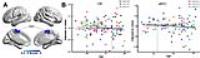
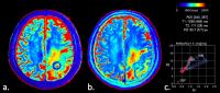
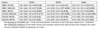
Effect of measured Hematocrit value on Glioma grading using Dynamic contrast enhanced  derived MR perfusion parameter
Prativa Sahoo1, Pradeep Kumar Gupta2, Ashish Awasthi3, Chandra Mani Pandey3, Rana Patir4, Sandeep Vaishya5, and Rakesh Kumar Gupta2
1Healthcare, Philips India ltd, Bangalore, India, 2Radiology and Imaging, Fortis Memorial Research Institute, Gurgaon, India, 3Biostatistics, Sanjay Gandhi Post Graduate Institute of Medical Sciences, Lucknow, India,4Neuorsurgury, Fortis Memorial Research Institute, Gurgaon, India, 5Neuorsurgury, Fortis Memorial Research Institute, Lucknow, India
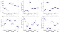
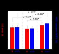
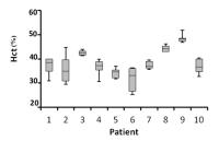
Quantification of  DCE-MRI assumes a constant blood hematocrite (Hct ) of 45% for adult human papulation. However Hct varies with disease condition and more with chemotherapy. Correction of the measured signal for blood Hct level is important as blood T1, quantification of contrast agent and arterial input function is dependent on it. Purpose of this study was to investigate the influence of Hct values on glioma grading using DCE-MRI derived perfusion parameters. Study suggest that even though grading of glioma not influenced by Hct values it does affect the kinetic parameters and  might be important for monitoring serial assessment of disease progressions.
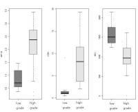

1345.   
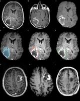
Can dynamic contrast enhanced MR perfusion metrics accurately discriminate different grades of Gliomas?
Jitender Saini 1, Pradeep Kumar Gupta2, Prativa Sahoo3, Rana Patir4, Sandeep Vaishya5, Arun Kumar Gupta1, Amey Savarderkar6, and Rakesh Kumar Gupta2
1Neuroimaging & Interventional Radiology, National Institute of Mental Health and Neurosciences, Bangalore, India, 2Radiology and Imaging, Fortis Memorial Research Institute, Gurgaon, India, 3Healthcare, Philips India ltd, Bangalore, India, 4Neuorsurgury, Fortis Memorial Research Institute, Gurgaon, India, 5Neuorsurgury, Fortis Memorial Research Institute, Lucknow, India, 6Neuorsurgury, National Institute of Mental Health and Neurosciences, Bangalore, India
Dynamic contrast enhanced MRI perfusion is a useful technique for assessment of glioma grading. This technique has been used in the past for discrimination of low from high grade gliomas. This study investigates the ability of DCE perfusion MRI to discriminate Grade II from Grade III and Grade III from Grade IV gliomas. Various DCE pharmacokinetic parameters were also analysed for their ability to distinguish the various grades of gliomas. 


1346.   
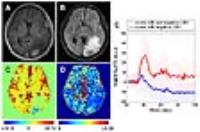
Investigation of hypoxia conditions using oxygenation enhance (OE)-MRI measurements in C6 glioma models
Yingwei Wu1, Yongming Dai2, Qi Fan1, and Xianfeng Tao1
1Shanghai Ninth People’s Hospital, School of Medicine, Shanghai Jiao Tong University, Shanghai, China, People's Republic of, 2Philips Healthcare, Shanghai, China, People's Republic of
We used oxygenation enhancement (OE)-MRI measurements to investigate hypoxia conditions of gliomas and to evaluate relationship between histopathology measurements and PSC. Oxygen amplitude maps of C6 glioma models were derived. ROI max and ROI non-max were defined. Time-SI curve from ROI areas was obtained and tissues from ROI areas was evaluated for microvessel density and expression of HIF-1a. We found that microvessel density in ROI non-max area were lower than those in ROI max area and expression of HIF-1α in ROI non-max area were higher than that in ROI max area. PSC had a linear positive correlation with vessel density.


1347.   
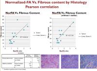
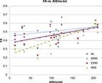
Quantitative DTI-FA Mapping in Prediction of Meningioma fibrous content, Consistency and grade.
Shanker Raja1,2, Wafa AlShawkeer3, Lama Mohammed Almudaimeegh3, Sadeq Al Dandan4, and Sharad P George5
1Radiology, Baylor College of Medicine, Bellaire, TX, United States, 2Radiology, KFMC, Riyadh, Saudi Arabia, 3King Khalid University Hospital, Riyadh, Saudi Arabia, 4King Fahad Medical City, Riyadh, Saudi Arabia,5Baylor College of Medicine, Houston, TX, United States
We utilized quantitative FA-maps derived from DTI to evaluate the fibrous content  consistency and  grade of meningioma.  Our results suggest that, quantitative FA mapping is promising in pre-operative prediction of  meningioma consistency pre-operatively, but only modestly correlates with histologic grading


1348.   
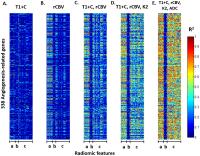
Imaging Angiogenesis Genotype of Glioblastoma by Radiomic Features of Multi-modality MRI
Chia-Feng Lu1,2,3, Fei-Ting Hsu4, Li-Chun Hsieh4, Yu-Chieh Jill Kao1,2, Hua-Shan Liu4,5, Ping-Huei Tsai2,4, Pen-Yuan Liao4, and Cheng-Yu Chen1,2,4
1Translational Imaging Research Center, College of Medicine, Taipei Medical University, Taipei, Taiwan, 2Department of Radiology, School of Medicine, Taipei Medical University, Taipei, Taiwan, 3Department of Biomedical Imaging and Radiological Sciences, National Yang-Ming University, Taipei, Taiwan, 4Department of Medical Imaging, Taipei Medical University Hospital, Taipei, Taiwan, 5Graduate Institute of Clinical Medicine, Taipei Medical University, Taipei, Taiwan
The multi-modality and multi-radomic-feature MRI may provide a more efficient regression model for imaging gene expressions than the conventional radiogenomic approach. 


1349.   
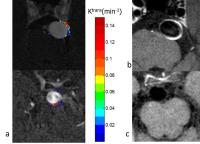
Optimization of glioma biopsy targeting applying T1-DCE MRI parameter maps – A double-blinded prospective study
Vera Catharina Keil1, Bogdan Pintea2, Gerrit H. Gielen3, Matthias Simon2, Juergen Gieseke1,4, Hans Heinz Schild1, and Dariusch Reza Hadizadeh1
1Department of Radiology, Universitätsklinikum Bonn, Bonn, Germany, 2Clinic for Neurosurgery and Stereotaxy, Universitätsklinikum Bonn, Bonn, Germany, 3Department of Neuropathology, Universitätsklinikum Bonn, Bonn, Germany, 4Philips Healthcare, Best, Netherlands
Many centers refrain from implementing semi-quantitative MRI techniques, such as T1w contrast-enhanced MRI (T1-DCE MRI), as a benefit for the patient is questioned. To elucidate if T1-DCE MRI has a benefit, we compared the standard neurosurgical biopsy target selection method (based on T1w contrast-enhanced or FLAIR maps) with a selection based on “hot spots” on Ktrans maps in a double-blinded, prospective setting with 27 glioma patients. 87 tissue samples were taken (55 Ktrans-based, 32 standard). Ktrans-based selection showed a strong tendency to be the more successful targeting method (glioblastoma: n=20/39 vs. n=11/20; p=0.085; WHO III/II: n=12/13 vs. n=6/11; p=0.061).


1350.   
Cerebrospinal fluid compression in cerebellum on treatment-naďve MRI might be an early indicator of poor survival in Glioblastoma: A preliminary study
Gavin Hanson1, Prateek Prasanna1, Jay Patel1, Anant Madabhushi1, and Pallavi Tiwari1
1Department of Biomedical Engineering, Case Western Reserve University, Cleveland, OH, United States
Glioblastoma Multiforme (GBM) is very aggressive form of primary brain tumor, and a key part of GBM pathogenesis is the mass effect of the tumor within the ridge container of the brain vault. Mass effect is strongly associated with mortality in patients with GBM. In this work, we seek to quantify the extent of mass effect throughout the brain volume as manifested on MRI to predict patient survival in GBM patients. We use a MRI-driven tensor based morphometry approach, combined with statistical mapping to allow the identification of regions where the deformation associated with mass effect is correlated with overall survival after diagnosis. 


1351.   
Gadolinium-DTPA-enhanced MR imaging of brain tumors: comparison with T1-Cube and 3D fast spoiled gradient recall acquisition in steady state sequences
Mungunkhuyag Majigsuren1,2, Takashi Abe2, and Masafumi Harada2
1Mongolian National University of Medical Sciences, Ulaanbaatar, Mongolia, 2The University of Tokushima, Tokushima, Japan
We compared the gadolinium enhancement characteristics of a heterogeneous population of brain tumors imaged by T1-Cube and 3D FSPGR at 3T MRI with time-dependent changes. A totally 91 lesions from 52 patients with brain tumors in 3T MRI. Fifty-one of the 91 lesions (56.04%) were depicted with T1-Cube first, and 40 lesions (43.96%), with 3D FSPGR first. 3D FSPGR images would be expected to exhibit greater enhancement than T1-Cube images. However, the overall mean CNR values were higher on T1-Cube images with both order sequences. We suggest the superiority of T1-Cube to 3D FSPGR for the detection of metastatic lesions.


1352.   
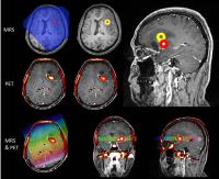
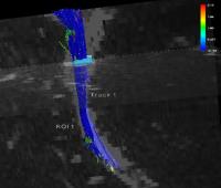
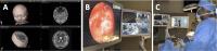
An MRS and PET guided biopsy tool for ultrasound-based intra-operative neuro-navigational systems.
Matthew Grech-Sollars1,2, Babar Vaqas3, Gerard Thompson4, Tara Barwick2,5, Lesley Honeyfield2, Kevin S O'Neill3, and Adam D Waldman1,2
1Division of Brain Sciences, Imperial College London, London, United Kingdom, 2Department of Imaging, Imperial College NHS Healthcare Trust, London, United Kingdom, 3Department of Neurosurgery, Imperial College NHS Healthcare Trust, London, United Kingdom, 4Department of Neuroradiology, Salford Royal NHS Foundation Trust, Salford, United Kingdom, 5Department of Surgery and Cancer, Imperial College London, London, United Kingdom
Glioma heterogeneity and the limitations of conventional structural MRI to identify agrressive tumour components limits targeting of stereotactic biopsy, and hence tumour characterisation. In vivo MR spectroscopy and PET allow for physiological characterisation of tumour and we here present a method for representing MRS and PET defined regions to biopsy using an ultrasound based neuronavigational system. Our method involves using colour-coded hollow spheres to represent the target biopsy regions, which can be easily identified during the surgery. This approach can be applied to target the most aggressive regions of a tumour and as a tool for imaging biomarker validation.


1353.   
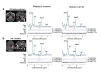
Translation of 2-hydroxyglutarate MR spectroscopy into clinics
Zhongxu An1, Sandeep Ganji1, Vivek Tiwari1, Edward Pan2,3,4, Bruce Mickey2,4,5, Elizabeth A. Maher2,3,5,6, and Changho Choi1,2,7
1Advanced Imaging Research Center, University of Texas Southwestern Medical Center, Dallas, TX, United States, 2Harold C. Simmons Cancer Center, University of Texas Southwestern Medical Center, Dallas, TX, United States, 3Department of Neurology and Neurotherapeutics, University of Texas Southwestern Medical Center, Dallas, TX, United States, 4Department of Neurological Surgery, University of Texas Southwestern Medical Center, Dallas, TX, United States, 5Annette Strauss Center for Neuro-Oncology, University of Texas Southwestern Medical Center, Dallas, TX, United States, 6Department of Internal Medicine, University of Texas Southwestern Medical Center, Dallas, TX, United States, 7Department of Radiology, University of Texas Southwestern Medical Center, Dallas, TX, United States
2-hydroxyglutarate (2HG) is an important biomarker for IDH-mutated gliomas. Thus in vivo measurement of 2-hydroxyglutarate can provide important information for brain tumor diagnosis and prognosis. Several techniques for in-vivo detection of 2HG were reported recently. However, due to limited access to scan parameters in clinical setup, translation of such techniques into clinics is limited. We report the reproducibility of a recently developed clinically-available PRESS-based 1H MRS method, for in vivo 2HG measurement at research and clinical scanners. 


1354.   
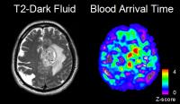
Tumor Classification Using Blood Arrival Histogram Obtained by Resting-state fMRI
Tianyi Qian1, Yinyan Wang2,3, Kun Zhou4, Yuanyuan Kang4, Shaowu Li2,5, and Tao Jiang2,5
1MR Collaborations NE Asia, Siemens Healthcare, Beijing, China, People's Republic of, 2Neurosurgery, Tiantan Hospital, beijing, China, People's Republic of, 3Beijing Neurosurgical Institute, Capital Medical University, Beijing, China, People's Republic of, 4Siemens Shenzhen Magnetic Resonance Ltd., APPL, Shenzhen, China, People's Republic of, 5Beijing Neurosurgical Institute, Capital Medical University, beijing, China, People's Republic of
In this study, a new post-processing pipeline of resting-statefMRI (rs-fMRI)was proposed for glioma grading, with the feasibility of extracting the timing information of brain perfusion from BOLD signal. The blood arrival time obtained from rs-fMRI shows unevenly distributed perfusion patterns in tumors. A histogram-based analysis method was employed to analyze the non-uniform distribution that could extract the patterns better than the routine method. The proposed pipeline was able to classify between low- and high-grade gliomas.


1355.   
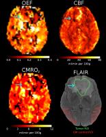
Mapping of brain tumor oxygen metabolism in native MRI
Patrick Borchert1, Lasse Dührsen2, Div S. Bolar3, Nils-Ole Schmidt2, Jan-Hendrik Buhk1, Jens Fiehler1, and Jan Sedlacik1
1Neuroradiology, UKE, Hamburg, Germany, 2Neurosurgery, UKE, Hamburg, Germany, 3Martinos Center, MGH, Boston, MA, United States
The QUIXOTIC method was tested in conjunction with ASL to map tumor oxygen metabolism in glioma patients. A higher oxygen extraction fraction was found for low grade gliomas, whereas lower cerebral blood flow was found for high grade gliomas. Both parameters were stable in healthy gray matter. These findings suggest, that the QUIXOTIC method is able to map tumor oxygen metabolism in conjunction with ASL. Furthermore, these findings may suggest, that low grade gliomas may maintain a more aerobic metabolism than high grade gliomas and that the uncontrolled tumor angiogenesis of high grade gliomas may cause hindered tumor perfusion.


1356.   
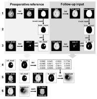
Validation of a semi-automatic coregistration of MRI scans in brain tumor patients during treatment follow-up
Jiun-Lin Yan1,2,3, Anouk van der Hoorn4,5, Timothy J Larkin6, Natalie R Boonzaier6, Tomasz Matys5, and Stephen J Price6
1Clinical Neuroscience, University of Cambridge, Cambridge, United Kingdom, 2Neurosurgery, Chang Gung Memorial Hospital, Keelung, Taiwan, 3Department of neurosurgery, Chang Gung University College of Medicine, Taoyuan, Taiwan, 4Department of radiology (EB44), University Medical Centre Groningen, Groningen, Netherlands, 5Department of radiology, University of Cambridge, Cambridge, United Kingdom, 6Brain tumour imaging laboratory, University of Cambridge, Cambridge, United Kingdom
Coregistration of lesional brain MRI between different time points is challenging. We aimed to propose a two staged semi-automatic coregistration methods to overcome the difficulty. Firstly, we calculated the transformation between presurgical tumor and postsurgical resection cavity by using the linear FLIRT co-registration. This creates a transformation matrix used for the progression and pseudoprogression area with optimal correction of variable brain shift. Then we applied this transformation matrix to a non-linear FNIRT transformation to coregister the brain. Validation by using registration target error showed smaller deviation can be achieved by using this method compared to direct non-linear registration. 


1357.   
Contrast-Enhanced Synthetic MRI for the Detection of Brain Metastases: Comparison Between Synthetic T1-weighted Inversion-recovery Image, Synthetic T1-weighted Image, and Conventional T1-weighted Inversion-recovery Fast Spin-Echo Image.
Misaki Nakazawa1,2, Akifumi Hagiwara2,3, Masaaki Hori2, Christina Andica2, Koji Kamagata2, Hideo Kawasaki2, Nao Takano2, Shuji Sato2, Nozomi Hamasaki2, Kouhei Tsuruta1,2, Sho Murata1,2, Ryo Ueda1,2, Shigeki Aoki2, and Atsushi Senoo1
1Graduate School of Human Health Sciences, Tokyo Metropolitan University, Tokyo, Japan, 2Department of Radiology, Juntendo University School of Medicine, Tokyo, Japan, 3Graduate School of Medicine, The University of Tokyo, Tokyo, Japan
The purpose of this study was to assess whether contrast-enhanced synthetic MRI is suitable for detecting brain metastases by comparing the lesion-to-white matter contrast, contrast-to-noise ratio, and number of brain metastases detected in synthetic and conventional magnetic resonance images. Synthetic T1IR images had better contrast compared with synthetic T1W or conventional T1IR images. Synthetic T1IR images enabled detection of more metastases than did synthetic T1W and conventional T1IR images even though statistical significance was not detected. Contrast-enhanced synthetic T1IR is useful for detecting brain metastases. Further optimization of contrast weighting is needed to maximize the ability to detect brain metastases. The purpose of this study was to assess whether contrast-enhanced synthetic MRI is suitable for detecting brain metastases by comparing the lesion-to-white matter contrast, contrast-to-noise ratio, and number of brain metastases detected in synthetic and conventional magnetic resonance images. Synthetic T1IR images had better contrast compared with synthetic T1W or conventional T1IR images. Synthetic T1IR images enabled detection of more metastases than did synthetic T1W and conventional T1IR images even though statistical significance was not detected. Contrast-enhanced synthetic T1IR is useful for detecting brain metastases. Further optimization of contrast weighting is needed to maximize the ability to detect brain metastases.


1358.   
Bayesian Estimation of Microstructural Parameters in Glioma Patients and Comparison with Genetic Analysis
Elias Kellner1, Marco Reisert1, Ori Staszewski2, Bibek Dhital1, Valerij G Kiselev1, Karl Egger3, Horst Urbach3, and Irina Mader3
1Department of Radiology, Medical Physics, University Medical Center Freiburg, Freiburg, Germany, 2Freiburg, Germany, 3Department of Neuroradiology, University Medical Center Freiburg, Freiburg, Germany
In a recent study, we proposed a method for fast and direct estimation of mictrostructural tissue parameters such as intra/extraaxonal volume fraction and diffusivities based on multishell DWI. In this study, we report the first method application to human gliomas and demonstrate connections of microstructural parameters with genetic markers IDH and 1p19q in a group of 32 patients.


1359.   
In Vivo Detection of 2-Hydroxyglutarate in Low-Grade Glioma Patients
Elizabeth D Phillips1, Llewellyn E Jalbert1, Yan Li1, Marisa M Lafontaine1, and Sarah J Nelson1
1Radiology and Biomedical Imaging, University of California, San Francisco, San Francisco, CA, United States
While the feasibility of utilizing 2HG as a magnetic resonance biomarker has been established ex vivo, several different approaches to obtaining in vivo data have been presented. This project aims to assess the concordance of 2HG detection using asymmetric echo PRESS MRSI with IDH1R132H mutation as identified via antibody staining in patients with LGG, and to investigate the relationship of other metabolites detected with this sequence to IDH status.  Further research is required before routine clinical implementation of these methods is recommended.


1360.   
1H Echo Planar Spectroscopic Imaging of 2-hydroxyglutarate in Gliomas at 7T in vivo
Zhongxu An1, Sandeep Ganji1, Vivek Tiwari1, Marco C. Pinho1,2, Edward Pan3,4,5, Bruce E. Mickey3,5,6, Elizabeth A. Maher3,4,6,7, and Changho Choi1,2,3
1Advanced Imaging Research Center, University of Texas Southwestern Medical Center, Dallas, TX, United States, 2Department of Radiology, University of Texas Southwestern Medical Center, Dallas, TX, United States,3Harold C. Simmons Cancer Center, University of Texas Southwestern Medical Center, Dallas, TX, United States, 4Department of Neurology and Neurotherapeutics, University of Texas Southwestern Medical Center, Dallas, TX, United States, 5Department of Neurological Surgery, University of Texas Southwestern Medical Center, Dallas, TX, United States, 6Annette Strauss Center for Neuro-Oncology, University of Texas Southwestern Medical Center, Dallas, TX, United States, 7Department of Internal Medicine, University of Texas Southwestern Medical Center, Dallas, TX, United States
2-hydroxyglutarate (2HG) is the first imaging biomarker for IDH-mutated gliomas. High-spatial resolution spectroscopic imaging of 2HG is clinically important. We propose a new EPSI read-out scheme to overcome the conventional limitation of EPSI spectral bandwidth at high field. With SNR and linewidth benefit at 7T, we demonstrated the in vivo feasibility of this new EPSI method in mapping of 2HG and other important brain metabolites in normal subject and glioma patients at 7T.


1361.   
Hybrid PET MRI of brain tumours: spatial relationship of tumour volume in FET PET and 3D MRSI
Jörg Mauler1, Karl-Josef Langen1,2, Andrew A. Maudsley3, Omid Nikoubashman4, Christian Filss1, Gabriele Stoffels1, and N. Jon Shah1,5
1Forschungszentrum Jülich, Jülich, Germany, 2Department of Nuclear Medicine, Faculty of Medicine, RWTH Aachen University, Aachen, Germany, 3Miller School of Medicine, University of Miami, Miami, FL, United States, 4Department of Neuroradiology, RWTH Aachen University, Aachen, Germany, 5Department of Neurology, Faculty of Medicine, JARA, RWTH Aachen University, Aachen, Germany
Gliomas are characterised by an elevated expression of amino acid transporters and cell turnover. The spatial overlap of the corresponding volumes was analysed in 46 subjects, based on O-(2-[18F]fluoroethyl)-L-tyrosine (FET) uptake, measured with PET and by means of the choline to N-acetyl-aspartate (Cho/NAA) ratio, determined by simultaneously acquired, 3D spatially resolved MR spectroscopic imaging data. The overlap between the respective volumes averaged out to (30±23) % with tumour volumes of (14±15) cm3 and (39±28) cm3 in case of FET uptake and increased Cho/NAA-ratio, respectively. Thus the imaging modalities may represent different metabolic properties of gliomas.


1362.   
Apparent diffusion coefficient in preoperative grading of gliomas: a comparison between ultra-high and conventional mono-b value diffusion-weighted MR imaging
YuChuan Hu1, LinFeng Yan1, ZhiCheng Liu1, YingZhi Sun1, DanDan Zheng2, TianYong Xu2, Wen Wang1, and GuangBin Cui1
1Department of Radiology, Tangdu Hospital, Fourth Military Medical University, Xi’an, China, People's Republic of, 2MR Research China, GE Healthcare China, Beijing, China, People's Republic of
The preoperative grading of gliomas, which is critical for determination of the most appropriate treatment, remains unsatisfactory. As an improved MRI technique, diffusion-weighted imaging (DWI) is considered the most sensitive for early pathological changes and therefore can potentially be useful in evaluating the glioma grades. Recently, apparent diffusion coefficient (ADC) values derived from the high (3000 sec/mm2) b values DWI were reported to improve the diagnostic performance of DWI in differentiating high- from low-grade gliomas5. But a mono-exponential model and relatively lower high-b values were used in this study.We used a tri-component model to calculate ultra-high ADC (ADCuh) in our research, aiming to retrospectively compare the efficacy of ultra-high and conventional mono-b value DWI in the glioma grading.


1363.   
18F-methylcholine PET/CT and magnetic resonance spectroscopy imaging and tissue biomarkers of cell membrane turnover in primary brain gliomas – a pilot study
Matthew Grech-Sollars1,2, Katherine Ordidge1,2, Babar Vaqas3, Lesley Honeyfield2, Sameer Khan2, Sophie Camp3, David Towey2, David Peterson3, Federico Roncaroli4, Kevin S O'Neill3, Tara Barwick2,5, and Adam D Waldman1,2
1Division of Brain Sciences, Imperial College London, London, United Kingdom, 2Department of Imaging, Imperial College NHS Healthcare Trust, London, United Kingdom, 3Department of Neurosurgery, Imperial College NHS Healthcare Trust, London, United Kingdom, 4Department of Neuropathology, Imperial College NHS Healthcare Trust, London, United Kingdom, 5Department of Surgery and Cancer, Imperial College London, London, United Kingdom
Choline elevation has been reported as a marker of aggressive glioma phenotype in numerous in vivo MRS studies, and more recently 18F-methylcholine-PET has been applied to glioma characterisation. This study examines the relationship between MRS and PET choline measures in defined tumour regions, in order to validate these against tissue biomarkers of choline metabolism and proliferation. Our initial results raise the possibility that imaging markers of choline metabolism are influenced by inflammatory and reactive processes for low grade tumours. 


1364.   
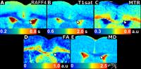
Assessment of Anti-EGFRvIII Chimeric Antigen Receptor (CAR) T cell Therapy for Patients with Glioblastomas using Diffusion, Perfusion and MR Spectroscopy
Sumei Wang1, Donald M O’Rourke2, Sanjeev Chawla1, Gaurav Verma1, Gabriela Plesa3, Carl H June3, Marcela V Maus4, Steven Brem2, Eileen Maloney2, Jennifer JD Morrissette5, Maria Martinez-Lage5, Arati Desai6, Ronald L Wolf1, Harish Poptani1,7, and Suyash Mohan1
1Radiology, University of Pennsylvania, Philadelphia, PA, United States, 2Neurosurgery, University of Pennsylvania, Philadelphia, PA, United States, 3Pathology and Laboratory Medicine, Center for Cellular Immunotherapies, University of Pennsylvania, Philadelphia, PA, United States, 4Center for Cancer Immunology, Massachusetts General Hospital Cancer Center, Charlestown, MA, United States, 5Pathology and Laboratory Medicine, University of Pennsylvania, Philadelphia, PA, United States, 6Hematology-Oncology, University of Pennsylvania, Philadelphia, PA, United States, 7Cellular and Molecular Physiology, University of Liverpool, Liverpool, United Kingdom
Chimeric Antigen Receptor (CAR) T cell therapy is a novel method of treating tumors. Since EGFRvIII is expressed in some glioblastomas, we evaluated the efficacy of anti-EGFRvIII CART for treating these tumors. Treatment response was assessed via serial MRI scans at 1 and 2 months after CAR-T cell therapy. The rCBVmax and Cho/Cr ratio decreased whereas MD and FA stayed relatively stable for most patients, indicating a positive response that can be assessed by these methods.


1365.   
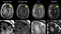
Fast Imaging Employing Steady-State Acquisition of Brain Metastasis: from mouse to woman
Donna H Murrell1,2, Keng Yeow Tay3, Eugene Wong2,3, Ann F Chambers2,3, Francisco Perera3, and Paula J Foster1,2
1Imaging Research Laboratories, Robarts Research Institute, London, ON, Canada, 2Department of Medical Biophysics, Western University, London, ON, Canada, 3London Health Sciences Centre, London, ON, Canada
Brain metastatic burden may be underestimated in the clinic because some tumors are impermeable to Gadolinium (Gd). Preclinical studies by our group demonstrated that fast imaging employing steady-state acquisition (FIESTA) was advantageous for detecting small Gd-impermeable tumors. Here, we show clinical translation of this imaging strategy. We present FIESTA images of human brain metastasis alongside standard clinical MRI and illustrate potential clinical utility of this sequence. Initial data suggests FIESTA can visualize intra-tumor heterogeneity where standard clinical MRI could not. Additional lesions were observed in FIESTA; we hypothesize some may be arachnoid cysts, though metastasis cannot be ruled out. 


1366.   
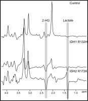
Distinguishing the Chemical Signature of Different IDH Mutations in Brain Tumor Patients at 7 Tesla
Uzay E Emir1, Sarah Larkin2, Nick de Pennington2, Puneet Plaha3, Natalie Voets1, James Mccullagh4, Richard Stacey3, Peter Jezzard1, Stuart Clare1, Christopher Schofield4, Tom Cadoux-Hudson3, and Olaf Ansorge2
1FMRIB Centre, University of Oxford, Oxford, United Kingdom, 2Nuffield Department of Clinical Neurosciences, University of Oxford, Oxford, United Kingdom, 3Department of Neurosurgery, John Radcliffe Hospital, Oxford University Hospitals NHS Trust, Oxford, United Kingdom, 4Department of Chemistry, University of Oxford, Oxford, United Kingdom
In this study, we show a proton magnetic resonance spectroscopy (1H-MRS) acquisition scheme at 7T, enabling discernible 2-HG in the spectra of IDH-mutant patients  acquired within 20s and quantify metabolic changes associated with the IDH mutation. Due to the increased sensitivity and specificity of this scheme at 7T, we demonstrate elevated 2-HG and Lactate accumulation in IDH2 R172K (mitochondrial) compared to the IDH1 R132H (cytosolic) mutant tumors in human brains noninvasively.


1367.   
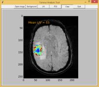
Developing a Semi-Automatised Tool for Grading Brain Tumours with Susceptibility-Weighted MRI
Maria Duvaldt1 and Tomas Jonsson1
1Dept. of Medical Physics, Karolinska University Hospital, Stockholm, Sweden
In order to make an adequate decision on the further treatment of a glioma cancer patient a tissue sample from the tumour is microscopically analysed and classified on a malignancy scale set by the WHO. In this project a software program with a graphical user interface is developed, where the malignancy grade of a tumour could be found by image analysis of susceptibility-weighted MR images. The parameters examined are the local image variance and intratumoural susceptibility signal and the results show the possibility of distinguishing high grade from low grade astrocytoma by image analysis only.


1368.   
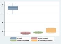
Non-Gaussian measurements of water diffusion in glioma as a tool for probing tumor heterogeneity and grade.
Fulvio Zaccagna1, Frank Riemer1, Mary McLean2, Andrew N. Priest3, James T. Grist1, Joshua Kaggie1, Sarah Hilborne1, Tomasz Matys1, Martin J. Graves1, Jonathan H. Gillard1, Stephen J. Price4, and Ferdia A. Gallagher1
1Department of Radiology, University of Cambridge, Cambridge, United Kingdom, 2Cancer Research UK Cambridge Institute, University of Cambridge, Cambridge, United Kingdom, 3Radiology, Cambridge University Hospitals NHS Foundation Trust, Cambridge, United Kingdom, 4Neurosurgery Unit, Department of Clinical Neurosciences, University of Cambridge, Cambridge, United Kingdom
Glioma grade and extent of local infiltration are used to guide surgical tumor management. Heterogeneity imaging is a way of assessing the tumor microenvironment, which may improve diagnosis and therapy planning. Diffusion Kurtosis Imaging (DKI) is a novel promising technique that estimates non-Gaussian water diffusion as a measure of heterogeneity. We investigate the use of DKI in glioma as a tool to improve tumor grading and to estimate infiltration. Our preliminary results show a mean kurtosis of 0.56±0.02 in glioblastoma and 1.14±0.07 in normal-appearing white matter. DKI may thus represent a useful tool for estimation of tumor heterogeneity in glioma.


1369.   
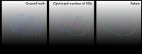
Impact of semi-automatic delineation of hotspots of contrast enhancing region in predicting the outcome of GBM patients after brain surgery
Adrian Ion-Margineanu1,2, Sofie Van Cauter3,4, Diana M Sima1,2, Frederik Maes2,5, Stefan Sunaert3, Stefaan Van Gool6, Uwe Himmelreich7, and Sabine Van Huffel1,2
1ESAT - STADIUS, KU Leuven, Leuven, Belgium, 2Medical IT, iMinds, Leuven, Belgium, 3Department of Radiology, University Hospitals of Leuven, Leuven, Belgium, 4ZOL - Ziekenhuis Oost-Limburg, Genk, Belgium, 5ESAT - PSI, KU Leuven, Leuven, Belgium, 6Department of Pedriatic Neuro-Oncology, University Hospitals of Leuven, Leuven, Belgium, 7Department of Imaging and Pathology, Biomedical MRI / MoSAIC, Leuven, Belgium
Delineating contrast enhancing (CE) tissue is an integral part of the RANO criteria for therapy response assessment in high-grade gliomas. We propose a semi-automatic delineation of hotspots of CE (HCE) in brain tumour follow-up of 29 glioblastoma multiforme patients after surgery. Based on multi-parametric magnetic resonance data we predict the post-operative evolution of the brain tumour by labelling each patient at each time point as responsive or progressive. The results obtained with our semi-automatic method are better in most of the cases than the results obtained with the original manual delineations. Moreover, our method can efficiently impute missing data.


1370.   
Automatic normalization of DCE-MRI derived cerebral blood volume (CBV) may improve glioma grading
Prativa Sahoo1, Indrajit Saha2, and Rakesh Kumar Gupta3
1Healthcare, Philips India ltd, Bangalore, India, 2Philips Healthcare, Philips India ltd, Gurgaon, India, 3Radiology and Imaging, Fortis Memorial Research Institute, Gurgaon, India
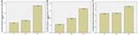
DCE-MRI derived relative blood volume (rCBV) correlates excellently with grade of glioma. Traditionally rCBV is calculated by dividing CBV value of tumor region with the CBV value from the corresponding contra-lateral region by identifying and placing region of interest (ROI). This technique is tedious needs user expertise.  The main aim of this study was to develop an automatic method to normalize CBV so that the user-induced biasness in glioma grading due to ROI placement can be reduced. Normalized CBV provides a better contrast between tumor and normal region


1371.   
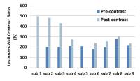
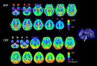
The Value of CBF Combined With Temporal Information in Grading High-Grade Astrocytomas: A Multi-Inversion-Time Arterial-Spin-Labeling Magnetic Resonance Study
Shuang Yang1, Tianyi Qian2, Jianwei Xiang1, Yingchao Liu3, Fei Gao1, Peng Zhao3, Josef Pfeuffer4, Guangbin Wang1, and Bin Zhao1
1Shandong Medical Imaging Research Institute, Shandong University, Jinan, China, People's Republic of, 2MR Collaborations NE Asia, Siemens Healthcare, Beijing, China, People's Republic of, 3Shandong provincial Hospital, Shandong University, Jinan, China, People's Republic of, 4Application Development, Siemens Healthcare, Erlangen, Germany
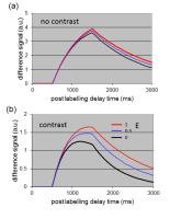
This study aimed to demonstrate the feasibility of multi-inversion-time arterial spin labeling (mTI-ASL) for differentiating between WHO III and WHO IV grade astrocytomas, as well as the added value of bolus arrival time (BAT) information in evaluating tumor perfusion. In the first part of this study, we evaluated the reproducibility of mTI-ASL in healthy subjects, and then mTI-ASL was used to evaluate 45 astrocytoma patients. There was no major variation between two consecutive mTI-ASL measurements in healthy volunteers. Furthermore, mTI-ASL provided valuable information for the classification of astrocytomas, while BAT added relevant information for grading by estimating the temporal dynamics of local tumor-mass perfusion.


1372.   
The effect of prophylactic cranial irradiation on brain diffusion and magnetization transfer
Mary A McLean1, Nicola L Ainsworth1,2, Anna M Brown1, Susan V Harden2, and John R Griffiths1
1CRUK Cambridge Institute, University of Cambridge, Cambridge, United Kingdom, 2Oncology, Addenbrooke's Hospital, Cambridge, United Kingdom
We investigated the effect of prophylactic cranial irradiation (PCI: 25 Gy in 10 fractions) on brain MRI at 3T. Six patients with small cell lung cancer were scanned at 4-month intervals: at diagnosis, following chemotherapy, and following PCI. Paired t-tests before and after PCI in right frontal white matter showed increased ADC and decreased FA and MTR following treatment. However, the parameters did not differ significantly from the scan at diagnosis, and other brain regions showed no significant changes on repeated-measures ANOVA. These observations are consistent with previous reports of more marked changes following higher-dose radiotherapy treatment.


1373.   
Differentiation of Glioblastoma Multiforme and Primary Cerebral Lymphoma with Diffusion-Weighted MR Imaging
Ching Chung Ko1,2, Yu Chang Lee3, Ming Hong Tai2, Tai Yuan Chen1, Yu Ting Kuo1, and Jeon Hor Chen3,4
1Department of Medical Imaging, Chi Mei Medical Center, Tainan, Taiwan, 2Institute of Biomedical Science, National Sun Yat-Sen University, Kaohsiung, Taiwan, 3Department of Radiology, I-Shou University and Eda Hospital, Kaohsiung, Taiwan, 4Center for Functional Onco-Imaging, University of California, Irvine, CA, United States
Atypical glioblastoma multiformes (GBMs) with solid enhancing tumor and without visible necrosis may mimic primary cerebral lymphomas (PCLs), and atypical PCLs with visible necrosis may mimic GBMs. This study aimed to differentiate these two brain tumors using qualitative DWI signals and quantitative ADC values acquired in tumoral necrosis, the most enhanced tumor area, and the peritumoral edema. The results showed GBMs tended to have significantly higher ADC in the enhanced tumor area, and lower ADC in the peritumoral edema area than PCLs.


1374.   
IDH-1 Mutation and Non-Enhancing Component of Glioblastoma
Daniel M Fountain1, Timothy J Larkin2, Natalie R Boonzaier2, Jiun-Lin Yan2, and Stephen J Price2
1The Brain Tumour Imaging Laboratory, Division of Neurosurgery, University of Cambridge, Cambridge, United Kingdom, 2Division of Neurosurgery and Wolfson Brain Imaging Centre, The Brain Tumour Imaging Laboratory, Cambridge, United Kingdom
IDH-1 mutated glioblastoma is associated with improved survival, and greater sensitivity to further resection of non-enhancing disease than IDH-1 wild-type. We used structural, diffusion tensor, perfusion and spectroscopic imaging data in a mixed model across the peritumoral region in 54 patients. Applying a mixed model methodology across three levels of data resolution, we demonstrated that IDH-1 mutated tumors demonstrated raised choline and lowered glutamate and glutamine compared to IDH-1 wild-type. The findings provided an AUC of 0.943 when combined with age. We hypothesised this results in greater sensitivity to treatment and reduced excitotoxicity, thus explaining their relatively superior prognosis.


1375.   
Evaluation of 7T MRI for endoscopic surgical planning and guidance for skull base tumors - preliminary experience
Hadrien A Dyvorne1, Thomas F Barrett2, Bradley N Delman3, Raj K Shrivastava2, and Priti Balchandani1
1Translational and Molecular Imaging Institute, Icahn school of Medicine at Mount Sinai, New York, NY, United States, 2Neurosurgery, Icahn School of Medicine at Mount Sinai, New York, NY, United States,3Radiology, Icahn school of Medicine at Mount Sinai, New York, NY, United States
Skull based tumors pose some of the most complex challenges in neurosurgery owing to their proximity to important structures such as optic nerves and arteries. For this reason, surgical planning heavily depends on high quality MR images. In this study we evaluated the performance of 7T imaging against standard scans at 3T and 1.5T for delineating such structures. Furthermore, the high-resolution scans were integrated in the neurosurgical workflow in order to evaluate improvements in surgical time and confidence of surgical decision-making.


1376.   
Semi-automatic segmentation of medulloblastoma using active contour method
Ka Hei Lok1, Lin Shi2,3, Queenie Chan4, and Defeng Wang5,6
1Department of Imaging and Intenvntional Radiology, The Chinese University of Hong Kong, Sha Tin, Hong Kong, 2Department of Medicine and Therapeutics, The Chinese University of Hong Kong, Sha Tin, Hong Kong,3Chow Yuk Ho Technology Centre for Innovative Medicine, The Chinese University of Hong Kong, Sha Tin, Hong Kong, 4Philips Healthcare, Hong Kong, Hong Kong, 5Research Center for Medical Image Computing, Department of Imaging and Intenvntional Radiology, The Chinese University of Hong Kong, Sha Tin, Hong Kong, 6Shenzhen Research Institute, The Chinese University of Hong Kong, Shenzhen, China, People's Republic of
Brain tumours are the second commonest form of childhood malignancy while medulloblastoma is the most common brain tumor in children. Accurate Segmentation of medulloblastoma is necessary for maximum tumor surgical removal. We proposed a novel method to segment medulloblastoma by modifying signed pressure function (SPF) function in Gaussians Filtering Regularized Level Set (SBGRLS) method. Quantitative validation is performed in this project. The method is proved to be clinical-oriented which is fast, robust, accurate with minimal user interaction.


1377.   
Withdrawn - Value of Amide Proton Transfer Imaging in Correlation with Histopathological Grades of Adult Diffuse Gliomas : Comparison and Incremental Value with Dynamic Susceptibility Contrast-Enhanced MRI and Diffusion Weighted Imaging
Seung-Koo Lee1, Yoon Seong Choi2, Sung Soo Ahn1, Ho-Joon Lee1, and Jinna Kim1
1Seoul, Korea, Republic of, 2Radiology, Severance Hospital, Yonsei University College of Medicine, Seoul, Korea, Republic of
We investigated the difference in APT values according to histopathological grades, and compared the diagnostic value of APT with relative cerebral blood volume (rCBV) from dynamic susceptibility contrast-enhanced (DSC) MRI and apparent diffusion coefficient (ADC) from diffusion weighted imaging (DWI) for histopathological grades in adult diffuse gliomas. We optimized APT imaging protocol for clinical setting and found that APT values were increased along with glioma grades, and APT values has incremental values over ADC values for glioma grading. We suggest that APT imaging can be a useful noninvasive imaging biomarker for glioma grading, in combination with ADC.


1378.   
Time-signal curves analysis of dynamic contrast-enhanced magnetic resonance imaging used in differential diagnosis of pituitary lesions.
shiyun tian1, Weiwei Wang1, and yanwei miao1
1Radiology Department, the First Affiliated Hospital of Dalian Medical University, Dalian, China, People's Republic of
Pituitary microadenomas are commonly visualized as well-defined lesions that enhance less than the normal pituitary gland, but it is not clear about the enhancement pattern of microadenomas. Our work is to evaluate the TIC type and the five parameters extracted from time-signal curves of DCE-MRI in the normal pituitary gland, microadenoma and the Rathke’s cleft cyst.


1379.   
MR based texture and location analysis of lower grade gliomas combined with genetic mutation information
Manabu Kinoshita1, Hideyuki Arita2, Mio Sakai3, Naoki Kagawa2, Yonehiro Kanemura4, Yasunori Fujimoto2, Katsuyuki Nakanishi3, and Toshiki Yoshimine2
1Neurosurgery, Osaka Medical Center for Cancer and Cardiovascular Diseases, Osaka, Japan, 2Neurosurgery, Osaka University Graduate School of Medicine, Suita, Japan, 3Radiology, Osaka Medical Center for Cancer and Cardiovascular Diseases, Osaka, Japan, 4National Hospital Organization Osaka National Hospital, Osaka, Japan
Extensive genetic analysis of WHO grade 2 and 3 gliomas (lower grade glioma) revealed that they comprise of several disease subtypes with different genetic or molecular backgrounds. The present investigation was conducted to elucidate the differences revealed on MR images including textures and locations of the tumors according to genetic mutation status (IDH and TERT promoter mutation) of lower grade gliomas. T2-entropy, a newly introduced image texture metric revealed that tumor heterogeneity is different depending on genetic status. Furthermore, classic oligodendroglial tumors located at the mid-base frontal lobe while astrocytic tumors occupied much lateral side of the brain.


1380.   
RADIOMICS  of advanced multiparmetric MRI in posterior fossa tumors is supreme to the domain wizards! A pilot study
Shanker Raja1, Sarah Farooq2, William Plishker3, Ali Daghriri4, Sadeq Wasil Al-Dandan5, Abdullah Ali Alrashed4, Muhammad Usman Manzoor6, and Sharad George7
1Baylor College of Medicine, Bellaire, TX, United States, 2King Fahad Medical City, Riyadh, Saudi Arabia, 3IGI Technologies, College Park, MD, United States, 4Medical Imaging, King Fahad Medical City, Riyadh, Saudi Arabia, 5Pathology and Laboratory Medicine, King Fahad Medical City, Riyadh, Saudi Arabia, 6Radiology, King Fahad Medical City, Riyadh, Saudi Arabia, 7Johns Hopkins Aramco Healthcare, Dhahran, Saudi Arabia
We uniquely  extracted textural features from multiple sequences of advanced FMRI  to preoperatively differentiate posterior fossa tumor histology.  Furthermore, as opposed to  recently published work (1,3,4) we found that in our series, textural feature subset derived from perfusion images is slightly superior to those of ADC maps. In addition, as expected, the  observations from this work concurs that RADIOMICS is definitely on par and probably  surpasses  domain experts in this endevour.  


1381.   
Evaluation of vascular permeability in gliomas by using parameter K2 from dynamic susceptibility contrast data-sets and histogram analysis.
Toshiaki Taoka1, Hisashi Kawai1, Toshiki Nakane1, Toshiteru Miyasaka2, and Shinji Naganawa1
1Radiology, Nagoya University, Nagoya, Japan, 2Radiology, Nara Medical University, Kashihara, Japan
 Permeability images can provide additional information to perfusion images in the clinical practice of brain tumors. However, permeability imaging by dynamic contrast enhancement methods requires a long acquisition time. K2 is an index that represents permeability and can be calculated from the dataset of perfusion images with the dynamic susceptibility contrast method, which requires a short acquisition time. In the current study, we calculated K2 for various grades of gliomas and found that K2 showed a significantly higher 20th percentile value in Grade IV compared to Grade III gliomas, providing useful information for grading of gliomas.


1382.   
Characterising tumour progression and pseudoprogression on preoperative multimodal MRI imaging
Jiun-Lin Yan1,2,3, Anouk van der Hoorn4, Timothy J Larkin5, Natalie Rosella Boonzaier5, Tomasz Matys6, and Stephen J Price5
1Clinical Neuroscience, University of Cambridge, Cambridge, United Kingdom, 2Department of neurosurgery, Chang Gung Memorial Hospital, Keelung, Taiwan, 3Department of neurosurgery, Chang Gung University College of Medicine, Taoyuan, Taiwan, 4Department of radiology (EB44), University of Groningen, Groningen, Netherlands, 5Brain tumour imaging laboratory, University of Cambridge, Cambridge, United Kingdom,6Department of radiology, University of Cambridge, Cambridge, United Kingdom
Glioblastoma is a highly malignant tumor which recur mostly within 2 cm around the resected contrast enhancement. However, it is difficult to identify tumor invasiveness pre-surgically especially in non-enhanced area. Thus, we aimed to identify possible imaging characteristics preoperatively using multimodal MR techniques in the peritumoral regions that eventually leads to tumor recurrence or progression. Our study showed lower isotorpic p, anisotopic q and ADC for progression compared to non-progression regions. In addition, MRS showed a not statistically significant trend of higher choline/NAA, higher choline and lower NAA in these progression area.


1383.   
Radiogenomic Mapping of Dysregulated Angiogenesis in Glioblastoma.
Kevin, Li-Chun Hsieh1, Fei-Ting Hsu1, Chia-Feng Lu1, and Cheng-Yu Chen1
1Translational Imaging Research Center, Taipei Medical University, Taipei, Taiwan
In this TCGA study, we identified several qualitative and quantitative radiomics imaging surrogates in glioblastoma, which can be used to differentiate whether this tumor have dysregulated angiogenesis at the molecular level. These features can also be used to predict disease survival.


1384.   
Differentiation of glioblastoma multiforme and single brain metastasis by the distribution pattern of intratumoral susceptibility sign derived from susceptibility-weighted imaging
Hyunkoo Kang1 and Keuntak Roh1
1Department of Radiology, Seoul Veterans Hospital, Seoul, Korea, Republic of
Susceptibility-weighted imaging (SWI) is an emerging magnetic resonance imaging (MRI) technique that exploits the susceptibility differences between the tissues. SWI provides the enhancement of small vessels and microhemorrhages and detection of iron in the brain. These characteristics permit SWI to show anatomical and functional heterogeneity of brain tumors by exquisite sensitivity to the blood products and venous vasculature. The aim of this study is to determine whether the distribution pattern of intratumoral susceptibility sign (ITSS) derived from SWI could differentiate glioblastoma multiforme (GBM) and single brain metastasis. We investigated the distribution patterns of ITSS of the tumors and applied an ITSS grading system based on the degree of the ITSS. Then, we compared the grade of the visibility of ITSS in the central portion of tumors (CITSS) and in the tumor capsular area (PITSS) on SWI in consensus. In clinical use, SWI is also useful for differentiating GBMs from metastases.


1385.   
Differentiating contrast-enhanced glioma from peritumoral edema using the intravascular fraction derived from IVIM MRI - a comparative study with DSC MRI
Yen-Shu Kuo1,2, Han-Min Tseng3, and Wen-Chau Wu4
1Graduate Institute of Biomedical Electronics and Bioinformatics, National Taiwan University, Taipei, Taiwan, 2Radiology, Cathay General Hospital, Taipei, Taiwan, 3Neurology, National Taiwan University Hospital, Taipei, Taiwan, 4Graduate Institute of Oncology, National Taiwan University, Taipei, Taiwan
In this study, we performed intravoxel incoherent motion (IVIM) MRI in 25 patients with histologically proven gliomas, and compared the intravascular fraction f with the cerebral blood volume derived from dynamic susceptibility-contrast (DSC) MRI (CBVDSC). Results showed that f was able to differentiate contrast-enhanced glioma from peritumoral edema by detecting elevated vascularity. Cross-modal comparison indicated that f correlated better with contrast-leakage-corrected CBVDSC than uncorrected value.


1386.   
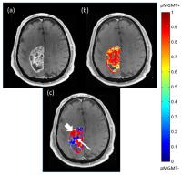
Normalization of Multi-contrast MRI and Prediction of Tumor Phenotypes
Yong Ik Jeong1, Charles Cantrell1, David Manglano1, Thomas Gallagher1, Jeffery Raizer1, Craig Horbinski1, and Timothy J Carroll1
1Northwestern University, Chicago, IL, United States
Genetic profiling of cancers has the potential to identify epigenetic changes that predict response to treatments. In this study, we try to overcome the limitations posed by heterogeneity of tumor phenotypes by using normalized quantitative MRI to predict local gene expression. We report the findings of retrospectively comparing T1, T1 post Gd, T2 and ADC to Verhaak subtypes and pMGMT methylation status in histologically confirmed GBM patients.


1387.   
Intra- and inter-individual association of FET-PET- and MR-Perfusion-parameters in untreated glioma
Jens Goettler1, Anne Kluge1, Mathias Lukas2, Stephan Kaczmarz1, Jens Gempt3, Florian Ringel3, Mona Mustafa2, Markus Schwaiger2, Claus Zimmer1, Stefan Foerster2, Christine Preibisch1,4, and Thomas Pyka2
1Department of Neuroradiology, Technische Universität München, Munich, Germany, 2Clinic for Nuclear Medicine, Technische Universität München, Munich, Germany, 3Clinic for Neurosurgery, Technische Universität München, Munich, Germany, 4Clinic for Neurology, Technische Universität München, Munich, Germany


18F-fluoroethyltyrosine (FET) PET and dynamic susceptibility contrast (DSC) perfusion weighted imaging are useful imaging techniques to diagnose glioma and to delineate tumor extension. However it is still unclear whether static and dynamic parameters of FET-PET and DSC are associated with each other. In this study we examined 45 patients with glioma in a hybrid PET-MR 3T scanner assessing FET time-activity-curves and DSC-parameters simultaneously. Static as well as dynamic PET-measures highly correlated with DSC-parameters such as relative cerebral blood volume (rCBV) and relative peak height (rPH). Results point to a complementary role of both modalities pre-therapeutically.


1388.   
Estimating damage to the blood-brain barrier from radiotherapy treatment
Magne Kleppestř1, Christopher Larsson1, and Atle Bjornerud1,2
1The Intervention Centre, Oslo University Hospital, Oslo, Norway, 2Department of Physics, University of Oslo, Oslo, Norway
Brain tumors are usually subjected to radiation therapy upon diagnosis. In this work, it is made an attempt at investigating if this therapy might cause injury to the non-cancerous parts of the brain. To this end dynamic contrast-enhanced MRI was used to estimate leakage across the blood-brain barrier.22 patients were imaged before and after undergoing a treatment schedule, and findings from the two examinations were compared to uncover any change. The data shows no significant variation in either permeability or blood plasma volume.


1389.   
MR appearance of Primary central nervous system lymphoma: as prognostic factors influencing the response to clinical treatment
jing Liu1 and shuixing Zhang1
1Radiology, Department of Radiology, Guangdong Academy of Medical Sciences/Guangdong General Hospital, Guangzhou, Guangzhou, China, People's Republic of
Currently, the treatment response in primary central nervous system lymphoma (PCNSL) is monitored by serial contrast-enhanced anatomic MR imaging, which often showing characteristic radio-morphological features such as lesion location, strong and homogenous contrast-enhancement, moderate edema and absence of necrosis. The purpose of our study was to investigate the objective response rate (ORR) and identify MR findings as predictors to evaluate the therapeutic response in PCNSL. Our result shows that tumor size, number, location, homogenous enhancement and the planned therapeutic strategy were independent factors correlated with treatment response in patients of PCNSL.

1390.   
Neural basis of the association between anxiety and depression symptoms in unmedicated major depressive disorder patients
Cancan He1, Liang Gong1, Chunming Xie1, and Yingying Yin1
1Department of Neurology, Affiliated ZhongDa Hospital, School of Medicine, Southeast University, Nanjing, China, People's Republic of
In this study, 75 unmedicated MDD patients and 42 cognitively normal(CN) subjects underwent the resting-state functional magnetic resonance imaging (R-fMRI) scan. We found that the MDD patients showed dysfunctional connectivity in wide-spread amygdala functional connectivity(AFC) networks, and these abnormal amygdala connectivity were influenced the trait property in MDD. Further analysis revealed that MDD patients with lower HAMA scores showed milder depressive symptom and greater AFC strength while MDD patients with higher HAMA scores showed more severe depressive s­­­­­ymptom and lower AFC strength. Beyond that, the mediation effects of AFC networks on the association between anxiety and depression all reached a significant level in MDD patients.


1391.   
Corpus callosum morphology and microstructure in late-life depression
Louise Emsell1,2, Christopher Adamson3, Filip Bouckaert1, Thibo Billiet2, Daan Christiaens4, Francois-Laurent De Winter1, Marc Seal3, Pascal Sienaert1, Stefan Sunaert2, and Mathieu Vandenbulcke1
1UPC-KU Leuven, Leuven, Belgium, 2Translational MRI, KU Leuven, Leuven, Belgium, 3Developmental Imaging, Murdoch Childrens Research Institute, Melbourne, Australia, 4ESAT/PSI, KU Leuven, Leuven, Belgium
Differences in corpus callosum (CC) morphology and microstructure have been implicated in late-life depression (LLD), however it is not clear to what extent microstructural alterations result from partial volume effects arising from macrostructural differences. Here we combined T1 morphological measures (thickness and area) with multiple diffusion MRI measures (fractional anisotropy, radial diffusivity and apparent fibre density (AFD)) to investigate the mid-sagittal CC in 51 patients with LLD and 52 healthy controls. LLD was associated with subtle, independent regional macro- (reduced area) and microstructural (reduced AFD) differences in the corpus callosum, unrelated to depression subtype or illness severity.


1392.   
7 T MRS Investigation of the Glutamatergic System in Depression
Clark Lemke1,2, Charles Masaki1, Uzay Emir2, Beata Godlewska1, and Phil Cowen1
1Department of Psychiatry, University of Oxford, Oxford, United Kingdom, 2FMRIB, University of Oxford, Oxford, United Kingdom
The glutamatergic system is believed to play a significant role in depression pathology. While many magnetic resonance spectroscopy (MRS) studies of depression have targeted the glutamatergic system, they have all been performed at magnetic field strengths of 4 T or lower – limiting their ability to differentiate between glutamate and glutamine. This study presents the first investigation of the glutamatergic system in depressed subjects at 7 T. Voxels were placed in the occipital cortex, anterior cingulate cortex, and putamen and metabolites were quantified using LCModel. Results indicate a significant decrease in glutamate in the occipital cortex and a significant increase of glutamine in the putamen.


1393.   
Disrupted reward circuits is associated with cognitive deficits, depression severity, and trait property in unmedicated major depressive disorder
Liang Gong1, Yingying Yin1, Cancan He1, Chunming Xie1, Yonggui Yuan1, Zhijun Zhang1, and Hongxing Zhang2
1Department of Neurology, Affiliated ZhongDa Hospital, School of Medicine, Southeast University, Nanjing, China, People's Republic of, 2Department of Psychiatry, Henan Provincial Mental Hospital, XingXiang, China, People's Republic of
we employed the resting-state fMRI technique with voxelwise multivariate regression analysis to identify that the disrupted topological organization within reward circuits was significantly associated with cognitive deficits, depression severity, and trait property in major depressive disorder (MDD) patients. Importantly, distinct and common neural pathways underlying cognitive deficit and depression were identified, and implied the independent and synergistic effects of cognitive deficits and depression severity on reward circuits in MDD patients.


1394.   
Brain grey matter volume alterations in treatment resistant depression -- systematic review and meta-analysis
Xin Xu1, Jia Liu2, and Qiyong Gong2
1Department of Psychiatry, Huaxi MR Research Center(HMRRC),West China Hospital of Sichuan Universityl, Chengdu, China, People's Republic of, 2Department of Radiology, Huaxi MR Research Center(HMRRC),West China Hospital of Sichuan Universityl, Chengdu, China, People's Republic of
To our knowledge, this is the first study to pool VBM studies for a meta-analysis of grey matter differences among the treatment-sensitive depression (TSD), treatment-resistant depression (TRD) and health control (HC) by using anisotropic effect-size signed differential mapping (AES-SDM). Although both TRD and TSD groups showed abnormal grey matter in frontal and temporal cortex, the exact brain regions were mostly different in both groups except the left inferior frontal gyrus (IFG). Furthermore, grey matter volume reductions in the bilateral cingulate cortex were only observed in the TRD group.


1395.   
DTI-based connectome analysis of adolescent depression reveals hypoconnectivity of the right caudate
Olga Tymofiyeva1, Colm G Connolly1, Tiffany C Ho1, Matthew D Sacchet2, Eva Henje Blom1,3, Kaja Z LeWinn1, Duan Xu1, and Tony T Yang1
1UCSF, San Francisco, CA, United States, 2Stanford University, Stanford, CA, United States, 3Karolinska Institutet, Stockholm, Sweden
The goal of this study was to perform DTI-based connectome analysis in a cohort of depressed adolescents and matched non-depressed controls. Our findings highlight the role of right caudate connectivity, in particular to frontal gyri, insula, and anterior cingulate, in this population.


1396.   
Dysfunction of the Cingulo-Opercular Network in First-Episode, Medication-Naive Patients with Major Depressive Disorder
xiaoping wu1, yanjun gao1, Pan Lin2, Junle Yang1, Rui Yang3, and Jian Yang4
1Department of Radiology, the Affiliated Xi’an Central Hospital of Xi’an Jiaotong University, Xi’an, China, People's Republic of, 2Key Laboratory of Biomedical Information Engineering of Education Ministry, Institute of Biomedical Engineering, Xi’an Jiaotong University, Xi’an, China, People's Republic of, 3Department of Psychiatry, the Affiliated Xi’an Central Hospital of Xi’an Jiaotong University, Xi’an, China, People's Republic of,4Department of Radiology, the First Affiliated hospital of Xi’an Jiaotong University, Xi’an, China, People's Republic of
Patients with MDD showed abnormalities in the connectivity of the CON. We found abnormal connectivity in MDD patients between the dACC and the bilateral middle frontal gyrus (MFG) and between the middle temporal gyrus (MTG) and precentral gyrus. Moreover, regression analysis showed that depression symptom severity (measured with the Hamilton Depression Rating Scale (HDRS), Hamilton Anxiety Scale (HAMA) and Automatic Thoughts Questionnaire scores (ATQ)) was significantly correlated with the FC values of the CON.


1397.   
Gray and white matter volume changes and the correlation with depression and anxiety in obese patients revealed by voxel-based morphometry
Jun-Cheng Weng1,2, Chi-Ju Lai1, Hse-Huang Chao3, Ming-Chou Ho4, and Vincent Chin-Hung Chen5
1Department of Medical Imaging and Radiological Sciences, Chung Shan Medical University, Taichung, Taiwan, 2Department of Medical Imaging, Chung Shan Medical University Hospital, Taichung, Taiwan, 3Tiawan Center for Metabolic and Bariatric Surgery, Jen-Ai Hospital, Taichung, Taiwan, 4Department of Psychology, Chung Shan Medical University, Taichung, Taiwan, 5Department of Psychiatry, Chang Gung Memorial Hospital, Chiayi, Taiwan
Obesity is an important health issue in modern society. The prevalence of obesity has been increasing in these years and morbid obesity is related to cardiovascular disease and overall mortality. Past reviews regarded binge eating as a manifestation of dysfunctional reward system and disinhibition. Some authors considered binge eating as a kind of addiction. Recent study demonstrated the more extensive involvement of brain pathways other than reward system. The mechanism of change is not clear. There is scanty of research regarding correlation between change of activation pattern in brain areas in functional MRI, binge eating and psychiatric illness. To gain insight into the correlation of physiological alteration and psychiatric illness and to develop subsequent detectable biomarker are crucial. The goal of our study was to investigate the morphological changes in gray and white matter between obese patients and healthy subjects using voxel-based morphometry (VBM). Our results suggested the changes of the volume in the brain structures may closely linked to the symptom and behavior of obese patients.  Apply these novel markers to monitoring and improving cormorbid psychiatric illness will be an essential part of multidiscipline integral care for obese patients.


1398.   
MR Imaging of Major Depressive Disorder: Effects of Sertraline Treatment
Hung-Wen Kao1,2, Chien-Yuan Eddy Lin3,4, Chu-Chung Huang5, Yi-Hui Lin6, I-Ling Chung7, Yu-Chuan Chang8, Guo-Shu Huang1, and Ching-Po Lin2,5,6,9
1Department of radiology, Tri-Service General Hospital, National Defense Medical Center, Taipei, Taiwan, 2Department of Biomedical Imaging and Radiological Sciences, National Yang-Ming University, Taipei, Taiwan,3GE Healthcare, Taipei, Taiwan, 4GE Healthcare MR Research China, Beijing, China, People's Republic of, 5Institute of Neuroscience, National Yang-Ming University, Taipei, Taiwan, 6Institute of Brain Science, National Yang-Ming University, Taipei, Taiwan, 7Institute of Biomedical Science, Academia Sinica, Taipei, Taiwan, 8Graduate Institute of Biomedical Electronics and Bioinformatics, National Taiwan University, Taipei, Taiwan,9Brain Research Center, National Yang-Ming University, Taipei, Taiwan
We hypothesized that a predictive MR imaging model of sertraline treatment could be established to help treatment planning for patients with Major depressive disorder (MDD). The voxel-based morphometry analysis showed increase volume of the lingual and occipital gyri in patients with MDD as compared with those in healthy controls and the size of the occipital gyrus decreased after 6-week treatment of sertraline. The findings support that patients with MDD might have a functional abnormality of visual areas and antidepressant treatment might shift the abnormal activity in the antidepressant-susceptible brain region to a normal level.


1399.   
Simultaneous Real-time fMRI and EEG Neurofeedback for Emotion Regulation Training in Depressed Patients
Vadim Zotev1, Raquel Phillips1, Masaya Misaki1, Ahmad Mayeli1,2, and Jerzy Bodurka1,3
1Laureate Institute for Brain Research, Tulsa, OK, United States, 2Electrical and Computer Engineering, University of Oklahoma, Tulsa, OK, United States, 3College of Engineering, University of Oklahoma, Tulsa, OK, United States
We have performed an exploratory study of emotion self-regulation training in major depressive disorder (MDD) patients using simultaneous real-time fMRI and EEG neurofeedback (rtfMRI-EEG-nf). MDD patients learned to upregulate two fMRI and two EEG target measures, relevant to MDD, using rtfMRI-EEG-nf during a happy emotion induction task.  The target measures included fMRI activities of the left amygdala and left rACC, as well as frontal EEG asymmetries in the alpha and high-beta bands. Our results demonstrate that MDD patients can learn to successfully upregulate all four measures simultaneously. These findings may lead to development of more efficient neurotherapies for MDD.

1401.   
Adaptive Tissue Cluster Tracking on Quantitative MRI for Fully Automatic Brain Segmentation on Young Children
Marcel Warntjes1,2, Suraj Serai3, James Leach3, and Blaise Jones3
1Center for Medical Imaging Science and Visualization, Linköping, Sweden, 2SyntheticMR AB, Linköping, Sweden, 3Department of radiology, Cincinnati, OH, United States
Brain tissue properties change rapidly during the first few years of life. This poses a problem for brain segmentation algorithms since adult tissue definitions for white matter and grey matter do not apply for young children. An automatic tissue cluster tracking algorithm was developed to determine WM and GM cluster positions in a 3-dimensional search-space of quantitative R1 relaxation rate, R2 relaxation rate and proton density. These positions are then used to segment the brain, independent of age.


1415.   
Longitudinal metabolite trajectories in the midfrontal gray matter in normally developing South African children
Martha J Holmes1, Frances C Robertson1, Francesca Little2, Mark F Cotton3, Els Dobbels3, Andre JW van der Kouwe4,5, Barbara Laughton3, and Ernesta M Meintjes1
1MRC/UCT Medical Imaging Research Unit, Department of Human Biology, University of Cape Town, Cape Town, South Africa, 2Department of Statistical Sciences, University of Cape Town, Cape Town, South Africa,3Children’s Infectious Diseases Clinical Research Unit, Department of Paediatrics & Child Health, Tygerberg Children’s Hospital and Faculty of Medicine and Health Sciences, Stellenbosch University, Cape Town, South Africa, 4A.A. Martinos Centre for Biomedical Imaging, Department of Radiology, Massachusetts General Hospital, Charlestown, MA, United States, 5Department of Radiology, Harvard Medical School, Boston, MA, United States
Magnetic resonance spectroscopy (MRS) measures changes in localized brain metabolism that occur alongside structural and functional development. Well-described trajectories of major metabolites with age provide a benchmark of normal brain maturation. In a longitudinal study, we examined the trajectories of NAA, choline and creatine in normally developing South African children at 5, 7 and 9 years. We found age-related increases in NAA and creatine levels, and constant choline levels in the midfrontal gray matter.  Since no gender or ethnicity effects were observed, these results are generalizable to a wide pediatric population against which pathology and abnormal development may be compared.


1403.   
Measuring Longitudinal Changes in Cerebral Blood Flow and Blood Volume in Neonates with the Intravoxel Incoherent Motion Method
Alex Cerjanic1,2, Ellen Grant3, Borjan Gagoski3, Marie Drottar3, Thea Francel3, Alana Matos3, Clarissa Carruthers3, Jonathan Litt4, Ryan Larsen2, and Bradley P Sutton1,2
1Bioengineering, University of Illinois at Urbana-Champaign, Urbana, IL, United States, 2Beckman Institute, University of Illinois at Urbana-Champaign, Urbana, IL, United States, 3Boston Children's Hospital, Boston, MA, United States, 4Beth Israel Deaconness, Boston, MA, United States
Diffusion weighted MRI was used on a cohort of 5 neonates to quantify cerebral blood flow and cerebral blood volume through the intravoxel incoherent motion (IVIM) model. Data at two time points, approximately 2 weeks and 14 weeks, were obtained. The obtained pseudodiffusion coefficient and the perfusion fractions were examined across white matter, gray matter, and the basal ganglia for all subjects. A significant longitudinal decrease in the perfusion fraction (-1.12%) was noted in the white matter between 2 and 14 weeks while the static diffusion coefficient of tissue decreased for all tissue classes between those time points. 


1400.   
Development of a new prototype body holder for MR examination in unanesthetized neonates
Iichiro Osawa1, Takako Aoki1, Takashi Ushimi1, Kaiji Inoue1, Junji Tanaka1, and Mamoru Niitsu1
1Radiology, Saitama Medical University Hospital, Saitama, Japan
To avoid motion artifacts, neonates often require anesthesia during MRI scans. However, this procedure increases the risk of adverse events such as respiratory depression. We developed a body holding device to minimize motion without anesthesia and examined nine low-birth-weight neonates, comparing MR image quality between unanesthetized and anesthetized conditions. The device is based on a modified spinal immobilizer and is easily handled with a short setup time. We obtained structural images during natural sleep uneventfully, preserving the image quality. In summary, the body holder can reduce the motion of neonates safely and improve image quality.  


1406.   
Perfusion and diffusion in the extremely preterm young adult thalamus
Andrew Melbourne1, Zach Eaton-Rosen1, Eliza Orasanu1, Joanne Beckmann2, Alexandra Saborowska3, David Atkinson3, Neil Marlow2, and Sebastien Ourselin1
1Medical Physics and Biomedical Engineering, University College London, London, United Kingdom, 2Institute for Women's Health, University College London, London, United Kingdom, 3University College Hospital, London, United Kingdom
This work investigates the appearance of the thalamus using multiple MR imaging contrasts between a population of extremely-preterm born adolescents and their term-born peers.


1414.   
BRAIN METABOLITE DIFFERENCES IN ONE-YEAR-OLD PRETERM INFANTS WITH INTRAUTERINE GROWTH RESTRICTION: ASSOCIATION WITH STRUCTURAL CHANGES AND NEURODEVELOPMENTAL OUTCOME
Rui Vasco Simoes1,2,3, Emma Muńoz-Moreno4, Nuria Bargallo5,6, Magdalena Sanz-Cortes7, and Eduard Gratacos1,2,3
1Fetal Medicine Research Center, BCNatal (Hospital Clinic and Hospital Sant Joan de Deu), Barcelona, Spain, 2Fetal Medicine Research Center, Institut d’Investigacions Biomediques August Pi i Sunyer (IDIBAPS), Barcelona, Spain, 3Center for Biomedical Research on Rare Diseases (CIBER-ER), Barcelona, Spain, 4Experimental MRI 7T Unit, Institut d’Investigacions Biomediques August Pi i Sunyer (IDIBAPS), Barcelona, Spain,5Medical Image platform, Institut d’Investigacions Biomediques August Pi i Sunyer (IDIBAPS), Barcelona, Spain, 6Dept. Radiology, Hospital Clinic, Barcelona, Spain, 7Dept. Obstetrics and Gynecology, Baylor College of Medicine, Houston, TX, United States
It is difficult to address the differential effects of Intrauterine growth restriction (IUGR)  and prematurity, as they represent two independent problems occurring simultaneously and can both contribute to impaired neurodevelopment. We have studied one-year-old preterm-IUGR infants and preterm and term appropriate for gestational age (AGA) infants, by MRI/MRS at 3T. Preterm-IUGR infants present metabolite profile changes in the frontal lobe, which are associated with brain structural and biophysical alterations, and poorer neurodevelopmental outcome at two years.


1402.   
Distinctive microstructural changes of association white matter tracts during preterm human brain development
Minhui Ouyang1, Austin Ouyang2, Qiaowen Yu2, Lina Chalak3, and Hao Huang1,4
1Department of Radiology, Children's Hospital of Philadelphia, Philadelphia, PA, United States, 2Advanced Imaging Research Center, University of Texas Southwestern Medical Center, Dallas, TX, United States,3Department of Pediatrics, University of Texas Southwestern Medical Center, Dallas, TX, United States, 4Department of Radiology, Perelman School of Medicine, University of Pennsylvania, Philadelphia, PA, United States
Association white matter tracts connecting different cortical regions underlie initial brain circuit formation from mid-fetal to normal time of birth. We examined the microstructure changes of association tracts and compare them to those of commissural, limbic and projection tracts with high resolution diffusion MRI of 10 fetal brains specimens at 20 postmenstrual weeks, 19 in vivo preterm brains at 35 weeks and 17 in vivo brains at 40 weeks. Distinctive microstructural developmental patterns were found in association tract groups compared to other tract groups during 35-40 weeks with DTI-derived metrics (including fractional anisotropy, mean, axial and radial diffusivity measurements). 


1408.   
Deformation-based morphometry identifies brain structural damages in 6 month-old infants with neonatal encephalopathy and predicts their developmental outcome
Hosung Kim1, Kevin Shapiro2, Maria Luisa Mandelli2, Hannah Clanley Glass2, Dawn Gano2, ELIZABETH Rogers3, Donna M Ferriero2, Anthony James Barkovich1, and Duan Xu1
1Radiology & Biomedical Imaging, University california San francisco, San Francisco, CA, United States, 2Neurology, University california San francisco, San Francisco, CA, United States, 3Pediatrics, University california San francisco, San Francisco, CA, United States
Neonatal encephalopathy (NE) is a major cause of mortality and permanent neurological disabilities in term infants.  Using t1w MRI and DBM, we found that neonatal seizure was related to WM atrophy in multiple locations.  Larger birth weight was associated with increased overall GM and WM volumes. A significant association was identified between language ability at 2 years old and increase in GM volume in Wernicke’s area.  This DBM approach has the potential for predicting early developmental outcome in infants with NE, as the volume of Wernicke’s area significantly correlated with the scores of language ability evaluated in early childhood.


1412.   
Maternal Obesity Affects Offspring’s Brain Resting-State Functional Connectivity
Xuehua Li1,2, Yilu Zhang2, Aline Andres1, R.T. Pivik1, Charles Glasier2, Raghu Ramakrishnaiah2, Thomas Badger1, and Xiawei Ou1,2,3
1Arkansas Children's Nutrition Center, Little Rock, AR, United States, 2Radiology, University of Arkansas for Medical Sciences, Little Rock, AR, United States, 3Arkansas Children's Hospital Research Institute, Little Rock, AR, United States
Recent studies have reported negative associations between maternal obesity during pregnancy and cognitive/neurodevelopmental outcome of children. It is speculated that neuro-programming differs in offspring of obese and normal weight women. In this study, we evaluated and compared the resting-state functional connectivity in 2-week-old infants born to normal weight or obese mothers, and we observed significant differences in brain connectivity associated with maternal obesity.


1409.   
Tract-based spatial statistics to assess the effect of histologic chorioamnionitis on white matter development in preterm infants
Devasuda Anblagan1,2, Rozalia Pataky2, Margaret J Evans3, Sarah Sparrow2, Chinthika Piyasena4, Emma J Telford2, Scott I Semple4,5, Alastair Graham Wilkinson6, Mark E Bastin1, and James P Boardman1,2
1Centre for Clinical Brain Sciences, University of Edinburgh, Edinburgh, United Kingdom, 2MRC Centre for Reproductive Health, University of Edinburgh, Edinburgh, United Kingdom, 3Department of Pathology, University of Edinburgh, Edinburgh, United Kingdom, 4Centre for Cardiovascular Sciences, University of Edinburgh, Edinburgh, United Kingdom, 5Clinical Research Imaging Centre, University of Edinburgh, Edinburgh, United Kingdom, 6Department of Radiology, Royal Hospital for Sick Children, Edinburgh, United Kingdom
Chorioamnionitis is associated with preterm birth in around 40% of cases. There are uncertainties about its contribution to diffuse white matter injury associated with preterm birth, and its importance in relation to other injurious exposures experienced by preterm infants. 90 preterm infants, 26 born with histopathological evidence of chorioamnionitis, were scanned at term equivalent age using a whole brain diffusion MRI protocol, and TBSS analysis was run. We found that chorioamnionitis is associated with lower fractional anisotropy, indicative of diffuse white matter injury in preterm infants, and this is independent of known predictors for abnormal brain development after preterm birth.


1407.   
Characterizing microstructure and shape of the extremely preterm 19 year-old corpus callosum
Andrew Melbourne1, Eliza Orasanu1, Zach Eaton-Rosen1, Joanne Beckmann2, Alexandra Saborowska3, David Atkinson3, Neil Marlow2, and Sebastien Ourselin1
1Medical Physics and Biomedical Engineering, University College London, London, United Kingdom, 2Institute for Women's Health, University College London, London, United Kingdom, 3University College Hospital, London, United Kingdom
This work investigates the appearance of the corpus callosum using multiple MR imaging contrasts between a population of extremely-preterm born adolescents and their term-born peers.


1410.   
Synchronous Aberrant Cerebellar and Opercular Development in Fetuses and Neonates with Congenital Heart Disease (CHD)
Alexandra Wong1, Thomas Chavez2, Jodie Votava-Smith2, David Miller2, Hollie Lai2, Sylvia delCastillo2, Lisa Paquette3, and Ashok Panigrahy2
1New York Medical College, Valhalla, NY, United States, 2Los Angeles, CA, United States, 3University of Southern California, Los Angeles, CA, United States
Children with congenital heart disease (CHD) demonstrate problems with multi-domain cognitive control of unknown etiology. Cingo-opercular and cerebellar brain networks are known to be critical in multi-domain cognitive control including language function. Little is known about the comparative structural growth trajectories of the cerebellum and operculum in CHD patients. To our knowledge, the literature only describes fetal opercular measurements by ultrasound.1 And, data from the neonatal period is scant, gathered from children suffering from “temporary neurologic dysfunction” or from cadaveric specimens.2,3 The fetal cerebellum has been described on MRI mostly in terms of its volume4,5 or area,6 although a few have used linear measurements as the basis of their fetal cerebellar growth illustration.7,8,9


1411.   
Disrupted Resting State Connectivity in Term Neonates with Complex CHD
Vincent Kyu Lee1,2, Vincent Schmithorst2, Shahida Sulaiman1, Lisa Paquette3, Jodie Votava-Smith3, and Ashok Panigrahy1,2
1Radiology, University of Pittsburgh, Pittsburgh, PA, United States, 2Radiology, Children's Hospital of Pittsburgh, Pittsburgh, PA, United States, 3Cardiology, Children's Hospital of Los Angeles, Los Angeles, CA, United States
The last trimester of brain development in fetuses with complex congenital heart disease (CHD) is abnormal.  In this study, we use ICA analysis on resting BOLD of CHD patients to characterize the neuronal activity and compare it to healthy controls.  A total of 117 BOLD images from CHD and healthy neonates were analyzed using Temporal concatenation ICA with MELODIC FSL.  Both CHDs and Controls exhibited common RSNs, but CHDs lacked additional RSNs observed in controls.  CHD group exhibited ICAs with less complexity than controls, which maybed due to global brain dysmaturation with disruption of cortical to subcortical connectivity.


1404.   
Quantitative MR relaxometry reveals subcortical T1 differences in very preterm children and adolescents
Ruth L O'Gorman1, Flavia Wehrle2, Tobias C Wood3, Andreas Buchmann4, Beatrice Latal4, Reto Huber4, Sean Deoni5, Gareth J Barker3, and Cornelia Hagmann2
1Center for MR Research, University Children's Hospital, Zurich, Switzerland, 2Neonatology, University Hospital, Zurich, Switzerland, 3Institute of Psychiatry, King's College London, London, United Kingdom,4Developmental Pediatrics, University Children's Hospital, Zurich, Switzerland, 5University of Colorado, Denver, CO, United States
Very preterm infants are at an increased risk of neurodevelopmental impairment later in life. This study investigates cerebral microstructural differences in 31 very preterm children and adolescents relative to their term-born peers, using quantitative MR relaxometry. The very preterm group showed significantly increased T1 in the caudate and thalamus and decreased T1 in insula and amygdala/hippocampus, but no significant differences in caudate, thalamus, or total brain volume. These results highlight the vulnerability of basal ganglia, thalamic and cortical structures to neonatal brain injury and underscore the role that quantitative relaxometry may play in evaluating microstructural changes associated with prematurity.


1405.   
Cortical thinning in young adolescents born preterm with very low birth weight
Tsung-Han Wu1, Tzu-chao Chuang1, Ming-Ting Wu2,3, and Pinchen Yang4
1Electrical Engineering, National Sun Yat-Sen University, Kaohsiung, Taiwan, 2School of Medicine, National Yang-Ming University, Taipei, Taiwan, 3Radiology, Kaohsiung Veteran General Hospital, Kaohsiung, Taiwan,4Psychiatry, Kaohsiung Medical University and Kaohsiung Medical University Hospital, Kaohsiung, Taiwan
By using a surface-based method (FreeSurfer), the cortical thickness measurement was performed on young adolescents born preterm with very low birth weight (n = 15, birth weight < 1500 g) and age-matched term born controls (n = 17).  The preterms, who present no brain injuries, showed a thicker cortex in parietal, occipital, and temporal regions compared to the controls, suggesting the delay of cortical thinning.


1413.   
Preliminary evaluation of altered brain microstructural in the emotion-cognitive region of children with hemophilia A: a diffusional kurtosis imaging study
Di Hu1, Ningning Zhang1, Huiying Kang1, Xiaolu Tang1, Yanqiu Lv1, Kaining Shi2, and Yun Peng1
1Beijing Children's hospital, Beijing, China, People's Republic of, 2Imaging Systems Clinical Science of Philips Healthcare, Beijing, China, People's Republic of
Our study is the first to evaluate relationship between emotion disorders and cognitive change in microstructure with hemophilia A, suggesting that the assessment of non-Gaussian directional diffusion using DKI provides more sensitive information about tissue microstructural changes than conventional image method and traditional psychological test.

1416.   
Arterial Spin Labeling Measured Choroidal Blood Flow is Reduced in Age-Related Macular Degeneration and Correlates with Severity Level
Weiying Dai1,2, Lauren O’Loughlin1, Gina Yu3, Li Zhao1, David Alsop1, and Jorge Arroyo3
1Radiology, Beth Israel Deaconess Medical Center & Harvard Medical School, Boston, MA, United States, 2Computer Science, State University of New York at Binghamton, Binghamton, NY, United States,3Opthalmology, Beth Israel Deaconess Medical Center & Harvard Medical School, Boston, MA, United States
Age-related macular degeneration (AMD) has been associated with reduced choroidal blood flow.  However, current methods do not provide spatial location of reduced choroidal blood flow. Here we explored the feasibility and capability of arterial spin labeling (ASL) magnetic resonance imaging (MRI) in observing reduced choroidal blood flow in AMD patients and the association with their severity levels. Choroidal blood flow was significantly reduced in patients with AMD compared to controls. Most importantly, choroidal blood flow was significantly correlated with the severity levels of AMD. This suggests that ASL may be a useful tool to study the role of choroidal blood flow in the pathogenesis of AMD.


1417.   
Physiological Fluctuations in the White Matter of Children with Sickle Cell Disease
Jackie Leung1, Zahra Shirzadi2, Bradley MacIntosh2,3, and Andrea Kassner1,4
1Physiology and Experimental Medicine, The Hospital for Sick Children, Toronto, ON, Canada, 2Medical Biophysics, University of Toronto, Toronto, ON, Canada, 3Sunnybrook Health Sciences Centre, Toronto, ON, Canada, 4Medical Imaging, University of Toronto, Toronto, ON, Canada
The pulsatility of the brain has been previously shown to be associated with cerebrovascular dysfunction. Recently, the use resting state BOLD imaging has been proposed to non-invasively assess this pulsatility by calculating the temporal variance in the white matter. This measure is known as physiological fluctuation in the white matter (PFwm). In this study, we compared PFwm acquired in children with sickle cell disease to healthy controls. The results show increased pulsatility in the disease group, providing evidence that this approach has the potential to be a clinically relevant tool in the assessment of cerebrovascular diseases.


1418.   
Cine Phase-contrast MRI in Pediatric Patients Undergoing Sedation: A Comparative Study With Combined Ketamine-Propofol vs Propofol.
Malek I Makki1, Philip Buhler2, Olivier Baledent3, Christian Kellenberger4, Ruth L O'Gorman5, Carola Sabandal2, Volker Ressel5, Markus Weiss2, Ianina Scheer4, and Achim Scmidt2
1MRI Research, University Children Hospital, Zurich, Switzerland, 2Anesthesia, University Children Hospital Zurich, Zurich, Switzerland, 3BioFlow Image, Universite de Picardie Jules Verne, Amiens, France, 4Radiology, University Children Hospital Zurich, Zurich, Switzerland, 5MRI Research, University Children Hospital Zurich, Zurich, Switzerland
The purpose of this investigation was to measure the brain blood flow differences between 2 MRI sedation techniques commonly used in pediatric radiology: propofol-based sedation technique and the combination of ketamine and propofol.  We performed retrospectively gated 2D cine phase-contrast MRI in 58 pediatric patients and measured the arterial and jugular blood flows and compared these values between the 2 groups.


1419.   
Non-Contrast Hybrid Arterial Spin Labeled (NoHASL) Imaging of the Intracranial Arteries
Farah Al-Rawi1, Elena Trajcevska1, Dinesh Gooneratne1, Windell Ang1, Yuliya Perchyonok1,2, Greg Fitt1, Andrew Kemp1, Shivraman Giri3, Davide Piccini4, Amy Brodtmann5, Helen Dewey6, Ioannis Koktzoglou7, and Ruth P Lim1,2
1Radiology, Austin Health, Melbourne, Australia, 2The University of Melbourne, Melbourne, Australia, 3Siemens Healthcare, Chicago, IL, United States, 4Siemens Healthcare, Lausanne, Switzerland, 5Neurology, Austin Health, Melbourne, Australia, 6Neurology, Eastern Health, Melbourne, Australia, 7Radiology, NorthShore University HealthSystem, Evanston, IL, United States
A Non-enhanced Hybrid Arterial Spin Labeling MRA (NoHASL) technique for assessment of the intracranial arteries was evaluated. 30 patients with known/suspected cerebral ischemia underwent time of flight MRA (TOF), NoHASL, and contrast enhanced MRA (CE-MRA). 21 arterial segments per patient were assessed by 2 neuroradiologists for image quality and haemodynamically significant stenosis. Overall image quality scores were diagnostic for all three sequences, with NoHASL and CE-MRA performing better for proximal intracranial segments, and TOF MRA performing better for smaller caliber arteries.


1420.   
Flow-Related Artifacts and Pitfalls in Magnetic Resonance Imaging/Angiography in Neuroradiology
Jae W Song1
1Radiology and Biomedical Imaging, Yale University, New Haven, CT, United States
Artifacts related to flow are common and can be a diagnostic pitfall for the interpreting neuroradiologist, if it is not recognized accurately. It is critical for the interpreting neuroradiologist to have a fundamental understanding of the physics that underlie image formation and the types of artifacts that emerge from magnetic resonance imaging and angiography.  We present a pictorial essay of commonly encountered flow-related artifacts and pitfalls in magnetic resonance imaging and angiography in neuroradiology and discuss the physics behind the formation of the artifact as well as how to minimize the artifact. Knowledge of these artifacts and pitfalls is essential to arrive at accurate diagnoses. 


1421.   
Preliminary application of arterial spin labeling and intravoxel incoherent motion in crossed cerebellar diaschisis
Hailong Luo1, Yong Zhang2, Xueying Ling1, Ying Wang1, and Li Huang1
1Medical Imaging Center, the First Affiliated Hospital of Jinan University, Guangzhou, China, People's Republic of, 2GE Healthcare MR Research China, Beijing, China, People's Republic of
In patients with crossed cerebellar diaschisis (CCD), the blood flow and glucose metabolism in the cerebellar was reduced. In this study, arterial spin labeling (ASL) and intravoxel incoherent imaging (IVIM) techniques were used to assess the micro-perfusion change in patients with CCD.


1422.   
Blood T1 and CBF Quantification in ASL MRI
Hua-Shan Liu1,2,3,4, Abbas F Jawad5, Nina Laney6, Erum A Hartung7, Allison M Port8, Ruben C Gur9, Stephen Hooper10, Jerilynn Radcliffe11, Susan L Furth6,12, and John A Detre13
1Department of Neurology, Perelman School of Medicine at the University of Pennsylvania, Philadelphia, PA, United States, 2Graduate Institute of Clinical Medicine, Taipei Medical University, Taipei, Taiwan,3Department of Medical Imaging, Taipei Medical University Hospital, Taipei, Taiwan, 4Translational Imaging Research Center, Taipei Medical University, Taipei, Taiwan, 5Department of Pediatrics, Children’s Hospital of Philadelphia, Perelman School of Medicine at the University of Pennsylvania, Philadelphia, PA, United States, 6Division of Nephrology, Children’s Hospital of Philadelphia, Philadelphia, PA, United States, 7Division of Nephrology, Department of Pediatrics, Children’s Hospital of Philadelphia, Perelman School of Medicine at the University of Pennsylvania, Philadelphia, PA, United States, 8Brain Behavior Laboratory, Department of Psychiatry, University of Pennsylvania, Philadelphia, PA, United States, 9Department of Psychiatry, University of Pennsylvania, Philadelphia, PA, United States, 10Department of Allied Health Sciences, University of North Carolina School of Medicine, Chapel Hill, NC, United States, 11Division of Developmental and Behavioral Pediatrics, Department of Pediatrics, Children's Hospital of Philadelphia, Perelman School of Medicine at the University of Pennsylvania, Philadelphia, PA, United States, 12Division of Nephrology, Departments of Pediatrics and Epidemiology, Perelman School of Medicine at the University of Pennsylvania, Philadelphia, PA, United States, 13Departments of Neurology and Radiology, Perelman School of Medicine at the University of Pennsylvania, Philadelphia, PA, United States
We evaluated three different approaches to blood T1 used to model ASL CBF measurements in a cohort of children with chronic kidney disease and controls. We observed significant changes in blood T1 depending on the approach used, leading to different results for both sex and group differences in CBF. Our results highlight the importance of blood T1 in ASL CBF quantification and suggest that hematocrit-based T1 may be the optimal approach if hematocrit can be measured at the time of the scan, especially for studies in patients with anemia.


1423.   
Automated extraction of arterial and venous function from time-resolved multiphasic MR angiography
Yoonho Nam1, Jinhee Jang1, Song Lee1, Bumsoo Kim1, and Myeong Im Ahn1
1Department of Radiology, Seoul St. Mary's Hospital, College of Medicine, The Catholic University of Korea, Seoul, Korea, Republic of
Time-resolved multiphasic MR angiography (TRMRA) has been suggested as a useful tool for assessment of anatomical and hemodynamic information of vascular structure. Although TRMRA with contrast agent injection gives huge amount of 4D data, most previous reports have been relied on visual inspection of time series of projection images. Hence, proper processing of acquired 4D data is required to enhance clinical utility of TRMRA. At this point, we propose an automatic extraction algorithm for arterial input function and venous output function in the neck region from 4D TRMRA data.


1424.   
ASL derived CBF Post Carotid Intervention Predicts Post-Operative Cognitive Impairment
Salil Soman1, Weiying Dai2, Elizabeth Hitchner3,4, Payam Massaband5,6, David Alsop1, Allyson C Rosen7,8, and Wei Zhou3,9
1Radiology, Harvard Medical School / BIDMC, Boston, MA, United States, 2Computer Science, State University of New York at Binghamton, Binghamton, NY, United States, 3Vascular Surgery, Stanford University, Stanford, CA, United States, 4Veterans Affairs Palo Alto Health Care System, Palo Alto, CA, United States, 5Radiology, Stanford University, Stanford, CA, United States, 6Radiology, Veterans Affairs Palo Alto Health Care System, Palo Alto, CA, United States, 7Psychology, Stanford University, Stanford, CA, United States, 8Psychology, Veterans Affairs Palo Alto Health Care System, Palo Alto, CA, United States, 9Vascular Surgery, Veterans Affairs Palo Alto Health Care System, Palo Alto, CA, United States
Carotid stenosis significantly increases the risk for stroke. Carotid revascularization surgeries have been shown to reduce this risk, but can also be associated with cognitive impairment that is not clearly linked to cardiovascular risk factors or perioperative complications. We performed baseline, 24 hours and 6 month post-surgery ASL brain CBF imaging, with baseline and 1 month post-operative neuropsychological testing to evaluate if CBF change patterns can predict cognitive impairment post-surgery. We found patterns of CBF change from baseline to 24 hours and 6 months post-surgery that predict decline in verbal learning and memory at 1 month.


1425.   
DCE derived kinetic perfusion indices predict seizure control in single calcified Neurocysticercosis
Alok Kumar Singh1, Ravindra Kumar Garg1, Prativa Sahoo2, Hardeep S Malhotra1, Pradeep Kumar Gupta3, Nuzhat Husain4, and Rakesh Kumar Gupta3
1Department of Neurology, KG Medical University, Lucknow, India, 2Healthcare, Philips India ltd, Bangalore, India, 3Radiology and Imaging, Fortis Memorial Research Institute, Gurgaon, India, 4Pathology, Ram Manohar Lohia Institute of Medical Sciences, Lucknow, India
The purpose of this study was to investigate the utility of DCE derived  kinetic parameters  and serum MMP-9 in predicting the control of  seizures in patients with calcified NCC while these are on AED therapy. We found that during follow up, Kep and Ktrans values decreased significantly in no recurrence group while increased in recurrence group. The serum MMP-9, a marker of BBB breakdown also supported the DCE derived kinetic metrics. Our results suggest that DCE derived kinetic parameters, might be able to predict the control of seizures in patients with single calcified NCC while these are on AED therapy


1426.   
Silent Magnetic Resonance Angiography with hybrid Arterial Spin Labeling Techniques
Jianxun Qu1, Bing Wu1, and Zhenyu Zhou1
1MR Research China, GE Healthcare, Beijing, China, People's Republic of
Continuous ASL (cASL) combined with zero TE readout is a promising MRA technique, immune to susceptibility, superb artery selectivity, and being silent. One drawback however, is with cASL along, hollowing artifacts or flow void is likely to appear. In this work, we incorporate and compare different hybrid ASL strategies to eliminate this effect, while keeping the silent nature of zTE MRA.


1427.   
Altered baseline cerebral blood flow and neurotransmitter levels in episodic and chronic migraine
Lars Michels1, Franz Riederer2,3, Jeanette Villanueva1, Andreas Gantenbein4, Peter Sandor4, Roger Luechinger5, Martin Wilson6, and Spyros Kollias1
1University Hospital Zurich, Zurich, Switzerland, 2Neurological Center Rosenhuegel and Karl Landsteiner Institute for Epilepsy Research and Cognitive Neurology, Vienna, Austria, 3University of Zurich, Zurich, Switzerland, 4RehaClinic, Bad Zurzach & Baden, Switzerland, 5Swiss Federal Institute of Technology, Zurich, Switzerland, 6University of Birmingham, Birmingham, United Kingdom
Although it has been described that cerebral blood flow and cortical excitability is altered in migraineurs, it is unknown if these processes may be differentially involved in chronic and episodic forms of the disease. We used arterial spin labeling MRI und magnetic resonance spectroscopy (GABA-editing) to address this problem. We found lower levels of combined glutamate and glutamine in chronic and episodic migraineurs relative to controls. Chronic patients showed hypoperfusion relative to controls and episodic migraineurs. Our results might indicate severe signs of cortical spreading depression in chronic migraineurs. The MRS findings suggest a disturbed excitation-inhibition balance in migraineurs.

1428.   
Disruptions of resting state functional MRI networks in comatose cardiac arrest patients
Ona Wu1, Brian L. Edlow2, Katherine Mott1, Gaston Cudemus-Deseda3, Ming Ming Ning2, Marjorie Villien1, William A. Copen4, James L. Januzzi5, Joseph T. Giacino6, Eric S. Rosenthal2, and David M. Greer7
1Athinoula A Martinos Center for Biomedical Imaging, Department of Radiology, Massachusetts General Hospital, Charlestown, MA, United States, 2Department of Neurology, Massachusetts General Hospital, Boston, MA, United States, 3Department of Anesthesia, Critical Care and Pain Medicine, Massachusetts General Hospital, Boston, MA, United States, 4Department of Radiology, Massachusetts General Hospital, Boston, MA, United States, 5Department of Cardiology, Massachusetts General Hospital, Boston, MA, United States, 6Department of Psychiatry, Spaulding Rehabilitation Hospital, Charlestown, MA, United States,7Department of Neurology, Yale School of Medicine, New Haven, CT, United States
Cardiac arrest patients who were comatose for more than 24 hours were prospectively studied to determine whether changes in the default mode network (DMN) and thalamocortical network (TCN) can be used to predict recovery of arousal. Arousal recovery was defined as either spontaneous eye opening or eye opening in response to stimuli prior to discharge. All patients had significantly altered DMN and TCN networks compared to healthy controls, with patients who failed to demonstrate eye opening having significantly greater disruption. Resting-state functional MRI may play an important role in predicting recovery and patient management decisions in comatose cardiac arrest patients.


1429.   
Can Resting-State fMRI Distinguish Healthy Tissues from Perfusion and Diffusion Lesions in Patients with Cerebrovascular Disease?
Thomas Christen1, Samantha Holdsworth1, Hesamoddin Jahanian1, Michael Moseley1, and Greg Zaharchuk1
1Radiology, Stanford University, Stanford, CA, United States
In this work, we acquired whole brain, high-temporal resolution resting-state BOLD fMRI in 10 healthy volunteers, 10 stroke patients, and 8 Moyamoya patients. Using information from co-registered perfusion and diffusion-weighted images, we defined 4 classes of tissue (healthy tissue, chronic perfusion deficit, acute diffusion core, and mismatch) and examined the spontaneous BOLD fluctuation patterns in these different regions. The results suggest that a single, short rs-fMRI sequence contains enough information to distinguish different tissue types in patients with cerebrovascular diseases, obviating the need for gadolinium and potentially dramatically shortening the duration of an acute stroke MR study. 


1430.   
Early extravasation of the experimental contrast agent GadoflurineM in ischemic stroke predicts infarct severity
Angelika Hoffmann1, Xavier Helluy1, Tassilo Dege1, Reiner Kunze2, Hugo Marti2, Sabine Heiland1, Martin Bendszus1, and Mirko Pham1
1Neuroradiology, University of Heidelberg, Heidelberg, Germany, 2Physiology and Pathophysiology, University of Heidelberg, Heidelberg, Germany
Very early post ischemic blood-brain barrier disruption has been difficult to detect in vivo. In this study we show that the experimental contrast agent Gadofluorine M visualizes very early blood-brain barrier disruption in a mouse model of ischemic stroke at 9.4T. Contrast agent leakage occurs multifocally along cortical and subcortical microvessels. The degree of leakage predicts final infarct severity and could therefore serve as a new predictive marker in ischemic stroke.  


1431.   
An analysis of fast and slow Neurite Orientation Dispersion and Density Index (NODDI) models
Kyler K. Hodgson1, Edward DiBella1, and Ganesh Adluru1
1University of Utah, Salt Lake City, UT, United States
This abstract reports on our analysis of two methods for computing the Neurite Orientation Dispersion and Density Index (NODDI). One method is markedly faster than the other and we demonstrate that the methods are highly similar in both normal and stroke studies. We perform statistical comparisons to draw conclusions regarding the data. Additionally, we report on our findings concerning the tuning of the faster NODDI method to reduce computation time and improve accuracy for specific microstructure maps. 


1432.   
Quantitative Perfusion, Oxygenation, and CMRO2 Imaging in Post-Acetazolamide Moyamoya Disease Patients
Wendy Ni1,2, Thomas Christen2, and Greg Zaharchuk2
1Department of Electrical Engineering, Stanford University, Stanford, CA, United States, 2Department of Radiology, Stanford University, Stanford, CA, United States
The acetazolamide challenge can be used to assess cerebrovascular reserve and oxygenation in patients with steno-occlusive diseases such Moyamoya, thus enabling evaluation of the quantitative blood oxygen-level dependent (qBOLD) approach of modeling tissue oxygenation.  In this study, we mapped post-acetazolamide oxygenation (with transverse relaxation rate R2’), CBF with arterial spin labeling (ASL), and CBV with dynamic susceptibility contrast (DSC).  We found that angiographically abnormal tissues are relatively hypoperfused and hypoxic.  Finally, we investigated a qBOLD biophysical model for quantitative tissue oxygenation which suggested no difference in the cerebral metabolic rate of oxygen consumption (CMRO2) between normal and affected regions.


1433.   
Non-parametric acute ischemic stroke penumbra delineation from dynamic DSC-MRI data with convex source separation
Sudhanya Chatterjee1, Dattesh D Shanbhag1, Uday Patil1, Venkata Veerendranadh Chebrolu1, and Rakesh Mullick1
1GE Global Research, Bangalore, India
In acute ischemic stroke (AIS), stroke volume is determined by DWI and volume at risk is identified by thresholding deconvolved Tmax map (> 6s). Tmax map is itself influenced by quality of AIF, its location, laterality and deconvolution algorithm. This can potentially impact estimation of "volume at risk”. In this work, we describe a CAMNS based source separation method with DSC concentration data to identify perfusion patterns without explicit parametrization of PWI data. We demonstrate that "volume at risk" estimation derived with CWSE may overcome the variability associated with the current methods based on Tmax maps only.


1434.   
Zero TE continuous ASL MRA in the characterization of cerebral aneurysm: a feasibility study
Song'an Shang1, Jianxun Qu2, Bing Wu2, Yingkui Zhang2, Xianfu Luo1, and Jingtao Wu1
1Department of Radiology, Subei People's Hospital of Jiangsu Province, Yangzhou, China, People's Republic of, 2MR Research China, GE Healthcare, Beijing, China, People's Republic of
Cerebral aneurysm is a high risk factor for cerebrovascular events. Although DSA is the standard reference, MRA is an alternative and repeatable technique for patients, especially those who are renal dysfunction. Hence, we introduce a novel MRA technique using zero TE and continuous ASL sequence on a clinical 3.0T MR scanner. 10 patients were recruited receiving zTE and TOF MRA acquisitions. Image quality and delineation of aneurysm were compared between two techniques. The results indicated that zTE possesses superiority than TOF, and shows a promise as being a replacement for TOF in imaging of cerebral aneurysm.


1435.   
Analysis Methods for Breath-Hold Based Cerebrovascular Reactivity in an Intraoperative Setup
Marco Piccirelli1, Christiaan Hendrik Bas van Niftrik2, Oliver Bozinov2, Athina Pangalu1, Antonio Valavanis1, Luca Regli2, and Jorn Fierstra2
1Department of Neuroradiology, University Hospital Zurich, Zurich, Switzerland, 2Department of Neurosurgery, University Hospital Zurich, Zurich, Switzerland
For the first time during neurosurgery, we determinate intraoperative CVR with Blood Oxygen-Level Dependent (BOLD) fMRI measurements with three cycles of apnea (mimicking BH) in mechanically ventilated neurosurgical patients. 
BOLD fMRI datasets of five neurovascular patients with unilateral hemispheric hemodynamic impairment were processed with various BH CVR analysis methods. Temporal lag (Phase), percent BOLD signal change (CVR) and explained variance (Coherence) maps were calculated using three different Sine models and two novel “Optimal Signal” model-free methods. 
Our analysis methods make the intraoperative determination of CVR possible, and increase sensitivity and reproducibility of BH derived BOLD fMRI CVR. 


1436.   
Co-existing Atherosclerotic Plaques in Intra- and Extra-cranial Arteries and Recurrent Stroke Risk: A 3D MR Vessel Wall Imaging Study
Yilan Xu1, Zechen Zhou2, Le He2, Donghua Mi3, Rui Li2, Chun Yuan2,4, and Xihai Zhao2
1Department of Radiology, Beijing Tsinghua Changgung Hospital, Beijing, China, People's Republic of, 2Center for Biomedical Imaging Research, Department of Biomedical Engineering, Tsinghua University, Beijing, China, People's Republic of, 3Department of Neurology, Beijing Tiantan Hospital, Beijing, China, People's Republic of, 4Department of Radiology, University of Washington, Seattle, WA, United States
This study investigated the characteristics of co-existing intra- and extra-cranial atherosclerotic plaques and their relationships with recurrent stroke by using 3D multicontrast vessel wall imaging techniques. We found that 77.6% of stroke patients had co-existing intra- and extra-cranial plaques. The number of co-existing plaques was significantly associated with recurrent stroke before (OR=2.42; 95% CI, 1.04-5.64; p=0.040) and after adjusted for traditional risk factors (OR=3.31; 95% CI, 1.09-10.08; p=0.035). Our findings suggest that the co-existing intra- and extra-cranial plaques are prevalent in stroke patients and the number of co-existing plaques might be an independent indicator for risk of recurrent stroke.  


1437.   
Intracranial aneurysm wall permeability: a potential risk predictor for rupture
Qi Haikun1, Peng Liu2, and Huijun Chen1
1Center for Biomedical Imaging Research, School of Medicine, Tsinghua University, Beijing, China, People's Republic of, 2Department of Neurosurgical, Beijing Neurosurgical Institute and Beijing Tiantan Hospital, Beijing, China, People's Republic of
The rupture risk prediction of unruptured intracranial aneurysm (IA) is very important in clinical practice and increased knowledge of predictors for IA rupture is needed. IA wall permeability has great potential for aneurysm rupture risk prediction, and can be quantified by DCE-MRI. In this study, we measured IA wall permeability using DCE-MRI, and compared it with established clinical/imaging risk metrics. We found IA wall permeability may be independent of aneurysm size and IA wall enhancement providing distinctive information for IA rupture risk prediction.


1438.   
Characterizing diffusion heterogeneity changes after acute ischemic stroke
Ona Wu1, Arne Lauer2, Gregoire Boulouis2, Lisa Cloonan2, Mark Etherton2, Abigail S. Cohen2, Pedro T. Cougo-Pinto2, Katherine Mott1, William A. Copen3, and Natalia S. Rost2
1Athinoula A Martinos Center for Biomedical Imaging, Department of Radiology, Massachusetts General Hospital, Charlestown, MA, United States, 2Department of Neurology, Massachusetts General Hospital, Boston, MA, United States, 3Department of Radiology, Massachusetts General Hospital, Boston, MA, United States
Diffusion kurtosis imaging (DKI) has been suggested to be a more sensitive marker for microstructural injury than diffusion tensor imaging (DTI). To investigate this hypothesis, we analyzed DKI data from acute ischemic stroke patients enrolled in a prospective serial MRI study (N=18). Axial diffusivity and axial kurtosis values within the ischemic core were significantly correlated with time-to-MRI. Regional differences in both diffusivity and kurtosis were observed as a function of tissue outcome suggesting DKI may provide complementary information to that obtained from DTI.


1439.   
Magnetic Resonance Imaging Detection of Multiple Ischemic Injury Produced by a Mild Transient Cerebral Ischemia Preceded by an Experimental Minor Stroke
Ursula I. Tuor1,2, Min Qiao1, David Rushforth2, and Tadeusz Foniok2
1Physiology and Pharmacology, University of Calgary, Calgary, AB, Canada, 2Experimental Imaging Centre, University of Calgary, Calgary, AB, Canada
A mild photothrombosis elicited an initial ischemic insult consisting of a small cortical infarct overlying a peri-infarct region of scattered necrosis.  One, 2 or 7 days later a relatively short transient middle cerebral artery occlusion was produced.  Peri-infarct regions were observed to be susceptible to the second (1 or 2 day later) ischemic event appearing as enhanced T2 increases and increased tissue damage.  With one week between insults, there was no T2 increase and less ischemic damage in the peri-infarct region. The results are relevant for improving diagnosis and management of patients with recurrent transient ischemic insults. 


1440.   
A marker for hyperacute ischemic stroke at ultra-low magnetic field
Mathieu Sarracanie1,2,3, Fanny Herisson4, Najat Salameh1,2,3, Cenk Ayata4, and Matthew Rosen1,2,3
1MGH/HST Athinoula A. Martinos Center for Biomedical Imaging, Charlestown, MA, United States, 2Harvard Medical School, Boston, MA, United States, 3Department of Physics, Harvard University, Cambridge, MA, United States, 4Department of Radiology, MGH/Neurovascular Research Lab, Boston, MA, United States
Ischemic stroke treatment with a thrombolytic agent given in the hyperacute phase can greatly impact the outcome for stroke patients, however stroke status monitoring with CT and MRI is generally only possible once patients are admitted to a hospital. Here, we demonstrate T1 contrast at ultra-low magnetic field strength in a rat model of stroke, with subtle changes noticeable as early as t=20min, and more clearly at t=3h and t=24h following stroke onset. We believe that the use of portable, ultra-low field MRI scanners as an early-detection methodology could have great impact on the treatment and monitoring of ischemic stroke.


1441.   
Dynamic Changes of Amide Proton Transfer (APT) and Multi-parametric MRI Signals in Transient Focal Ischemia in Rats
Dong-Hoon Lee1, Xuna Zhao1, Hye-Young Heo1, Yi Zhang1, Shanshan Jiang1, and Jinyuan Zhou1
1Department of Radiology, Johns Hopkins University School of Medicine, Baltimore, MD, United States
APT MRI is a novel imaging technique to provide in vivo image contrasts related with the changes of endogenous mobile amide proton concentration and/or tissue pH. In this abstract, based on the quantified APT signals and multi-parametric MR images, we attempted to evaluate signal changes in transient focal ischemia in rat models. Our results clearly showed that the APT imaging can be a useful technique to predict the ischemia reperfusion status, and to provide the quantitative results more accurately.


1442.   
Quantitative in vivo MRI study of Dahl and Sprague-Dawley rat brains in response to salt loading
Kenneth W Fishbein1, Mikayla L Hall1, Mustapha Bouhrara1, Yulia Grigorova1, Jeffrey Long1, Christopher A Morrell1, Edward G Lakatta1, Peter Rapp1, Alexei Y Bagrov1, Richard G Spencer1, and Olga V Fedorova1
1National Institute on Aging, National Institutes of Health, Baltimore, MD, United States
Dahl salt-sensitive rats are a common preclinical model for hypertension. We compared brain morphology and MRI contrast parameters (T2, T2*, MTR and diffusion) in Dahl and Sprague-Dawley rats on low-salt and high-salt diets. Two of five Dahl rats on a high-salt diet exhibited stroke lesions on T2 and diffusion-weighted images. Dahl rats had smaller brain and hippocampus volumes and larger percent ventricular volume relative to Sprague-Dawley rats, regardless of diet. Dahl rats on the high-salt diet had thinner cortex, and longer T2 and shorter T2* in whole brain (excluding lesions and ventricles). Dahl rat brains therefore exhibit distinct morphological and contrast features on MRI, some of which are independent of salt loading.


1443.   
High resolution MRI and DTI in a Genetic Mouse Model of Neonatal Hypoxia-Ischemia Injury
Cynthia Yang1, Daniele Procissi1, and Maria L Dizon2,3
1Radiology, Northwestern University, Chicago, IL, United States, 2Pediatrics, Northwestern University, Chicago, IL, United States, 3Neonatology Division, Prentice Women's Hospital, Chicago, IL, United States
White matter injury in the neonatal brain is characterized by lifelong abnormalities in motor control and plasticity.  In vivo assessment of experimental interventions are necessary for the development of novel preventive therapies which are currently lacking. We tested multi direction (64 directions) and multiple b-values (0, 900, 1800 sec/mm2) DTI as a means to monitor changes and progression of neurological disorders and reorganization following HI injury  in a mouse model overexpressing microRNA-21.


1444.   
Relaxation-normalized fast diffusion kurtosis imaging for semi-automatic segmentation of acute stroke lesion
Iris Yuwen Zhou1, Yingkun Guo1,2, Yu Wang3, Emiri Mandeville4, Suk-Tak Chan1, Mark Vangel1, Eng H Lo4, Xunming Ji3, and Phillip Zhe Sun1
1Athinoula A. Martinos Center for Biomedical Imaging, Department of Radiology, Massachusetts General Hospital and Harvard Medical School, Charlestown, MA, United States, 2Department of Radiology, Key Laboratory of Obstetric & Gynecologic and Pediatric Diseases and Birth Defects of Ministry of Education, West China Second University Hospital, Sichuan University, Chengdu, China, People's Republic of,3Cerebrovascular Diseases Research Institute, Xuanwu Hospital of Capital Medical University, Beijing, China, People's Republic of, 4Neuroprotection Research Laboratory, Department of Radiology and Neurology, Massachusetts General Hospital and Harvard Medical School, Charlestown, MA, United States
Kurtosis augments DWI for defining irreversible ischemic injury. However, long acquisition time of conventional DKI limits its use in the acute stroke setting. Moreover, the complexity of cerebral structure/composition makes kurtosis map heterogeneous, limiting the specificity of kurtosis hyperintensity to acute ischemia. With strongest correlation found between mean kurtosis and R1, we proposed the relaxation-normalized fast DKI approach to mitigate the kurtosis heterogeneity in normal brain with substantially reduced scan time. We further demonstrated that this approach enabled semi-automatic lesion segmentation and enhanced stratification of the heterogeneous DWI lesion, aiding the translation of fast DKI to the acute stroke setting.


1445.   
Diffusion and Multi-delay Arterial Spin Labeling Imaging of Cerebral Blood Flow, Cerebrovascular Reserve, and Transit Time in Moyamoya Disease Before and After Acetazolamide Challenge
Christian Federau1, Soren Christensen1, Zungho Zun2, Sun-Won Park3, Wendy Ni1, Michael Moseley1, and Greg Zaharchuk1
1Stanford University, Stanford, CA, United States, 2Children's National Medical Center, Washington, DC, United States, 3Seoul National University, Seoul, Korea, Republic of
We assessed the changes in arterial spin labeling cerebral blood flow (CBF) and arterial transit time (ATT), as well as in apparent diffusion coefficient (ADC), before and after acetazolamide challenge in preoperative Moyamoya patients as function of the severity of feeding vessel stenosis. We found a significant increase after acetazolamide challenge in CBF (mL/min/100g) in territories of normal (50.9±19.0 to 66.8±19.3, p<0.0001) and mildly stenosed (52.9±18.8 to 66.2±23.4, p < 0.0001) vessels, but not in severely stenosed/occluded vessels (57.8±31.7 to 58.1±23.4, NS). ATT significantly decreased but no change in ADC was identified after acetazolamide.


1446.   
High lesion-to-wall contrast ratio in intracranial arterial wall imaging using whole-brain IR-SPACE: A potential approach to stroke etiology assessment without the need for MR contrast media
Zhaoyang Fan1, Qi Yang1,2, Shlee Song3, Xiuhai Guo4, Wouter Schievink5, Xiaoming Bi6, Gerhard Laub6, Patrick Lyden3, and Debiao Li1,7
1Biomedical Imaging Research Institute, Cedars-Sinai Medical Center, Los Angeles, CA, United States, 2Radiology, Xuanwu Hospital, Beijing, China, People's Republic of, 3Neurology, Cedars-Sinai Medical Center, Los Angeles, CA, United States, 4Neurology, Xuanwu Hospital, Beijing, China, People's Republic of, 5Neurosurgery, Cedars-Sinai Medical Center, Los Angeles, CA, United States, 6MR R&D, Siemens Healthcare, Los Angeles, CA, United States, 7Bioengineering, University of California, Los Angeles, CA, United States
Variable-flip-angle 3D fast spin-echo (SPACE) has emerged as a promising imaging technique to assess intracranial wall abnormalities. Gadolinium-based MR contrast medium is usually used to highlight wall lesions which are sometimes unclear on pre-contrast vessel wall images in part due to suboptimal lesion-to-wall contrast. A whole-brain inversion-recovery-prepared SPACE sequence has recently been developed to improve vessel wall delineation by substantially enhanced T1 contrast weighting and cerebrospinal fluid attenuation. To test the hypothesis that the sequence may be used for noncontrast wall evaluation, we evaluated the lesion-to-wall contrast on pre-contrast images from a group of stroke and transient ischemic attack patients.


1447.   
Whole-Brain CBF and BAT Template Measured by Multi-TI Arterial Spin-Labeling Technique and Its Application in Cerebellar Infarction
Yelong Shen1, Bin Zhao1, Guangbin Wang1, Shuang Yang1, Shan Li2, Josef Pfeuffer3, and Tianyi Qian4
1Shandong Medical Imaging Research Institute, School of Medicine, Jinan, China, People's Republic of, 2Department of Neurology, Provincial Hospital Affiliated to Shandong University, Jinan, China, People's Republic of, 3Siemens Healthcare, Application Development, Erlangen, Germany, Erlangen, Germany, 4Siemens Healthcare, MR Collaborations NE Asia, Beijing, China, People's Republic of
Single-TI ASL usually underestimates the cerebral blood flow in areas with longer blood arrival time, especially in the cerebellum. In this study, we built a template of whole-brain cerebral blood flow and blood arrival time based on multi-inversion time-ASL (mTI-ASL). No significant differences were found when comparing young vs. old groups and female vs. male groups. The application in cerebellar infarction patients demonstrates that mTI-ASL performs better than sTI-ASL especially in areas with longer BAT. The CBF/BAT template created based on normal subjects could be used to better identify perfusion deficits.

1448.   
4D Spiral Flow in MR compatible Spinal Canal Phantom with and without Occlusion
Matthew Lee Dobson1, Bryan Gootee1, Michael Kendrick2, Robert Bert3, MJ Negahdar1, and Amir Amini1
1Electrical and Computer Engineering, University of Louisville, Louisville, KY, United States, 2VA Medical Center, Louisville, KY, United States, 3Department of Radiology, University of Louisville, Louisville, KY, United States
A 3’ clear polycarbonate tube together with a dowel rod extending the entire length of the tube, centered in the middle of the tube, were used to model the spinal canal and the spinal cord. Normal saline solution was used to mimic the Cerebrospinal (CSF) fluid.  The dowel rod was centered with winged support structures that were 3D printed from a CAD model. A spinal canal occlude was also 3D printed. 4D flow MR imaging was performed and results indicate that the flow phantom has utility for validation and testing of MR methods for measurement of CSF flow.


1449.   
A probabilistic framework to learn average shaped tissue templates and its application to spinal cord image segmentation
Claudia Blaiotta1, Patrick Freund1,2, Armin Curt2, Jorge Cardoso3, and John Ashburner1
1Wellcome Trust Centre for Neuroimaging, University College London, London, United Kingdom, 2Spinal Cord Injury Center Balgrist, University of Zurich, Zurich, Switzerland, 3Centre for Medical Image Computing, University College London, London, United Kingdom
Magnetic resonance imaging of the spinal cord has a pre-eminent role for understanding the physiopathology of neurological disorders; nevertheless it is confronted with numerous technical challenges, which currently limit its applicability. In this work we focus on the problem of automatically extracting and segmenting the cord, a crucial processing step for neuroimaging studies. We present a novel computational framework that allows delineating structures within the cord, thus providing a reliable and fast alternative to manual segmentation. We test the method on a data set of high-resolution cervical scans and demonstrate the consistency of our results with expert manual annotation.


1450.   
Diffusional Kurtosis Tractography of Cervical Spinal Cord White Matter with Multi-band EPI Technique
Masaaki Hori1,2, Ryuji Nojiri2, Yasuaki Tsurushima2, Katsutoshi Murata3, Keiichi Ishigame2, Kouhei Kamiya4, Yuichi Suzuki4, Koji kamagata1, and Shigeki Aoki1
1Radiology, Juntendo University School of Medicine, Tokyo, Japan, 2Tokyo Medical Clinic, Tokyo, Japan, 3Siemens Japan, Tokyo, Japan, 4Radiology, The University of Tokyo, Tokyo, Japan
We investigate the effect of multi-band reduction factor (MBf) on tractography methods, diffusional kurtosis tractography (DKI) -based and diffusion tensor imaging (DTI) –based, and quantitative diffusion metrics in the cervical spinal cord white matter in vivo. The numbers of WM tracts increased in DKI tractography, compared with DTI tractography for the same position. Moreover, the numbers of WM tracts decreased in MBf of 3 data, compared with MBf of 2. Unchanged diffusion metrics values were observed on any conditions. DKE-based method seem to be preferable and MBf of 2 is recommended for spinal cord WM tractography.


1451.   
Quantification and Visualization of CSF flow in the Cervical Spine using 4D Spiral flow MRI
MJ Negahdar1, Robert Bert2, and Amir Amini1
1Electrical and Computer Engineering, University of Louisville, Louisville, KY, United States, 2Department of Radiology, University of Louisville, Louisville, KY, United States
To determine feasibility of 4D spiral flow in measurement and visualization of CSF flow in the cervical spine, 5 normal volunteers underwent both a 4D spiral flow and a 4D conventional flow. Results indicate that 4D spiral flow achieved highly accurate flow waveforms with a substantial reduction in total scan time. 


1452.   
Regional measures of water diffusion associated with impairment in chronic SCI
Ann S Choe1,2,3, Cristina L Sadowsky3,4, Seth A Smith5,6, Peter C.M. van Zijl1,2, Visar Belegu3,7, and James J Pekar1,2
1Russell H. Morgan Department of Radiology and Radiological Science, Johns Hopkins University School of Medicine, Baltimore, MD, United States, 2F.M. Kirby Research Center for Functional Brain Imaging, Kennedy Krieger Institute, Baltimore, MD, United States, 3International Center for Spinal Cord Injury, Kennedy Krieger Institute, Baltimore, MD, United States, 4Physical Medicine and Rehabilitation, Johns Hopkins University School of Medicine, Baltimore, MD, United States, 5Radiology and Radiological Sciences, Vanderbilt University, Nashville, TN, United States, 6Vanderbilt University Institute of Imaging Science, Vanderbilt University, Nashville, TN, United States, 7Department of Neurology, Johns Hopkins University School of Medicine, Baltimore, MD, United States
Prior studies have shown that DTI allows for noninvasive assessment of the severity of spinal cord injury (SCI). The present study investigated whether subject-specific demarcation of injury (vs. anatomically-driven ROI placement) could enhance the specificity of diffusion measures, specifically, fractional anisotropy (FA). Results showed that FA averaged over the region inferior to the injury epicenter demonstrated significant associations with impairment, suggesting that FA measures in the region are sensitive to Wallerian degeneration in the descending ventrolateral motor columns. We conclude that in chronic SCI, regional analysis of water diffusion using subject-specific injury demarcation may be more specific to impairment.


1453.   
Quality assessment of a semi-automated spinal disc volume segmentation method
Johanna Kramme1, Michael Diepers2, Matthias Günther1,3, Simone Steinert4, and Johannes Gregori1
1mediri GmbH, Heidelberg, Germany, 2Kantonsspital Aarau, Aarau, Switzerland, 3Fraunhofer MeVis, Bremen, Germany, 4TETEC AG, Reutlingen, Germany
Quality assessment of a semi-automated spinal disc volume segmentation method for use in lumbar herniated disc studies. To demonstrate reliability of an interpolation method which relies on a reduced number of delineated regions of interest (ROI), thereby reducing time and effort by up to 65%.


1454.   
Assessing neurodegeneration across the spinal axis using high-resolution MRI
Gergely David1, Eveline Huber1, Armin Curt1, Nikolaus Weiskopf2,3, Siawoosh Mohammadi3,4, and Patrick Freund1,2,3
1Spinal Cord Injury Center, Balgrist, University Hospital Zurich, University of Zurich, Zurich, Switzerland, 2Department of Neurophysics, Max Planck Institute for Human Cognitive and Brain Sciences, Leipzig, Germany,3Wellcome Trust Centre for Neuroimaging, UCL Institute of Neurology, London, United Kingdom, 44Department of Systems Neuroscience, University Medical Center Hamburg-Eppendorf, Hamburg, Germany
Traumatic spinal cord injury (SCI) affects both grey and white matter and may result in atrophy due to anterograde/retrograde degeneration of the motor and sensory tracts. Several studies have investigated the cervical spinal cord in SCI patients, but little is known about the degeneration occurring below the lesion site. In this study, we utilize high-resolution magnetic resonance imaging to demonstrate the feasibility of measuring spinal cord, grey matter and dorsal column area at the cervical and lumbar enlargement. Investigating volumetric differences at both spinal levels allows for a more comprehensive assessment of neurodegeneration in SCI patients.


1455.   
Improvement of visualization of intracranial blood vessel uniformity on MR angiography using a Silent scan
Yasuhiro Fujiwara1 and Yoshiyuki Muranaka2
1Department of Medical Imaging, Faculty of Life Sciences, Kumamoto University, Kumamoto, Japan, 2Radiological Center, Fukui Prefectural Hospital, Fukui, Japan
We evaluated the uniformity of the intracranial vascular signal using a Silent MR angiography (MRA). Experiments with phantoms and healthy subjects revealed that this sequence improved the uniformity of the vascular signal under the condition of complex flow. Silent MRA improved contrast, coefficient of variation, and accuracy for intracranial blood vessels with turbulent flow compared with time-of-flight MRA. The signal intensities obtained by Silent MRA were independent of flow conditions. Although it has limited spatial resolution and requires additional imaging time, this sequence may have the potential to improve the image quality of intracranial blood vessels.


1456.   
Combing fMRI and probabilistic DTI tractography to improve corticospinal tract visualization in patients with brain tumor
Chen Niu1, Xin Liu2, Pan Lin2, Zhigang Min1, Wenfei Li1, Liping Guo1, Maode Wang1, Qi Li1, and Ming Zhang1
1Department of Medical Imaging, The First Affiliated Hospital of Xi'An Jiaotong University, Xi'an, China, People's Republic of, 2Institute of Biomedical Engineering, Xi'an Jiaotong University, Xi'an, China, People's Republic of
we evaluated a combinatorial approach that used functional activation and anatomical landmark data to define multiple ROIs for CST fiber tracking in patients with a brain tumor. Our results suggest that a combination of fMRI and DTI fiber tracking may provide a more comprehensive analysis of the CST pathway, which would be beneficial for characterizing spatial relationships between the CST pathway and the tumor. A dual ROI CST fiber tracking approach has the potential to play a critical role in preoperative planning to optimize surgical treatment and improve post-surgical outcome.


1457.   
Accelerated 3D black blood imaging using quadruple inversion recovery technique
Kohei Yuda1, Takashige Yoshida1, Yuki Furukawa1, Masami Yoneyama2, Seishi Takoi1, and Nobuo Kawauchi3
1Radiology, Tokyo Metropolitan Police Hospital, Tokyo, Japan, 2MR Clinical Scientist, Philips Electronics Japan,Ltd, Tokyo, Japan, 3Radiology of division, Tokyo Metropolitan Police Hospital, Tokyo, Japan
Black blood imaging (BBI) for atherosclerotic plaque compartment usually have evaluated by pre-pulse sequence such as dual inversion recovery(DIR) turbo spin echo (TSE) with cardiac synchronization. This study aimed to evaluate the scan time reduction sequence of zoomed imaging quadruple inversion recovery using VISTA (zQIR-VISTA) in black blood MR imaging of cerebral artery and compare with a conventional VISTA.

1458.   
Diffusion Tensor Imaging sheds light on microstructural brain changes related to the process of vocal learning in juvenile zebra finches.
Julie Hamaide1, Geert De Groof1, Johan Van Audekerke1, Marleen Verhoye1, and Annemie Van der Linden1
1Bio-Imaging Lab, University of Antwerp, Antwerp, Belgium
Vocal learning in songbirds has until now mainly been studied by invasive methods such as histology and molecular testing. Here we use in vivo Diffusion Tensor Imaging to map the structural development of the zebra finch brain which might help unveil brain areas implicated in the process of song learning and brain areas subject to a downregulation of plasticity characterizing the end of the critical periods which results in song crystallization.


1459.   
Comparative Analysis by Magnetic Resonance Imaging of Extracellular Space Diffusion in the Young and Adult Rats
Shuangfeng Yang1, Hongbin Han2,3, Yan Wang1, and Yun Peng1
1Imaging Center, Beijing Children's Hospital, Beijing, China, People's Republic of, 2Department of Radiology, Peking University Third Hospital, Beijing, China, People's Republic of, 3Beijing Key Lab of Magnetic Resonance Imaging Device and Technique, Beijing, China, People's Republic of
The brain extracellular space is an irregular and tortuous space among neural cells and capillaries. Its normal development is important to maintain electrical signal conduction between cells, material transport and so on, especially in the early stage after birth, during which angiogenesis is not yet complete. ECS may provide the main pathway for metabolites. In the present study, gadolinium-diethylenetriaminepentaacetic acid tracer-based magnetic resonance imaging was employed to realize dynamic imaging and quantitative analysis of the diffusion and clearance of substances in the rat brain in vivo. With this method the differences of diffusion parameters in the young and adult rats can be detected.


1460.   
In vivo longitudinal 1H MRS comparison of hippocampal and cerebellar changes due to Chronic Hepatic Encephalopathy, a rat model study
Veronika Rackayova1, Olivier Braissant2, Corina Berset3, Jocelyn Grosse4, Rolf Gruetter1,3, Valérie A. McLin5, and Cristina Cudalbu3
1Laboratory of Functional and Metabolic Imaging, Center for Biomedical Imaging, Ecole Polytechnique Fédérale de Lausanne (EPFL), Lausanne, Vaud, Switzerland, Lausanne, Switzerland, 2Service of Biomedicine, University Hospital of Lausanne, Lausanne, Vaud, Switzerland, Lausanne, Switzerland, 3Center for Biomedical Imaging, Ecole Polytechnique Fédérale de Lausanne (EPFL), Lausanne, Vaud, Switzerland, Lausanne, Switzerland, 4Laboratory of behavioral genetics, Ecole Polytechnique Fédérale de Lausanne (EPFL), Lausanne, Vaud, Switzerland, Lausanne, Switzerland, 5Swiss Center for Liver Disease in Children, Department of Pediatrics, University Hospitals Geneva, Geneva, Switzerland, Geneva, Switzerland
Chronic liver disease leads to Hepatic Encephalopathy - spectrum of neuropsychiatrical disorders. We investigated potential neurometabolic differences between two key brain regions (hippocampus and cerebellum). Cerebellum shows similar increase of glutamine but lower tNAA, Tau, Cr, Asc and different osmotic response indicating that these regions are influenced unequaly.


1461.   
In Vivo Imaging of Rapid Structural Brain Plasticity Following Environmental Enrichment in Mice
Jan Scholz1, Kaitlyn Easson2, and Jason P Lerch1,3
1Mouse Imaging Centre, Hospital for Sick Children, Toronto, ON, Canada, 2Department of Biomedical and Molecular Sciences, Queen's University, Toronto, ON, Canada, 3Department of Medical Biophysics, University of Toronto, Toronto, ON, Canada
The time course of the MRI changes associated with learning and experience is still unclear. Here we show with rapid in vivo imaging that brief periods of environmental enrichment of 24-48 h are associated with volumetric increases in a network of distinct brain areas. For the first time we show that the size of these changes is directly related to the length of the enrichment. Our results indicate that the volumetric increases might plateau after about 48 h. This suggests that studies of human brain plasticity, which have often imaged after several weeks of training, might have underestimated the speed of these structural changes.


1462.   
Neuroanatomical abnormalities in a PAX6 deficient mouse model studied by Voxel Based Morphometry
Khan Hekmatyar1, Anastasia M Bobilev2, Kenji K Johnson2, and James D Lauderdale2
1BioImaging Research Center, University of Georgia, Athens, GA, United States, 2Department of Genetics, University of Georgia, Athens, GA, United States
Heterozygous PAX6 mutations confines not only defects in eye, but also in the brain.  Our MRI study reveals the structural abnormalities in the brain of mouse model of Small EyeNeu (PAX6 Sey Neu/+) and compare with clinical form of this disease using voxel based morphometry using magnetic resonance imaging.


1463.   
3D Map of Perivascular Network in the Rat Brain
Magdoom Kulam1, Alec Brown2, Michael A King3, Thomas H Mareci2,4,5, and Malisa Sarntinoranont1,5
1Department of Mechanical & Aerospace Engineering, University of Florida, Gainesville, FL, United States, 2Department of Physics, University of Florida, Gainesville, FL, United States, 3Department of Pharmacology & Therapeutics, University of Florida, Gainesville, FL, United States, 4Department of Biochemistry & Molecular Biology, University of Florida, Gainesville, FL, United States, 5Department of Biomedical Engineering, University of Florida, Gainesville, FL, United States
In the absence of lymphatic vessels in the brain, metabolic wastes were known to be cleared out of the brain along perivascular spaces which are annular gaps between blood vessels and the parenchyma. Abnormalities in the perivascular transport have been implicated in neurodegenerative disorders such as Alzheimer’s and syringomyelia. In this study, we have obtained a high resolution 3D reconstruction of the perivascular network in the rat brain for the first time. Combining the reconstructed vascular and perivascular networks using the current method with physical models may shed light into mechanisms underlying perivascular transport in normal and pathological states. 


1464.   
Effects of High-Fat Diet on White Matter Integrity: A Diffusion Tensor Imaging Study in Wistar Rats.
Andrzej R. Gazdzinski1, Yu Zhang2, Jaroslaw Orzel3,4, Bartosz Kossowski5, Piotr Bogorodzki5, Zuzanna Setkowicz6, and Stefan P. Gazdzinski7
1Military Institute for Aviation Medicine, Warsaw, Poland, 2University of California San Francisco, San Francisco, CA, United States, 3Faculty of Electronics and Information Technology, Warsaw University of Technology, Warsaw, Poland, 4Mossakowski Medical Research Centre Polish Academy of Sciences, Warsaw, Poland, 5Warsaw University of Technology, Warsaw, Poland, 6Neuroanatomy, Jagiellonian University, Krakow, Poland, 7CNS Lab, Military Institute for Aviation Medicine, Warsaw, Poland
Human DTI studies have demonstrated lower fractional anisotropy and higher mean diffusivity in obese humans. In animal models, high-fat diet is commonly used to induce obesity. However, we observed increase in hippocampal volumes and hippocampal metabolite concentrations in our study of long term effects of high fat diet on brain morphology, function, and behavior in Wistar rats. The rusults of this DTI study are partially consistent with our previous results. Unchanged or increasing mean diffusivity in certain brain regions likely reflects increased concentration of water. It would lead to lower concentration of metabolites, which is contradictory to our earlier findings.


1465.   
In vivo Parametric T 1/R 1 Imaging Correlation with Myelin Density and Microstructure Properties of Rat Corpus Callosum
Xiao Wang1,2,3, Xiao-hong Zhu1, Yi Zhang1, and Wei Chen1
1Center for Magnetic Resonance Research, University of Minnesota Medical School, Minneapolis, MN, United States, 2Diagnostic Radiology, University of Minnesota Medical School, Minneapolis, MN, United States,3Transitional Year Residency Program, Hennepin County Medical Center, Minneapolis, MN, United States
Corpus callosum (CC) is a prominent white matter commissure of the brain bridging two cerebral hemispheres and communicating between the cortical and subcortical neurons. It is known that the fiber composition and microstructure of CC varies anteriorly to posteriorly (1, 2). Due to different spatial scale, co-register of macro-morphologic MR image with micro-morphologic histology transmission electron microscopy (TEM) of CC is extremely strenuous and challenging yet necessary and important. In the present study, we performed an extensive and near point to point comparison between MR T1/R1 imaging in vivoand histological TEM of the entire CC in normal rat. It shows that there is a significantly positive correlation between R1 and myelin density and negative correlation between R1 and the axon diameter in normal rat corpus callosum. The overall results indicate that T1/R1 images are tightly correlated to myelin density and provide robust assessment of myelin density and  axon size in vivo, thus,  should provide valuable information of the microstructure properties of the tissue.  Moreover, all measures are highly inhomogeneous in CC.


1466.   
Longitudinal MRI characterizes the impact of prenatal irradiation on ageing
Tine Verreet1,2, Janaki Raman Rangarajan3,4,5, Kristof Govaerts5,6, Frederik Maes3,4, Sarah Baatout1, Lieve Moons2, Mohammed A Benotmane1, and Uwe Himmelreich5,6
1Radiobiology Unit, Molecular and Cellular Biology, Belgian Nuclear Research Centre, SCK•CEN, Mol, Belgium, 2Laboratory of Neural Circuit Development and Regeneration, University of Leuven (KU Leuven), Leuven, Belgium, 3Electrical Engineering (ESAT-PSI), University of Leuven (KU Leuven), Leuven, Belgium, 4Medical IT, iMinds, Leuven, Belgium, 5Molecular Small Animal Imaging Center (MoSAIC), Faculty of Medicine, University of Leuven (KU Leuven), Leuven, Belgium, 6Biomedical MRI Unit, Department of Imaging and Pathology, Faculty of Medicine, University of Leuven (KU Leuven), Leuven, Belgium
Prenatal exposure to ionising radiation can severely compromise brain development, leading to functional impairment of the brain. Behavioral deficits and/or morphological alterations have been reported, but the consequences of prenatal irradiation at older age remains unexplored. We irradiated pregnant mice with different doses (0.05 to 1.0Gy) at embryonic day 11 and investigated structural sequelae at an old age using in vivo longitudinal MRI. Apart from small brain size, we noticed predominant regional changes and increase in brain volume as the mice aged (unlike humans). Hippocampus seems to be affected by exposure to even low-doses and relates to impaired spatio-cognitive performance.


1467.   
Gd-enhanced Susceptibility Weighted Imaging in Neonatal Rats
Yu-Chieh Jill Kao1,2, Chia-Feng Lu1,2,3, Hua-Shan Liu4,5, Fei-Ting Hsu4, Ping-Huei Tsai2,4, Li-Chun Hsieh4, Pen-Yuan Liao4, and Cheng-Yu Chen2,4
1Translational Research Imaging Center, College of Medicine, Taipei Medical University, Taipei, Taiwan, 2Department of Radiology, School of Medicine, Taipei Medical University, Taipei, Taiwan, 3Department of Biomedical Imaging and Radiological Sciences, National Yang-Ming University, Taipei, Taiwan, 4Department of Medical Imaging, Taipei Medical University Hospital, Taipei, Taiwan, 5Graduate Institute of Clinical Medicine, Taipei Medical University, Taipei, Taiwan
Gd-enhanced susceptibility weighted imaging in neonatal rats, which highlights the penetrating vessels in the neonatal brain, may provide a new imaging protocol to investigate pediatric neurological disorders.


1468.   
Developing a Rat Model of Brainstem Coma: Initial MRI and MRA Investigations of Basilar Artery Occlusion
Patricia Pais Roldán1, Brian Edlow2, and Xin Yu1
1Translational Neuroimaging and Neural Control Research Group, High Field Magnetic Resonance Department, Max Planck Institute for Biological Cybernetics, Tübingen, Germany, 2Department of Neurology, Massachusetts General Hospital, Harvard Medical School, Boston, MA, United States
The ascending reticular activating system (ARAS) of the brainstem mediates arousal, which is an essential component of consciousness. In order to infarct the ARAS and induce an unarousable state in rats, we performed a two-point basilar artery (BA) occlusion.  We used high resolution MRI to map the neuroanatomical distribution of the resulting brainstem infarction and MRA to map the penetrating branches of the BA.  BA occlusion reproducibly caused medial-ventral brainstem infarction but did not create an unarousable state, suggesting that a larger region of ARAS infarction will be needed to create a rat model of brainstem coma.


1469.   
Detection of neuronal activities concerning the retrieval of the conditioned taste aversion with lipopolysaccharide
Chizuko Inui-Yamamoto1, Fuminori Sugihara1, Yuki Mori1, Ting Chen1, Zhenyu Cheng1, Yutaka Komai2, and Yoshichika Yoshioka1
1Biofunctional Imaging, WPI IFReC, Osaka University, Suita, Japan, 2Single Molecule Imaging, WPI IFReC, Osaka University, Suita, Japan
It is well known lipopolysaccharides (LPS) is produced by infected bacteria and triggers several acute phase responses after infection. Some reports show that rodents can acquire aversion to the taste stimulus paired with LPS. However, the brain mechanisms in the conditioned taste aversion (CTA) with LPS and in its retrieval remain obscure. To elucidate the brain mechanism in the retrieval of CTA with LPS (LPS-CTA), we tried to visualize the brain activities by using the manganese enhanced MRI (MEMRI). In consequence, we found the activation of DMH in relation with the regulation of body temperature in the retrieval of LPS-CTA cause body.
 

The International Society for Magnetic Resonance in Medicine is accredited by the Accreditation Council for
Continuing Medical Education to provide continuing medical education for physicians.