|
1599.
 |
Comparison of bi-exponential and mono-exponential model of
diffusion weighted imaging in evaluation of pulmonary nodules or
masses: preliminary experience
Xinchun Li1, Qi Wang1, Yingjie Mei2,
Jiaxi Yu1, Qiao Zou1, Yingshi Deng1,
and Yudong Yu1
1Department of Radiology, The First Affiliated
Hospital of Guangzhou Medical University, Guangzhou, China,
People's Republic of, 2Philips
Healthcare, Guangzhou, China, People's Republic of
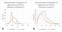
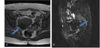
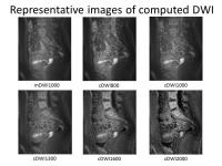
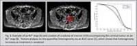
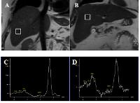
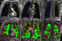
The differential diagnosis of benign and malignant focal
lesions of the lung is a hot and difficult problem in daily
chest imaging. The purpose of our study was to evaluate the
potential of intravoxel incoherent motion (IVIM)–derived
parameters as well as apparent diffusion coefficient (ADC)
in differentiating solitary pulmonary lesions. The results
demonstrate that IVIM-DWI could be more helpful for
distinguishing malignant from benign lesions in lung. D has
the best diagnostic efficiency.
|
|
1600.
 |
MR imaging of saline flooded lung – A feasibility study in a
large animal model
Frank Wolfram1, Thomas Lesser1, Harald
Schubert2, Joachim Böttcher3, Jürgen R
Reichenbach4, and Daniel Güllmar4
1Department of Thoracic and Vascular Surgery, SRH
Wald-Klinikum Gera, Teaching Hospital of Friedrich Schiller
University of Jena, Gera, Germany, 2Institute
of Laboratory Animal Sciences and Welfare, Jena University
Hospital - Friedrich Schiller University Jena, Jena,
Germany, 3Institute
of Diagnostic and Interventional Radiology, SRH Wald-Klinikum
Gera, Teaching Hospital of Friedrich Schiller University of
Jena, Gera, Germany,4Medical Physics Group / IDIR,
Jena University Hospital - Friedrich Schiller University
Jena, Jena, Germany
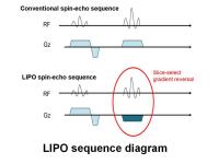
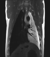
MR imaging of ventilated lung is a challenging task. The low
proton density with extremely short T2* and local field
inhomogeneities on tissue-air interfaces are sub-optimal for
MRI. Unilateral lung flooding replaces air content of one
lung wing with saline. This experimental method enables
sonographic guidance as well as therapeutic ultrasound
ablation. The untoward properties of lung might change to
ideal conditions with a homogen and high proton density
after flooding. The aim of the study was to investigate the
feasibility of in-vivo unilateral lung flooding in MR
environment and to evaluate the MR imaging capabilities of
flooded lung in a large animal model.
|
|
1601.
 |
Monitoring therapeutic response in anatomy and functions on
pulmonary fibrosis by ultra-short echo time (UTE) MRI in an
orthotopic mouse model 
Masaya Takahashi1, Keisuke Ishimatsu1,
Shanrong Zhang1, Hua Lu2, and Connie
C.W. Hsia2
1Advanced Imaging Research Center, University of
Texas Southwestern Medical Center, Dallas, TX, United
States, 2Pulmonary
and Critical Care Medicine, University of Texas Southwestern
Medical Center, Dallas, TX, United States
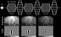
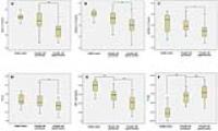
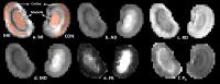
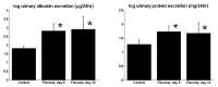
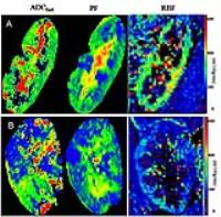
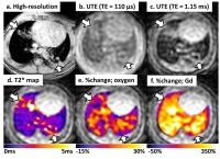
The purpose of this study was to investigate the ability of in
vivo ultra-short
echo time (UTE)-MRI for assessment of pulmonary
microstructure and functions of ventilation-perfusion in an
animal model of pulmonary fibrosis in comparison with
high-resolution MRI, physiological global measures and
histomorphology.
|
|
1602.
 |
Optimized four channel phased array coil for mice lung imaging
at 11.7 T
Marta Tibiletti1, Dominik Berthel2,
Michael Neumaier3, Dorothee Schüler2,
Detlef Stiller3, and Volker Rasche1,4
1Core Facility Small Animal MRI, Ulm University,
Ulm, Germany, 2Rapid
Biomedical GmbH, Rimpar, Germany, 3Target
Discovery Research, In-vivo imaging laboratory, Boehringer
Ingelheim Pharma GmbH & Co. KG, Biberach an der Riss,
Germany, 4Department
of Internal Medicine II, Ulm University, Ulm, Germany
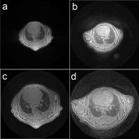
Lung imaging with MRI is challenging, due to the low proton
density in the tissue, short T2* values due to multiple
air-tissue interfaces, and respiratory and cardiac motion. A
major step for providing sufficient signal to noise ratio
(SNR) is the availability of dedicated coils optimized for
the specific application. In this work, we present a
4-channel mouse phased-array coil optimized for the thoracic
anatomy of mice. Depending on the field-of-view an average
two- to threefold gain in SNR was observed in direct
comparison to a conventional transmitt/receive quadrature
volume coil at 11.7 T.
|
|
1603.
 |
Three dimensional inversion recovery dual-echo ultrashort echo
time imaging with k-space reordering for effective suppression
of longer T2 species in lung parenchyma imaging
Neville D Gai1, Ashkan A Malayeri1,
and David A Bluemke1
1Radiology & Imaging Sciences, NIH, Bethesda, MD,
United States
Effective imaging of short T2 species requires efficient
suppression of longer T2 tisues to maximize short T2
contrast and dynamic range. While inversion with segmented
k-space acquisition in Cartesian schemes is straightforward,
inversion with segmented k-space UTE radial acquisition
offers some challenges since the center of k-space is
sampled with each acquisition resulting in magnetization
modulation related artifacts. Here we perform 3D inversion
recovery dual-echo UTE imaging of lung parenchyma using a
reordered k-space radial scheme to perform artifact free
high contrast imaging of native lung parenchyma.
|
|
1604.
 |
A new CF-specific MRI-Score: can it predict loss of lung
function?
Ilias Tsiflikas1, Matthias Teufel1,
Sabrina Fleischer1, Dominik Hartl2,
Konstantin Nikolaou1, and Juergen F Schaefer1
1Diagnostic and Interventional Radiology,
University Hospital of Tuebingen, Tübingen, Germany, 2Pediatrics
I - CF Center, University Hospital of Tuebingen, Tübingen,
Germany
The study successfully evaluated a new developed CF-specific
MRI score. Our results show that the MRI-Score can predict
the loss of pulmonary function. Thus, our findings may help
that MRI can serve as a novel predictive marker for loss of
lung function in CF and thereby help to tailor
individualized monitoring and treatment strategies.
|
|
1605.
 |
Free Breathing Multi-parametric quantitative Assessment of
Mesothelioma with MRI
Ravi Teja Seethamraju1, Noreen Dunham2,
Donna Oka2, Aida Faria2, and Ritu
Randhawa Gill2
1MR R&D, Siemens Healthcare, Boston, MA, United
States, 2Radiology,
Brigham and Women's Hospital, Boston, MA, United States
We demonstrate that free breathing multi-parametric
quantitative assessment of mesothelioma with MRI is
feasible. DCE imaging of the thorax with 3D Radial stack of
stairs gradient echo (radial VIBE) sequence can be acquired
while free breathing and the resulting pharmacokinetic maps
are of higher diagnostic value than current standard of 2D
or 3D FLASH acquisitions without the need for
co-registration. Similarly DWI with readout segmented EPI
(RESOLVE) provides similar diagnostic value with a free
breathing acquisition. These two biomarkers help improve the
evaluation of tumor in mesothelioma patients.
|
|
1606.
 |
Matrix pencil decomposition of time-resolved proton MRI for
robust and improved assessment of lung ventilation and perfusion
Grzegorz Bauman1,2 and
Oliver Bieri1,2
1Radiological Physics, University Hospital of
Basel, Basel, Switzerland, 2Department
of Biomedical Engineering, University of Basel, Basel,
Switzerland
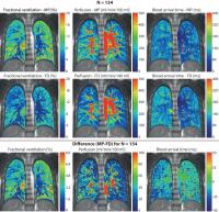
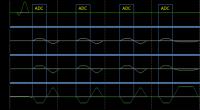
In the contemporary Fourier decomposition lung MRI,
time-resolved registered 2D image series are Fourier
transformed to identify in a power spectrum the underlying
respiratory and cardiac frequencies. Subsequently, the
amplitudes corresponding to the respiratory and cardiac
motion are extracted voxel-wise to eventually produce
ventilation and perfusion images. However, the analysis of
truncated oscillatory signals and the peak search in the
Fourier spectrum is usually very unstable and inaccurate.
Here, we propose to use a robust and fully-automated method
of signal analysis using a matrix pencil decomposition in
combination with a linear least squares analysis for
improved quantitative pulmonary function assessment.
|
|
1607.
 |
19F Ventilation Imaging of Cystic Fibrosis Patients
Yueh Lee1, Esther Akinnagbe-Zusterzeel1,
Jennifer Goralski1, Scott Donaldson1,
Hongyu An2, H. Cecil Charles3, and
Richard Boucher1
1The University of North Carolina at Chapel Hill,
Chapel Hill, NC, United States, 2Washinton
University, St. Louis, MO, United States, 3Duke
University, Durham, NC, United States
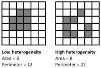
19F MRI Ventilation imaging of cystic fibrosis patients
demonstrates the disease heterogeneity using a
straightforward dynamic protocol.
|
|
1608.
 |
Non-cartesian SENSE reconstruction of 3D UTE Cones for fast MR
lung imaging
Konstantinos Zeimpekis1,2, Klaas Pruessmann2,
Florian Wiesinger3, Patrick Veit-Haibach1,
and Gaspar Delso4
1Nuclear Medicine, University Hospital Zurich,
Zurich, Switzerland, 2Information
Technology and Electrical Engineering, ETHZ, Zurich,
Switzerland, 3GE
Global Research, Munich, Germany, 4GE
Healthcare, Waukesha, WI, United States
This study is about a first attempt to use CG-SENSE parallel
reconstruction for non-cartesian 3D Ultra-short Echo Time
Cones sequence for lung imaging. Primary goal is to test
under-sampled data that reduce the scan time effectively to
one quarter of the fully sampled acquisition and check if
the reconstruction manages to capture lung density signal to
be used for accurate PET Attenuation Correction on a PET/MRI
since conventional sequences that are currently used do not
capture any. We test also the possibility for high
resolution lung imaging from the undersampled data
reconstructed with CG-SENSE algorithm.
|
|
1609.
 |
A Segmentation Pipeline for Measuring Pulmonary Ventilation
Suitable for Clinical Workflows and Decision-making
Fumin Guo1, Khadija Sheikh1, Rachel
Eddy1, Dante PI Capaldi1, David G
McCormack2, Aaron Fenster1, and Grace
Parraga1
1Robarts Research Institute, The University of
Western Ontario, London, ON, Canada, 2Department
of Medicine, The University of Western Ontario, London, ON,
Canada
Clinical translation of hyperpolarized 129Xe
MRI for large-scale and multi-centre applications requires
image analysis tools that can provide clinically-acceptable
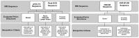
measurements of pulmonary information. Here we proposed a
pipeline that consists of 1H-129Xe
registration, segmentation and ventilation defects
generation for regional and quantitative evaluation of 129Xe
ventilation. 1H-129Xe
registration was performed using a state-of-art registration
approach. 1H
MRI segmentation was performed using primal-dual analysis
methods and modern convex optimization techniques with
incorporation of region information from 129Xe
MRI. We applied the pipeline across a range of pulmonary
abnormalities and this computationally efficient pipeline
demonstrated high agreement with reference standard,
suggesting its suitability for efficient clinical workflows.
|
|
1610.
 |
Quantitative Aerosol Deposition in Mechanically-Ventilated
Healthy and Asthmatic Rats using UTE-MRI
Hongchen Wang1, Georges Willoquet1,
Catherine Sebrié1, Sébastien Judé2,
Anne Maurin2, Rose-Marie Dubuisson1,
Luc Darrasse1, Geneviève Guillot1,
Ludovic de Rochefort1, and Xavier Maître1
1Imagerie par Résonance Magnétique Médicale et
Multi-Modalités (UMR8081) IR4M, CNRS, Univ. Paris-Sud,
Orsay, France, 2Centre
de Recherches Biologiques, CERB, Baugy, France
Asthma is the most common chronic respiratory disease
treated with inhaled therapy. However, aerosol deposition
patterns are complex and imaging methods are needed to
improve delivery efficiency. 3D UTE-MRI combined with
aerosolized Gd-DOTA had been formerly applied onto
spontaneous nose-only-breathing animals. Here, a mechanical
administration system was developed to ventilate and
nebulize rats. Resulting aerosol distribution and kinetics
were compared with free-breathing in healthy and asthmatic
animals.
|
|
1611.
 |
Investigation of the Multiple T2* Compartments in Lung
Parenchyma using a 3D Multi-Echo Radial sequence
Aiming Lu1, Xiangzhi Zhou1, Mitsue
Miyazaki1, Masao Yui2, Masaaki Umeda2,
and Yoshiharu Ohno3,4
1Toshiba Medical Research Inst., Vernon Hills,
IL, United States, 2Toshiba
Medical System Corp, Otawara, Japan, 3Advanced
Biomedical Imaging Research Center, Kobe University Graduate
School of Medicine, Kobe, Japan, 4Division
of Functional and Diagnostic Imaging Research, Department of
Radiology, Kobe University Graduate School of Medicine,
Kobe, Japan
T2* mapping with a single-exponential model have been
demonstrated to be useful in accessing pulmonary functional
loss. However, the model does not fully explain the signal
evolution at longer TEs. We propose to improve the T2*
characterization in the lung parenchyma with a
bi-exponential model. Using a 3D multi-echo radial sequence,
our results demonstrated that short T2* values and the
volume fractions of the two compartments could be obtained
on a clinical 3T scanner. In addition to the improved
accuracy of the short T2* measurement, the added fraction
values could also potentially be used as biomarkers.
|
|
1612.
 |
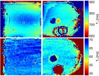
T1 relaxation time in lungs of asymptomatic smokers
Daniel Alamidi1, Simon Kindvall2,
Penny Hubbard Cristinacce3, Deirdre McGrath3,
Simon Young4, Josephine Naish3, John
Waterton3, Per Wollmer5, Sandra Diaz5,
Marita Olsson6, Paul Hockings7,8,
Kerstin Lagerstrand1, Geoffrey Parker3,9,
and Lars E Olsson2
1Department of Radiation Physics, Institute of
Clinical Sciences, Sahlgrenska Academy, University of
Gothenburg, Gothenburg, Sweden, 2Department
of Medical Physics, Lund University, Translational Sciences,
Malmö, Sweden, 3Centre
for Imaging Sciences and Biomedical Imaging Institute,
Manchester Academic Health Sciences Centre, University of
Manchester, Manchester, United Kingdom, 4AstraZeneca
R&D, Alderley Park, United Kingdom, 5Department
of Translational Medicine, Lund University, Malmö, Sweden, 6AstraZeneca
R&D, Mölndal, Sweden, 7Medtech
West, Chalmers University of Technology, Gothenburg, Sweden,8Antaros
Medical, BioVenture Hub, Mölndal, Sweden, 9Bioxydyn
Ltd, Manchester, United Kingdom
Tobacco smoking is the primary cause of COPD. MRI may
improve the characterization of COPD where T1 of the lungs
is a potential biomarker. We investigated whether smoking
affects lung T1 in individuals with no known lung disease.
Lung T1 measurements were performed in asymptomatic current
and never smokers. T1 was shortened with age and an
indication of shortened T1 in smokers was observed that most
likely reflects early signs of smoking-induced lung
pathology. Our results may be of utility to power future
prospective studies with larger cohorts and improved
regional analysis.
|
|
1613.
 |
Retrospective reconstruction using recorded cardiac and
respiration data of 3D radial acquisition of a human torso
Daniel Güllmar1, Georg Hille2, Martin
Krämer1, Karl-Heinz Herrmann1, Jürgen
R Reichenbach1, and Jens Haueisen2
1Medical Physics Group / IDIR, Jena University
Hospital - Friedrich Schiller University Jena, Jena,
Germany, 2Institute
of Biomedical Engineering and Informatics, Faculty of
Computer Science and Automation, Technical University
Ilmenau, Ilmenau, Germany
The aim of the study was to acquire 3D radially sampled
k-space data of a human torso without breath hold and
prospective cardiac triggering. Respiration and cardiac
pulsation were continuously recorded simultaneously with MR
imaging over a time frame of 1 h. Retrospective data motion
triggering was used to reconstruct 8 up to 10 different
respiration phases and 12 up to 20 different cardiac cycle
phases, resulting in 96 up to 200 different phase
combinations. Image quality was evaluated based on SNR, CNR
and under sampling artifacts.
|
|
1614.
 |
Asymmetric line broadening in lung tissue
Lukas Reinhold Buschle1, Felix Tobias Kurz1,2,
Thomas Kampf3, Heinz-Peter Schlemmer1,
and Christian Herbert Ziener1
1E010 Radiology, German Cancer Research Center
(DKFZ), Heidelberg, Germany, 2Department
of Neuroradiology, Heidelberg University Hospital,
Heidelberg, Germany, 3Department
of Experimental Physics 5, University of Würzburg, Würzburg,
Germany
We analyze the local line shape in human lung tissue in
dependence of the underlying microscopic tissue parameters
such as diffusion coefficient, alveolar size and
susceptibility difference. The interplay between
susceptibility- and diffusion-mediated effects is discussed
in several dephasing regimes. In vivo measurements for human
lung tissue show an excellent agreement with simulations of
the dephasing process. This allows an improved quantitative
diagnosis of early pulmonary fibrosis and emphysema.
|
|
1615.
 |
Dephasing and diffusion on the alveolar surface
Lukas Reinhold Buschle1, Felix Tobias Kurz1,2,
Thomas Kampf3, Heinz-Peter Schlemmer1,
and Christian Herbert Ziener1
1E010 Radiology, German Cancer Research Center
(DKFZ), Heidelberg, Germany, 2Department
of Neuroradiology, Heidelberg University Hospital,
Heidelberg, Germany, 3Department
of Experimental Physics 5, University of Würzburg, Würzburg,
Germany
In lung tissue, the susceptibility difference between
air-filled alveoli and surrounding tissue causes a strong
dephasing of spin-bearing particles. The particles
experience an averaged magnetic field due to diffusion
effects. Thus, the dephasing process slows down. Both
diffusion and susceptibility effects are described by the
Bloch-Torrey equation that is solved for the local
magnetization on the surface of alveoli. The analytical
solution of the free induction decay is compared to in vivo
measurements in human lung tissue.
|
|
1616.
 |
Evaluation of three different VIBE Sequences for Pulmonary
Lesions Detection in Patients with Lung Cancer
Hong Wang1, Xing Tang1, Panli Zuo2,
Shun Qi1, and Hong Yin1
1Department of Radiology, Department of
Radiology,Xijing Hospital, Xi'an, China, People's Republic
of, 2Siemens
Healthcare, MR Collaborations NE Asia, Beijing, China,
People's Republic of
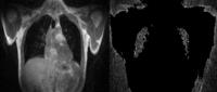
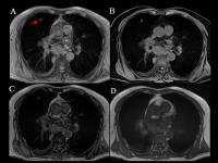
MR imaging is limited by poor evaluation of lung parenchyma
due to rapid single decay, low tissue portion density and
substantial respiratory motion. In this study, we evaluate
three different approaches of VIBE sequences in pulmonary
lesions detection, which including a short-TE breath-hold
VIBE, DIXON VIBE and a free-breathing Radial VIBE. We found
Breath-hold short-TE VIBE and Dixon VIBE sequences have
better performance in lesion detection than radial VIBE.
|
|
1617.
 |
Title: Breath-Hold Peripheral Pulse-Gated Black-Blood
T2-Weighted Lung Magnetic Resonance Imaging with the Variable
Refocusing Flip Angle Technique
Ryotaro Kamei1, Yuji Watanabe2, Koji
Sagiyama1, Satoshi Kawanami2, Atsushi
Takemura3, Masami Yoneyama3, and
Hiroshi Honda1
1Department of Clinical Radiology, Kyushu
University Graduate School of Medical Sciences, Fukuoka,
Japan, 2Department
of Molecular Imaging and Diagnosis, Kyushu University
Graduate School of Medical Sciences, Fukuoka, Japan, 3Healthcare,
Philips Electronics Japan, Tokyo, Japan
Breath-hold black-blood magnetic resonance imaging of the
lung provides promising results in focal lesion screening.
Using peripheral pulse gating, we intended to improve the
image quality obtained with previously reported methods.
Black-blood fat-saturated T2-weighted images were acquired
for healthy volunteers at various time points during the
pulse cycle. The degree of attenuation of the intraluminal
signals was quantified. The longest trigger delay, which
corresponds to the systolic phase, provided superior
black-blood effects and was considered optimal for signal
acquisition. Thus, peripheral pulse gating enabled
convenient and effective attenuation of the signals within
pulmonary vessels.
|
|
1618.
 |
Dynamic Contrast-Enhanced Perfusion MR Imaging at 3T System:
Influence of Contrast Media Concentration to Capabilities of
Pulmonary Perfusion Parameter and Functional Loss Evaluations as
Compared with Dynamic Contrast-Enhanced Perfusion Area-Detector
CT
Yoshiharu Ohno1,2, Yuji Kishida2,
Shinichiro Seki2, Hisanobu Koyama2,
Shigeru Ohyu3, Masao Yui3, Takeshi
Yoshikawa1,2, Katsusuke Kyotani4, and
Kazuro Sugimura2
1Advanced Biomedical Imaging Research Center,
Kobe University Graduate School of Medicine, Kobe, Japan, 2Radiology,
Kobe University Graduate School of Medicine, Kobe, Japan, 3Toshiba
Medical Systems Corporation, Otawara, Japan, 4Center
for Radiology and Radiation Oncology, Kobe University
Hospital, Kobe, Japan
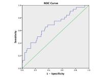
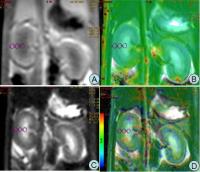
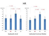
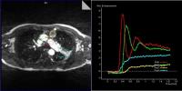
Quantification of perfusion parameter from dynamic
CE-perfusion MRI at 3T system may be more difficult than
that at 1.5T system, and contrast media concentration may
have larger influence to measurement error of perfusion
parameter on a 3T system. We hypothesized that a bolus
injection protocol with appropriately small contrast media
volume can provide accurate pulmonary perfusion parameter on
dynamic CE-perfusion MRI at a 3T system. The purpose of
this study was to determine the appropriate contrast media
volume for quantitative assessment of dynamic CE-pulmonary
MRI, when compared with dynamic CE-area-detector CT (ADCT)
for quantitative evaluation of perfusion within whole lung.
|
|
1619.
 |
High-resolution 3D ultra-short echo-time imaging of the lung in
young children at 3T without sedation
Wingchi Edmund Kwok1,2, Clement Ren3,
Gloria Pryhuber4, Mitchell Chess1, and
Jason C. Woods5
1Department of Imaging Sciences, University of
Rochester, Rochester, NY, United States, 2Rochester
Center for Brain Imaging, University of Rochester,
Rochester, NY, United States, 3Department
of Pediatrics, University of Rochester, Rochester, NY,
United States, 4Departments
of Pediatrics and Environmental Medicine, University of
Rochester, Rochester, NY, United States, 5Departments
of Pediatrics and Radiology, Cincinnati Children’s Hospital
Medical Center, Cincinnati, OH, United States
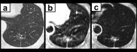
Our purpose was to study the feasibility of high-resolution
lung ultra-short TE imaging of young children at 3T without
sedation and tackle potential challenges. Two subjects aged
7 and 8 with mild cystic fibrosis were recruited. They were
supported by a child life specialist and the use of a mock
magnet. Siemens work-in-progress UTE and PETRA_D sequences
were used for lung imaging. The images depicted the lung
parenchyma, airways and vessels, and revealed abnormalities
such as bronchial wall thickening. The techniques should be
useful for the monitoring of lung development and evaluation
of lung diseases in children.
|
|
1620.
 |
Investigating the Correlation between Alveolar Surface-to-Volume
Ratio and Apparent Diffusion Coefficient with Hyperpolarized
Xenon-129 MRI
Kai Ruppert1,2, Kun Qing2, Talissa A.
Altes2,3, and John P. Mugler III2
1Cincinnati Children's Hospital, Cincinnati, OH,
United States, 2University
of Virginia, Charlottesville, VA, United States, 3University
of Missouri, Columbia, MO, United States
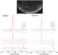
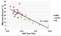
Chemical Shift Saturation Recovery (CSSR) MR Spectroscopy is
a method for monitoring the uptake of hyperpolarized
xenon-129 (HXe) by lung parenchyma. The purpose of this
study was to investigate the correlation between the
alveolar surface-to-volume ratio (S/V) as assessed by CSSR
spectroscopy and apparent diffusion coefficient measurements
in subjects with chronic-obstructive pulmonary disease,
healthy smokers and age-matched normals. Only for very short
delay times (5 ms or less) a good correlation was
established. Surprisingly, the best correlation, and
presumably most accurate S/V value, was obtained by using
just the red-blood cell peak at the shortest measured delay
time of 3ms.
|
|
1621.
 |
Hyperpolarized Xenon-129 Lung 3D SB-CSI at 1.5 and 3 Tesla
Steven Guan1, Kun Qing1, Talissa Altes1,
John Mugler III1, Borna Mehrad1,
Michael Shim1, Quan Chen1, Paul Read1,
James Larner1, Iulian Ruset2,3, Grady
Miller1, James Brookeman1, William
Hersman2,3, and Jaime Mata1
1University of Virginia, Charlottesville, VA,
United States, 2University
of New Hampshire, Duhram, NH, United States, 3XeMed,
Duhram, NH, United States
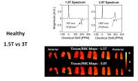
3D Single-Breath Chemical Shift Imaging (3D SB-CSI) is
capable of non-invasively assessing regional lung
ventilation and gas uptake/exchange within a single
breath-hold, typically less than 13 seconds. From this
study, we present preliminary clinical results of 3D SB-CSI
from healthy, cystic fibrosis (CF), interstitial lung
disease (ILD), and lung cancer (LC) subjects at 1.5T and
3T. Having novel information on regional changes in
ventilation and gas uptake/exchange allows for a better
understanding of lung physiology, disease progression, and
treatment efficacy.
|
|
1622.
 |
Spatial Fuzzy C-Means thresholding for semi-automated
calculation of percentage lung ventilated volume from
hyperpolarised gas and 1H
MRI
Paul J.C. Hughes1, Helen Marshall1,
Felix C. Horn1, Guilhem J. Collier1,
and Jim M. Wild1
1Academic Unit of Radiology, University of
Sheffield, Sheffield, United Kingdom
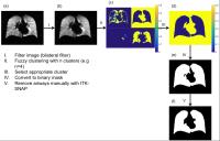
Automating image analysis is
key to accelerate quantitative image metric calculation and
increase consistency between observers. This work presents a
custom-built software to calculate percentage lung
ventilated volume (%VV) from hyperpolarised gas and 1H
MRI using spatial fuzzy c-means thresholding. The software
developed reduced analysis time and user input resulting in
significantly decreased interobserver variability when
postprocessing image data.
|
|
1623.
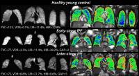 |
Differentiating Early Stage and Later Stage Idiopathic Pulmonary
Fibrosis using Hyperpolarized 129Xe
Ventilation MRI
Mu He1, Scott H. Robertson2, Jennifer
M. Wang3, Craig Rackley4, H. Page
McAdams5, and Bastiaan Driehuys5
1Electrical and Computer Engineering Department,
Duke University, Durham, NC, United States, 2Medical
Physics Graduate Program, Duke University, Durham, NC,
United States, 3School
of Medicine, Duke University, Durham, NC, United States, 4Pulmonary,
Allergy and Critical Care Medicine, Duke University Medical
Center, Durham, NC, United States, 5Radiology,
Duke University Medical Center, Durham, NC, United States
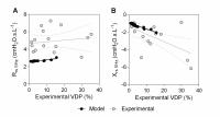
The use of 129Xe
MRI to characterize ventilation has received little
attention in IPF because these patients exhibit few
ventilation defect regions (VDR) compared to those with
other obstructive lung diseases. Here, we evaluate other
aspects of the ventilation distribution by optimized linear
binning. Ventilation distributions were quantified to
provide not only VDR, but also low and high-intensity
regions (LIR, HIR). We found that HIR was reduced by 3-fold
in patients with late versus early stage disease, as
measured by GAP IPF stage. Thus, loss of HIR may be a useful
marker of disease progression in IPF.
|
|
1624.
 |
A Dual Loop T/R-Xenon Coil for Homogenous Excitation with
Improved Comfort and Size
Wolfgang Loew1, Robert Thomen1, Randy
Giaquinto1, Ron Pratt1, Zackary
Cleveland1, Laura Walkup1, Charles
Dumoulin1, and Jason Woods1
1Imaging Research Center, Cincinnati Children’s
Hospital Medical Center, Cincinnati, OH, United States
Hyperpolarized gas MRI of lungs requires homogeneous RF
excitation and high SNR for proper spin-density mapping with
low flip angles. A dual loop T/R 129Xe
coil was designed and constructed to provide flexibility for
a wide range of patient sizes while maintaining high
transmit/receive homogeneity for hyperpolarized 129Xe
imaging and therefore provide high-quality images for
identifying and quantifying functional pulmonary
deficiencies. Electromagnetic field simulations were used to
analyze excitation profiles.
|
|
1625.
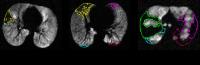 |
Lobar Ventilation Heterogeneity in Asthma and Cystic Fibrosis
Assessed with Hyperpolarized Helium-3 MRI and Computed
Tomography
Wei Zha1, Jeffery N Kammerman1, David
G Mummy2, Alfonso Rodriguez1, Robert V
Cadman1, Scott K Nagle1,3,4, Ronald L
Sorkness4,5,6, and Sean B Fain1,2,3
1Department of Medical Physics, University of
Wisconsin-Madison, Madison, WI, United States, 2Department
of Biomedical Engineering, University of Wisconsin-Madison,
Madison, WI, United States, 3Department
of Radiology, University of Wisconsin-Madison, Madison, WI,
United States, 4Department
of Pediatrics, University of Wisconsin-Madison, Madison, WI,
United States, 5Medicine-Allergy,
Pulmonary & Critical Care, University of Wisconsin-Madison,
Madison, WI, United States, 6Pharmacy,
University of Wisconsin-Madison, Madison, WI, United States
Seven cystic fibrosis (CF) and 69 asthma subjects with
different severities of disease underwent hyperpolarized
helium-3 MRI and multidetector computed tomography (MDCT).
Lobar segmentation was performed on proton MRI by
referencing corresponding MDCT. The lobar ventilation defect
percent (VDP) was measured by adaptive K-means.
Pairwise comparison showed that lobar VDP variation patterns
were different in CF vs. asthma, although patterns were
similar in severe vs. non-severe asthma. Disease-related
lobar VDP variation patterns may provide a sensitive
indicator for early detection and patterns of progression in
obstructive lung disease.
|
|
1626.
 |
Severity Evaluation in Cystic Fibrosis Using Oxygen-enhanced
MRI: Comparison to Hyperpolarized Helium-3 MRI
Wei Zha1, Stanley J Kruger1, Robert V
Cadman1, Kevin M Johnson1,2, Andrew D
Hahn1, Scott K Nagle1,2,3, and Sean B
Fain1,2,4
1Department of Medical Physics, University of
Wisconsin-Madison, Madison, WI, United States, 2Department
of Radiology, University of Wisconsin-Madison, Madison, WI,
United States, 3Department
of Pediatrics, University of Wisconsin-Madison, Madison, WI,
United States, 4Department
of Biomedical Engineering, University of Wisconsin-Madison,
Madison, WI, United States
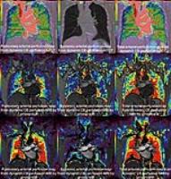
Oxygen-enhanced MRI using 3D radial ultrashort echo time
sequence (OE-MRI) is a promising alternative to evaluate
ventilation and defects with wider accessibility and better
affordability. Eleven cystic fibrosis (CF) subjects with
different severities of disease underwent OE-MRI and HP-MRI.
The disease severity ranks on the percent signal enhancement
map (PSE) derived from OE-MRI was compared to the whole lung
ventilation defect percent (VDP) measured from HP-MRI as a
reference standard using Spearman rank correlation. The
moderate association between VDP and PSE suggest OE-MRI
shows promise for differentiating disease severity in CF.
|
|
1627.
 |
Can the Forced Oscillation Technique and a Computational Model
of Respiratory System Mechanics Explain Asthma Ventilation
Defects?
Megan Fennema1, Sarah Svenningsen1,
Rachel Eddy1, Del Leary2, Geoffrey
Maksym3, and Grace Parraga1
1Robarts Research Institute, The University of
Western Ontario, London, ON, Canada, 2Environmental
and Radiological Health Sciences, Colorado State University,
Fort Collins, CO, United States, 3School
of Biomedical Engineering, Dalhousie University, Halifax,
NS, Canada
In patients with asthma, MRI has provided evidence of
ventilation-defects and heterogeneity. The etiology of
ventilation-heterogeneity is not well-understood, and
neither is its relationship with clinically-relevant
respiratory-system-impedance measurements. We evaluated the
potential relationships between MRI ventilation-defects and
respiratory-system-impedance measured in
vivo using
oscillometry and in
silico using
a computational airway-tree-model, in subjects clinically
diagnosed with asthma. Both experiments suggested a
significant relationship between MRI ventilation-defects and
respiratory-system-reactance. In
vivoexperimental data presented here reinforced the
validity of our computational airway-tree-model.
MRI-derived ventilation-defects in asthmatics can be
explained by lung impedance, specifically reactance,
measured experimentally and using a computational model.
|
|
1628.
 |
Quantitative Gas Exchange using Hyperpolarized 129Xe
MRI in Idiopathic Pulmonary Fibrosis
Ziyi Wang1, Scott Haile Robertson2,
Jennifer Wang3, Elianna Ada Bier2, Mu
He4, and Bastiaan Driehuys1,2,5
1Biomedical Engineering, Duke University, Durham,
NC, United States, 2Medical
Physics Graduate Program, Duke University, Durham, NC,
United States, 3School
of Medicine, Duke University, Durham, NC, United States, 4Electrical
and Computer Engineering, Duke University, Durham, NC,
United States, 5Radiology,
Duke University Medical Center, Durham, NC, United States
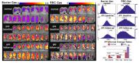
Hyperpolarized 129Xe
MRI exploits solubility and chemical shift to image regional
alterations in gas exchange. These properties have been
particularly promising for sensitive detection and
monitoring of idiopathic pulmonary fibrosis (IPF). Here we
seek to refine our analysis of regional gas exchange
impairment by mapping the 129Xe
uptake in blood and barrier tissues relative to gas-phase
signal intensity. This work shows that gas exchange
impairment is dominated by increased 129Xe
uptake in barrier tissues.
|
|
1630.
 |
Evaluation of esophageal cancer: comparison of MRI and CT
Wei Wang1, Wei Li1, Xueqian Shang1,
and Xiaoying Wang1
1Peking University First Hospital, Beijing,
China, People's Republic of
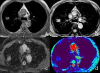
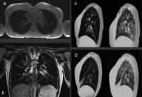
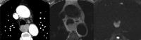
The study preliminary compared the ability of
non-contrast-enhanced MRI and contrast-enhanced CT in
detection, characterization and staging of esophageal
cancer. Ten patients’ chest CT and MR images were
subjectively evaluated. We found that MR was not inferior to
CT, and showed superior capacity in detecting early
unapparent cancer, delineating the tumor precisely and
depicting perfect contrast of surrounding structures. Also
MR was relatively safe than contrast-enhanced CT. MR may be
a potential useful tool for evaluation esophageal cancer.
|
|
1629.
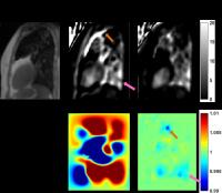
 |
In situ pH effects within Mycobacterium tuberculosis Infected
Mice revealed by UTE-CEST MRI
Jiadi Xu1, Vincent DeMarco2, Supriya
Pokkali 2,
Alvaro Ordonez2, Mariah Klunk2,
Marie-France Penet3, Zaver Bhujwalla3,
Peter van Zijl1,3, and Sanjay Jain2,4
1F. M. Kirby Center, kennedy Krieger Institute,
Baltimore, MD, United States, 2Center
for Infection and Inflammation Imaging Research, Center for
Tuberculosis Research, Johns Hopkins University School of
Medicine, Baltimore, MD, United States, 3Russell
H. Morgan Department of Radiology and Radiological Science,
Johns Hopkins University School of Medicine, Baltimore, MD,
United States, 4Department
of Pediatrics, Johns Hopkins University School of Medicine,
Baltimore, MD, United States
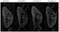
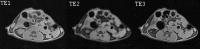
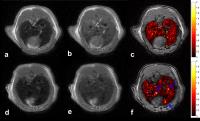
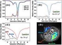
A UTE-CEST scheme was developed to acquire CEST spectrum on
M. Tuberculosis lesions in mouse lung. The scheme repeats a
selective saturation pulse together with an appropriate
mixing time; MRI images are acquired using the UTE technique
during the mixing times. The UTE readout is able to suppress
the respiratory motion artifacts commonly seen in lung MRI.
The pattern of the MTRasym spectra in the TB lesion, which
is dominated by protein signals, was used to assess lesion
pH.
|
 |
1631.
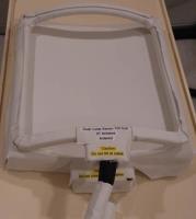
 |
Design of a multimodal (1H MRI/23Na MRI/CT) anthropomorphic
thorax phantom: Initial results at 3 T
Wiebke Neumann1, Florian Lietzmann1,
Lothar R. Schad1, and Frank G. Zöllner1
1Computer Assisted Clinical Medicine, Medical
Faculty Mannheim, Heidelberg University, Mannheim, Germany
Anthropomorphic phantoms are an essential tool for the
validation of image registration algorithms of multimodal
data and are important for quantification experiments in 1H
and 23Na
MR imaging. A human thorax phantom was developed with
insertable lung, liver, rib cage modules and tracking
spheres. Evaluation regarding the tissue-mimicking
characteristics with 1H
and 23Na
MR and CT imaging shows that the modules possess T1,
T2 and
HU values comparable to those of human tissues. This work
presents an MR- and CT-compatible phantom which allows
experimental studies for quantitative evaluation of
deformable, multimodal image registration algorithms and
realistic multi-nuclei MR imaging techniques.
|
|