ISMRM 24th Annual Meeting & Exhibition • 07-13 May 2016 • Singapore

Traditional Poster Session: Diffusion

1963 -1990 Diffusion: Clinical Applications
1991 -2032 Diffusion: Microstructure
2033 -2061 Diffusion Analysis & Tractography
2062 -2090 Diffusion: Analysis


1963.   
Prostate Cancer: Correlation of Intravoxel Incoherent Motion MR Parameters with Gleason Score
Dal Mo Yang1, Hyun Cheol Kim1, Sang Won Kim1, and Geon-Ho Jahng1
1Radiology, Kyung Hee University Hospital at Gangdong, Seoul, Korea, Republic of
Accurate assessment of prostate cancer aggressiveness is important for deciding treatment strategy. Functional MRI sequences such as DWI and DCE have been shown to provide information about tumor aggressiveness. Patients with high Gleason scores exhibited lower ADC values. In this study, we evaluated the potential of IVIM imaging to predict histologic prognostic parameters by investigating whether various IVIM parameters correlate with the Gleason score. The result indicates D is the best IVIM parameter for discriminating prostate cancers with low GS from prostate cancers with intermediate or high GS.


1964.   
Application of the inhomogeneous variable flip angle (I-VFA) scheme in hyperpolarized 129Xe DWI
Jianping Zhong1,2, Weiwei Ruan1, Xianping Sun1, Chaohui Ye1,2, and Xin Zhou1
1State Key Lab Magnet Resonance & Atom & Mol Phys, Wuhan Inst Phys & Math, Chinese Acad Sci, Wuhan, China, People's Republic of, 2School of Physics, Huazhong University of Science and Technology, Wuhan, China, People's Republic of
SNR and resolution are two important parameters for the quantitative assessment of MR images. In k-space, the central part contributes SNR, whereas the edges contribute details. The accuracy of the apparent diffusion coefficient (ADC) calibration was significantly affected by SNR. For hyperpolarized GRE sequences, the homogeneous variable flip angle scheme is sub-optimal and leads to low SNR images. We propose a simple method termed inhomogeneous variable flip angle (I-VFA) to derive ADC of hyperpolarized gases. Higher SNR images and more stable results can be achieved by this simple method.


1965.   
Is it well-thought-out to scan the preterm neonates at term-equivalent gestational age?
Yanyan Li1, Chao Jin1, Xianjun Li1,2, Miaomiao Wang1, Jie Gao1, Qinli Sun1, and Jian Yang1
1Department of Radiology, the First Affiliated Hospital of Xi’an Jiaotong University, Xi'an, China, People's Republic of, 2Department of Biomedical Engineering, School of Life Science and Technology, Xi'an Jiaotong University, Xi'an, China, People's Republic of
Considering the extra-environmental associated effects on brain development, it may be unreasonable to scan preterm infants at term-equivalent GA. To clarify this, we aim to explore the effects of postnatal days on neonatal WM maturation by DTI. Results indicate that postnatal days at-scan may be a considerable factor to investigate the WM maturation: during a close to in-uterine period, the absent effects of postnatal days may suggest the reasonability of performing neonatal MR-scans in such period; while as postnatal days increases, observed FA changes may imply the bias of comparing the preterm neonates at term-equivalent GA to term ones.


1966.   
Exploring the impact of common sequence variations on ADC reliability of lung lesions prior to protocol implementation in multi-centre clinical trials
Marianthi-Vasiliki Papoutsaki1, Alex Weller1, Matthew R Orton1, and Nandita M de Souza1
1Radiotherapy and Imaging, The Institute of Cancer Research, Sutton, London, United Kingdom
Standardization of diffusion-weighted (DW) protocols in multi-centre clinical trials is challenging. Prior to protocol development, the effect of inter-vendor related sequence variations on the apparent diffusion coefficient (ADC) reliability should be explored. In this study, the reliability of ADC estimates of lung lesions using two optimised DW protocols was assessed by mimicking vendor-related sequence variations. Patients with lung lesions were scanned twice using two DW protocols with different fat suppression techniques, diffusion gradient modes and TEs. These key variations increased the coefficient of variation of the ADC estimates of lung lesions, although absolute values did not differ significantly.


1967.   
Evaluation of Chemotherapeutic Effects in Patients with Lung Cancer using iShim-integrated Whole-Body Diffusion-Weighted Imaging
Xing Tang1, Hong Wang2, Panli Zuo3, Shun Qi4, and Hong Yin4
1Department of Radiology,Xijing hospital, Xi‘an, China, People's Republic of, 2Department of Radiology,Xijing Hospital, Xi'an, China, People's Republic of, 3Siemens Healthcare, MR Collaborations NE Asia, Beijing, China, People's Republic of, 4Department of Radiology, Department of Radiology, Xijing Hospital, Xi'an, China, People's Republic of
Whole-body diffusion-weighted imaging (WB-DWI) is now increasingly utilized for evaluation of the patient’s response to treatment. The purpose of this study is to evaluate the feasibility of WB-DWI with integrated slice-by-slice shimming (iShim) in patients with lung cancer.We found the SCLC is more sensitive to the chemotherapy than NSCLC using WB DWI.


1968.   
Diffusion Kurtosis Imaging and Tensor Imaging for Evaluation of Renal Changes in Diabetic Nephropathy: Preliminary study
Fan Mao1, Lihua Chen1, Yu Zhang2, Tao Ren1, Chenglong Wen1, and Wen Shen1
1Tianjin First Center Hospital, Tianjin, China, People's Republic of, 2Philips healthcare, Beijing, China, People's Republic of
Diffusion Tensor Imaging (DTI) as a noninvasive technique can provide valuable information based on the Brownian motion of water. However, the diffusion of water molecules in biological tissue like kidney does not follow a Gaussian distribution. Diffusion Kurtosis Imaging (DKI) can reflect the degree of restriction of hydrogen diffusion movement, and might detect the diffusion changes of kidney diseases more sensitive than DTI. Our study compared diffusion changes of DKI with DTI in kidneys of DN and healthy controls. The result showed DKI can be used for detecting renal changes in diabetes nephropathy with higher sensitivity compared to DTI.


1969.   
Intravoxel incoherent motion MRI in primary rectal cancer: correlation with histologic prognostic factors
Zhe Han1,2, Juan Chen2, Min Chen2, Chen Zhang2, and Lizhi Xie3
1Chinese Academy of Medical Science and Peking Union Medical College, Beijing, China, People's Republic of, 2Department of Radiology, Beijing Hospital, Beijing, China, People's Republic of, 3GE Healthcare, MR Research China, Beijing, China, People's Republic of
In this study we compared the association of intravoxel incoherent motion (IVIM) derived parameters with the histologic grade, N-stage, EGFR expression and K-RAS gene mutation of primary rectal cancer. Significant correlations were found between D values and differentiation grade, D* values and N-stage, f values and N-stage. IVIM derived parameters may be a promising imaging biomarker of tumor aggressiveness and prognosis.


1970.   
Liver metastasis from colorectal cancer; a comparison of reproducibility of ADC between multiple sites and vendors, at 1.5 T and 3 T
Ryan Pathak1, Neil A Thacker2, David M Morris2, Philippe Garteiser3, Sabrina Doblas3, Bernard E. Van Beers3, Houshang Amiri4, Arend Heerschap4, and Alan Jackson1
1The Wolfson Molecular Imaging Centre, University of Manchester, Manchester, United Kingdom, 2Centre for Imaging Sciences, University of Manchester, Manchester, United Kingdom, 3Laboratory of imaging biomarkers, INSERM, Paris Diderot University, Paris, France, 4Radboud University Medical Center, Nijmegen, Netherlands
ADC, calculated from diffusion-weighted MRI, is a potential quantitative imaging biomarker for detection of early treatment response. Imaging in the liver suffers from poor reproducibility, mainly as a result of respiratory motion. In this study we compare reproducibility in a multi-site, multi vendor setting at both 1.5 T and 3 T field strengths, for patients histologically diagnosed with colorectal cancer, who have radiological evidence of liver metastasis.


1971.   
Differential Diagnosis of Intrahepatic Cholangiocarcinoma and Hepatocellular Carcinoma by Using Diffusion-tensor Imaging
chen lihua1, liu ailian1, song qingwei1, wang heqing1, sun meiyu1, li ye1, chen anliang1, and xie lizhi2
1The Affiliated Hospital of Dalian Medical University, Dalian, China, Dalian, China, People's Republic of, 2GE Healthcare, MR Research China, Beijing, Beijing, China, People's Republic of
The advent of functional MR imaging has facilitated an increased role for imaging in risk stratification and treatment planning. In this study, DTI and DWI MR measurements were performed to investigate the correlation of the FA and ADC values in ROIs of the intrahepatic cholangiocarcinoma (ICC) and hepatocellular carcinoma (HCC), and in further the sensitivity, specificity and accuracy of the parameters for the diagnosis. DTI working at present scanning hardware are more capable to detect the pathophysiological changes unattainable compare to conventional MRI techniques.


1972.   
Evaluation of pathological stage and grade of endometrial carcinoma using sagittal DWI
Shifeng Tian1, Ailian Liu1, Ye Li1, and Jinghong Liu1
1The First Affiliated Hospital of Dalian Medical University, Dalian, China, People's Republic of
It has been reported that DWI has a high accuracy in the evaluation of the depth of EC, and ADC can predict the pathological grade of EC.The preoperative staging of sag DWI with EC was similar to sag T2WI. ADC value can be used to identify different pathological grades and some different stages of EC, with the increase of the pathological grade the ADC value decreased.


1973.   
To evaluate renal dysfunction using diffusion weighted magnetic resonance imaging based on intra-voxel incoherent motion (IVIM) and a mono-exponential model – a comparison study
Jiule Ding1, Jie Chen1, Zhenxing Jiang1, Hua Zhou2, Jia Di2, Wei Xing1, and Yongming Dai3
1Department of Radiology, Third Affiliated Hospital of Suzhou University, Changzhou, China, People's Republic of, 2Department of Nephrology, Third Affiliated Hospital of Suzhou University, Changzhou, China, People's Republic of, 3Philips Healthcare, Shanghai, China, People's Republic of
 The IVIM model was compared with the mono-exponential model to be used to differentiate sRI from non-sRI in this study. Results indicated that IVIM contributed little to improving the differentiation, therefore the mono-exponential model based ADC, a combination of fast and slow diffusion, might be more suitable as a biomarker image for assessing renal dysfunction.


1974.   
Estimation of pseudo-diffusion coefficient D* using different settings of low b-values in liver IVIM imaging
Meng-Chieh Liao1, Cheng-Ping Chien1, Shih-Han Hung1, Feng-Mao Chiu2, and Hsiao-Wen Chung1
1Graduate Institute of Biomedical Electronics and Bioinformatics, National Taiwan University, Taipei, Taiwan, 2Philips Healthcare, Taipei, Taiwan
The pseudo-diffusion coefficient (D*) in the liver estimated using intravoxel incoherent motion (IVIM) MRI currently suffers from inconsistent values reported in the literature. This study investigated the effect of low b-value settings on the estimation of D*. Data from healthy subjects with sixteen b-values were analyzed, with b-values of 0, 5, 10, and 15s/mm2 selectively removed and D* computed using a bi-exponential model. Results show progressive increases in D* estimations, with difference in values by a factor of two, which strongly suggest that the IVIM signals in the low b-value range do not obey single exponential decaying behavior.


1975.   
Structural Changes of the Superior Longitudinal Fasciculus and Cingulate Gyrus in Post Stroke Depression
Chenfei Ye1, Heather T Ma1, Jun Wu2, Xuhui Chen2, and Changle Zhang1
1Department of Electronic and Information Engineering, Harbin Institution of Technology Shenzhen Graduate School, Shenzhen, China, People's Republic of, 2Department of Neurology, Peking University Shenzhen Hospital, Shenzhen, China, People's Republic of
This study aim to investigate the relationship between depression after onset of stroke and superior longitudinal fasciculus and cingulate gyrus with multi-parameter DTI comparisons. These two brain structures distal to infarct regions were obtained by automatic segmentation and four parameters based on intensity distribution (mean, standard deviation, skewness and kurtosis) were quantitatively measured on each structure. Significant difference in these two structures was found among major depression subjects, mild depression subjects and the normal control. Our results verified that PSD patients latently exhibit neuroanatomical changes in superior longitudinal fasciculus and cingulate gyrus.


1976.   
High Resolution Cervical Spine DTI in Axial View using Non-triggered Multi-shot Acquisition and SYMPHONY Reconstruction
Xiaodong Ma1, Zhe Zhang1, Yuhui Xiong1, Erpeng Dai1, Yishi Wang1, Le He1, Chun Yuan1,2, and Hua Guo1
1Center for Biomedical Imaging Research, Department of Biomedical Engineering, School of Medicine, Tsinghua University, Beijing, China, People's Republic of, 2Vascular Imaging Laboratory, Department of Radiology, University of Washington, Seattle, WA, United States
In this study, 2D-navigated multi-shot EPI is used to achieve high resolution DTI in the cervical spine without cardiac triggering. A k-space reconstruction method, SYnergistic iMage reconstruction with PHase variatiOn and seNsitivitY (SYMPHONY), is used to correct the ghost artifacts caused by phase variations among different shots. The proposed technique is validated using quantitative analysis in healthy volunteers. Because no cardiac triggering is used, the scan time can be reduced. The improved spatial resolution and scan efficiency are beneficial for the quantitative evaluation of cervical spine in both neuroscience research and clinical diagnosis.


1977.   
Comparison of different mathematical models for IVIM in healthy human kidneys
Zhongwei Chen1, Youfan Zhao1, Zhenhua Zhang1, Haiwei Miu1, and Qiong Ye1
1Department of Radiology, The First Affiliated Hospital of Wenzhou Medical University, Wenzhou, China, People's Republic of
Various mathematical models have been applied in IVIM. Even with the same data, derived results change with the model used. Our study compared four popular mathematical models of IVIM in healthy human kidneys to explore this technique.


1978.   
Effects of variations in gestational age and birth anthropometric indicators on diffusion metrics of term neonatal white matter: a cohort study
Chao Jin1, Yanyan Li1, Xianjun Li1,2, Miaomiao Wang1, Jie Gao1, Qinli Sun1, and Jian Yang1
1Department of Radiology, the First Affiliated Hospital of Xi’an Jiaotong University, Xi'an, China, People's Republic of, 2Department of Biomedical Engineering, School of Life Science and Technology, University of Xi'an Jiaotong, Xi'an, China, People's Republic of
During the life span, brain development would be affected by numerous intra- and inter- factors in a short- or/and long-term period. To reveal typical birth indicators’ short-term effects, the effects of gestational age (GA), birth weight, crown-heel length and head circumference on term neonatal white matter were investigated by DTI. Results indicate that term neonates born with higher GA, birth weight and crown-heel length may hint better maturation of brain microstructure; among four birth indicators, GA was the main factor that influenced DTI-metrics. Particularly, longer crown-heel length with leftward superiority in corona radiata may presumably support early motor function.


1979.   
Assessment of fractional anisotropy of heart using ECG gating and second moment nulling pulse
Tomoya Nakamura1, Shuhei Shibukawa2, Yuma Sainokami2, Tomohiko Horie1, Isao Muro2, Terumitsu Hasebe1, Yutaka Imai2, and Tetsuo Ogino3
1Tokai University Hachioji Hospital, Hachioji, Japan, 2Tokai University Hospital, Isehara, Japan, 3Philips Healthcare Asia Pacific, Shinagawa, Japan
The purpose of this study is to assess the fractional anisotropy (FA) of heart using ECG gating and second moment nulling pulse which is intrinsically insensitive to motion. The FA at motion correction (MC) gradient was significantly higher than at acceleration motion correction (aMC) gradient, therefore, cardiac motion artifact resluts in an overestimation of FA. In conclusion, the use of second order motion correction gradient enables the quantification of FA at heart and has the potential to contribute to clinical cardiac imaging.


1980.   
Diffusion MRI of neuro-plasticity following complex motor learning
Maya Faraggi1, William D Richardson2, Derek K Jones3, and Yaniv Assaf4,5
1Neurobiology, Tel Aviv University, Tel Aviv, Israel, 2Wolfson Institute for Biomedical Research, University College London, London, United Kingdom, 3CUBRIC, Cardiff University, Cardiff, United Kingdom, 4Tel Aviv University, Tel Aviv, Israel, 5EMRIC, Cardiff University, Cardiff, United Kingdom
Neuroplasticity is the capacity of the nervous system to modify its organization as a result of a dynamic internal or external environment. In this study we aim to use DTI to characterize plasticity dynamics in the mouse brain as a result of a task with two degrees of difficulty.  In order to achieve that goal, we assessed motor learning ability using a running wheel with irregularly spaced rungs ("complex wheel"). Diffusion MRI revealed significant micro-structural changes in multiple brain areas expected to be affected by this task including the motor domain, sensory perception regions and white matter tracts .


1981.   
Fractional diffusion as a probe of microstructural change in a mouse model of Duchenne Muscular Dystrophy
Matt G Hall1, Paola Porcari2, Andrew Blamire2, and Chris A Clark1
1Institute of Child Health, University College London, London, United Kingdom, 2Newcastle Magnetic Resonance Centre, Newcastle University, Newcastle, United Kingdom
We apply a fractional diffusion model to preclinical data from a mouse model of Duchenne Muscular Dystrophy, and compare to histological measurements of the underlying tissue. We find that the alpha exponent of the model provides contrast which is indicative of the microstructural changes associated with DMD. We observe contrast between the wild type and mdx mouse model.


1982.   
Optimising Image Quality of Diffusion-Weighted Imaging of the Thyroid at 3.0 Tesla by Using iShim Sequence with iShim on
yin-chun liu1, meng-chao zhang2, hong zeng2, and lin liu2
1Ji Lin University sino-Japan hospital, chang chun, China, People's Republic of, 2chang chun, China, People's Republic of
My name is yin-chun liu.I am from Ji Lin University.


1983.   
Effects of Broad SPAIR Pulse, Continuous Fat Suppression Mode, Flow Compensation on Image Quality and Apparent Diffusion Coeffcient Reproducibility in iShim Diffusion-Weighted Imaging of the Abdomen at 3.0 T
He Sui1, Mengchao Zhang1, Hong Zeng1, and Lin Liu1
1Jilin University SINO-JAPAN Hospital, Changchun, China, People's Republic of
To detect the effects of Broad SPAIR Pulse, Continuous Fat Suppression Mode, Flow Compensation on Image Quality and Apparent Diffusion Coef?cient Reproducibility in iShim Diffusion-Weighted Imaging of the Abdomen at 3.0 T

The continuous fat suppression technique and Broad SPAIR Pulse can increase the fat saturation efficiency and decrease the ghost artifacts, when combined with flow compensation technique, the image qualit y can be further improved without affect ADC values.

High quality of the iShim Diffusion-Weighted Imaging can give a great help of detection of disease.


1984.   
Age-related changes of white matter diffusion anisotropy measures in old age observed with Double Diffusion Encoding
Marco Lawrenz1 and Juergen Finsterbusch1
1Systems Neuroscience, University Medical Center Hamburg-Eppendorf, Hamburg, Germany
With the help of double diffusion encoding experiments with two weighting periods applied successively microscopic tissue parameter can be gained. Rotationally invariant measures of the microscopic diffusion anisotropy such as the MA index may yield additional information complementary to DTI. Recent studies showed that MA can be determined in the living human brain, and normal values and their variation in groups of young and old healthy volunteers have been reported. In this study, the age-correlation of the diffusion anisotropy measures in terms of MA and FA values in a group of old volunteers (> 60 y) is discussed.


1985.   
Utility of histogram analysis of apparent diffusion coefficient value for distinguishing pituitary atypical adenomas from typical adenomas
Mariko Doai1, Naoko Tsuchiya1, Hisao Tonami1, and Osamu Tachibana2
1Radiology, Kanazawa Medical University, Kahoku, Japan, 2Neurosurgery, Kanazawa Medical University, Kahoku, Japan
The purpose of this study is to evaluate the utility of histogram analysis of apparent diffusion coefficient (ADC) value for distinguishing pituitary atypical adenomas from typical adenomas. The ADC maps were reviewed using Ziostation2, and placed a 3D volume-of interest on the tumor. The entire tumor were computed. Histogram parameters were then compared between atypical adenomas (n=3) and typical adenomas (n=11). Skewness and kurtosis of ADC histogram were significantly lower for atypical adenoma as compared with typical adenoma. ADC histogram analysis on the basis of the entire tumor volume can be useful in distinguishing atypical adenomas from typical adenomas.


1986.   
DW-MRI for evaluating lesions classified as responding and non-responding on RECIST criteria in patients with relapsed epithelial ovarian and primary peritoneal cancer re-challenged with platinum-based chemotherapy
Jennifer C Wakefield1,2, Jessica M Winfield1,2, Veronica Morgan2, Alison MacDonald2, Susana Banerjee1,2, Andrew N Priest3, Rebecca A Quest4, Susan Freeman3, Andrea G Rockall4, and Nandita M deSouza1,2
1Division of Radiotherapy and Imaging, Cancer Research UK Cancer Imaging Centre, The Institute of Cancer Research, London, United Kingdom, 2The Royal Marsden Hospital, Sutton, United Kingdom, 3Department of Radiology, Addenbrooke’s Hospital, Cambridge University Hospitals NHS Foundation Trust, Cambridge, United Kingdom, 4Imaging Department, Imperial College Healthcare NHS Trust, London, United Kingdom
The utility of Diffusion-weighted MRI (DW-MRI) in defining response by volume reduction or for determining the time-course of apparent diffusion coefficient (ADC) changes indicative of response has not been evaluated in patients with relapsed ovarian or peritoneal cancer. We evaluated post-treatment change in volume and ADC in lesions classified by RECIST criteria as responders and non-responders. We found responding lesions show greater change in volume and equivalent change in ADC to non-responding lesions after one cycle of chemotherapy. In non-responding lesions, the change in these parameters continued at the same rate post-first cycle of chemotherapy, indicating a delayed response.


1987.   
Hemodynamic-independent fluctuation MRI using self-correction in idiopathic normal pressure hydrocephalus
Naoki Ohno1, Tosiaki Miyati1, Marina Takatsuji2, Mitsuhito Mase3, Tomoshi Osawa3, and Yuta Shibamoto4
1Faculty of Health Sciences, Institute of Medical, Pharmaceutical and Health Sciences, Kanazawa University, Kanazawa, Japan, 2Division of Health Sciences, Graduate School of Medical Science, Kanazawa University, Kanazawa, Japan, 3Department of Neurosurgery and Restorative Neuroscience, Graduate School of Medical Sciences, Nagoya City University, Nagoya, Japan, 4Department of Radiology, Nagoya City University, Nagoya, Japan
Apparent diffusion coefficient (ADC) of the brain significantly changed during the cardiac cycle because of the water-molecule fluctuation. Moreover, this information assists in the diagnosis of idiopathic normal pressure hydrocephalus (iNPH). However, these changes (ΔADC) are affected by cerebral blood flow. Therefore, we corrected the effect of blood flow by using the diffusion data to evaluate hemodynamic-independent water fluctuation in iNPH. Corrected-ΔADC was significantly higher in iNPH group compared with control and atrophic ventricular dilatation groups. Hemodynamically independent analysis for water fluctuation MRI makes it possible to obtain more detailed information on biomechanical properties in iNPH.


1988.   
Diffusion weighted imaging of lymphedema post breast cancer treatment
Ned Charles1, Elizabeth Dylke1, David O'Brien1, Angela Borella2, Daniel Moses2, Sharon Kilbreath1, and Roger Bourne1
1University of Sydney, Sydney, Australia, 2Spectrum Medical Imaging, Sydney, Australia
Diffusion weighted imaging was performed in vivo in three patients with forearm lymphedema following lymphadenectomy for breast cancer.  The honeycomb-like structure of lymphedema was clearly visible on proton density images.  Parameter estimates from fitting monoexponential and kurtosis models to DWI data showed a shift in model parameters corresponding with the areas where lymphedema was present.  The parameter shifts suggest an increase in the partial volume of freely diffusing water consistent with edema, and suggest areas of increased interstitial water not visible in proton density images.


1989.   
The diagnostic value of Diffusion-weighted imaging in benign breast inflammatory lesions
Lina Zhang1, Jinli Meng2, Jianxun Qu 3, Jianguo Chu1, Ailian Liu1, Yanwei Miao1, Qingwei Song1, Zhijin Lang1, Jianyun Kang1, Qiang Wei1, and Bin Xu1
1The 1st affiliated hospital of Dalian Medical University, Da lian, China, People's Republic of, 2Chengban Branch of West China Hospital, Chengdu, China, People's Republic of, 3GE Healthcare, MR Research China, Beijing, Beijing, China, People's Republic of
To evaluate the diagnosis value of conventional MRI and Diffusion-weighted imaging in different subtypes of benign inflammation breast lesions. From the result we can see that morphological features as while as MR manifestations especially ADC findings may be of significant value for diagnosing different benign breast inflammatory lesions.


1990.   
High-resolution Multi-Station Diffusion imaging using accelerated Multi-Shot Acquisition Mode.
Arnaud Guidon1, Maggie M Fung2, Lloyd Estkowski3, Mei-Lan Chu4, Nan-Kuei Chen4, and Ersin Bayram5
1Global MR Applications & Workflow, GE Healthcare, Boston, MA, United States, 2Global MR Applications & Workflow, GE Healthcare, New York City, NY, United States, 3Global MR Applications & Workflow, GE Healthcare, Menlo Park, CA, United States, 4Brain Imaging and Analysis Center, Duke University Medical Center, Durham, NC, United States, 5Global MR Applications & Workflow, GE Healthcare, Houston, TX, United States
This study investigates the feasibility of a multishot acquisition method for high-resolution Whole-Body Diffusion Weighted Imaging (WBDWI) as compared to the standard single-shot EPI.  An accelerated Multishot acquisition mode is proposed to reduce the scan time of the high-resolution scan by half.

1991.   
Reducing acquisition time for axon diameter mapping using global optimization in the spatial-angular-microstructure space
Anna Auria1, David Romascano1, Erick J. Canales-Rodriguez2, Tim B. Dyrby3, Daniel C. Alexander4, Jean-Philippe Thiran1,5, Yves Wiaux6, and Alessandro Daducci1,5
1LTS5, École polytechnique fédérale de Lausanne (EPFL), Lausanne, Switzerland, 2Centro de Investigacion Biomedica en Red de Salud Mental (CIBERSAM), Barcelona, Spain, 3Danish Research Centre for Magnetic Resonance, Copenhagen University Hospital Hvidovre, Hvidovre, Denmark, 4Department of Computer Science and Centre for Medical Image Computing, University College London, London, United Kingdom,5University Hospital Center (CHUV) and University of Lausanne (UNIL), Lausanne, Switzerland, 6Institute of Sensors, Signals, and Systems, Heriot-Watt University, Edimburgh, United Kingdom
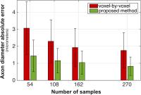
State-of-the-art microstructure imaging methods usually fit biophysical models to the diffusion MRI data on a voxel-by-voxel basis using non-linear procedures that require both long acquisitions and processing time. We recently introduced AMICO, a framework to reformulate these techniques as efficient linear problems and enable faster 
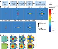
reconstructions. Here, we propose an extension that enables robust reconstructions from a reduced number of diffusion measurements, thus leading to faster acquisitions, too. Our novel formulation estimates simultaneously the microstructure configuration in all voxels as a global optimization problem, exploiting information from neighboring voxels that cannot be taken into account with existing techniques.


1992.   
Characterization of Brain White Matter Tissue Structure with Double-Diffusion-Encoded MRI
Yasar Goedecke1 and Jürgen Finsterbusch1
1Systems Neuroscience, University Medical Center Hamburg-Eppendorf, Hamburg, Germany
Double-diffusion-encoding (DDE) or double-wave-vector (DWV) experiments show a signal behavior that is specific for restricted diffusion. Thus, these experiments could provide more direct insight into tissue microstructure than conventional experiments, especially when targeting axon diameters. In this study, a previous DDE-based approach to estimate axon diameters is extended (i) to be applicable without prior knowledge of the fiber orientation, (ii) by considering a more complex tissue composition including spherical cells and an unrestricted compartment to model glial cells and extracellular space, and (iii) using the multiple correlation function framework that provides a more accurate approximation of the MR signal.


1993.   
Numerical Simulations Comparing Pore Imaging Methods Based on Diffusion-Weighted MR Imaging
Yasar Goedecke1 and Jürgen Finsterbusch1
1Systems Neuroscience, University Medical Center Hamburg-Eppendorf, Hamburg, Germany
In a conventional diffusion-weighted MRI experiment, the signal amplitude depends on the squared magnitude of the Fourier transformation of the pore or cell geometry, i.e. the underlying cell or pore geometry cannot be reconstructed. Several approaches have been proposed that determine the otherwise missing phase information and, thus, can image the pore or cell geometry directly. Here, the performance of these methods is compared with respect to their applicability in practice, e.g. considering the impact of the noise level, mixtures of pore sizes, orientations, and shapes, and gradient pulse durations and diffusion times achievable on standard MRI systems.


1994.   
The effect of axon shape and myelination on diffusion signals in a realistic Monte Carlo simulation environment
Michiel Kleinnijenhuis1, Jeroen Mollink1, Errin E Johnson2, Vitaly L Galinsky3, Lawrence R Frank3, Saad Jbabdi1, and Karla L Miller1
1Oxford Centre for Functional MRI of the Brain, University of Oxford, Oxford, United Kingdom, 2Sir William Dunn School of Pathology, University of Oxford, Oxford, United Kingdom, 3Center for Scientific Computation in Imaging, University of California San Diego, La Jolla, CA, United States
The cylindrical models often used in Monte Carlo diffusion simulations do not resemble the shape of axons very well. In this work, a more realistic substrate derived from electron microscopy data is used to investigate the influence of axon shape and myelination on the diffusion signal. In the DifSim simulation environment, diffusion signals from EM-derived substrates are compared to those from cylindrical substrates matched for volume fraction. Furthermore, the effect of removing the impermeable myelin sheath from the substrate is assessed.


1995.   
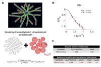
Modelling of diffusion in cultured epithelial cell spheroids
Sisi Liang1, Madiha Yunus2, Eleftheria Panagiotaki 3, Byung Kim4, Timothy Stait-Gardner5, Mikhail Zubkov5, Brian Hawkett4, William Price5, Carl Power6, and Roger Bourne2
1College of Engineering and Science, Victoria University, Melbourne, Australia, 2Discipline of Medical Radiation Sciences, Faculty of Health Sciences, University of Sydney, Sydney, Australia, 3Center for Medical Image Computing, University College London, London, United Kingdom, 4Key Centre For Polymer Colloids, University of Sydney, Sydney, Australia, 5Nanoscale Organisation and Dynamics Group, School of Science and Health, Western Sydney University, Sydney, Australia, 6Mark Wainright Analytical Centre, The university of New South Wales, Sydney, Australia
Cultured epithelial cell spheroids demonstrate many of the physiological properties of glandular epithelia and provide an ideal experimental model for investigation of the distinctive structural properties that may contribute to the reported low water mobility in prostate, breast, and gut epithelia. The structural connections are very similar to those in intact tissue and thus they provide a more realistic model of tissue than previously investigated models based on pelleted yeast or erythrocyte cells. We report an investigation of the correlation between known cell sizes in a spheroid culture and restriction radius estimated by a model of diffusion MRI signals.


1996.   
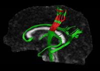
Imaging Three Dimensional Temporal Diffusion Spectrum Dispersion Profiles in the Brain
Dan Wu1, Frances J Northington2, and Jiangyang Zhang1,3
1Radiology, Johns Hopkins University School of Medicine, BALTIMORE, MD, United States, 2Pediatrics, Johns Hopkins University School of Medicine, BALTIMORE, MD, United States, 3Radiology, New York University School of Medicine, New Yourk, NY, United States
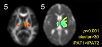
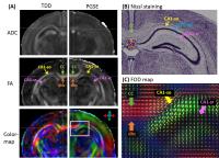
The dispersion profile of the temporal diffusion spectrum has been linked to key properties of tissue microstructures, however, its directional variance has not been shown. In this study, we extended the conventional one-dimensional dispersion profile to three-dimensional profile, and characterized its directionality with a tensor representation. The temporal diffusion dispersion (TDD) tensor demonstrated unique contrasts that reflected distinct microstructural organization in the mouse brain, and the high anisotropy from TDD tensors correlated with anisotropic structural arrangements, e.g., in the crossing fiber regions. The TDD contrasts are also sensitive to disrupted microstructures in a neonatal mouse model of hypoxic-ischemic injury.


1997.   
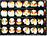
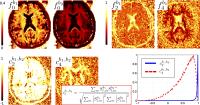
Spatiotemporal dynamics and patterns of cortical mean kurtosis and fractional anisotropy in the preterm brains
Tina Jeon1, Aristeidis Sotiras2, Minhui Ouyang1, Min Chen3, Lina Chalak4, Christos Davatzikos2, and Hao Huang1,5
1Radiology Research, Children's Hospital of Philadelphia, Philadelphia, PA, United States, 2Center for Biomedical Image Computing and Analytics, University of Pennsylvania, Philadelphia, PA, United States,3Department of Mathematical Sciences, University of Texas at Dallas, Richardson, TX, United States, 4Department of Pediatrics, University of Texas Southwestern Medical Center, Dallas, TX, United States, 5Perelman School of Medicine, University of Pennsylvania, Philadelphia, PA, United States
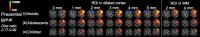
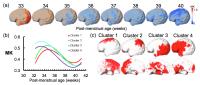
From early 3rd trimester to around birth, the cerebral cortex undergoes dramatic microstructural changes including dendritic arborization that disrupts the radial scaffold, a well-organized columnar organization. Decrease of cortical fractional anisotropy (FA) derived from DTI has been well documented. In this study, we hypothesized that non-Gaussian water diffusion properties (e.g. mean kurtosis or MK) from diffusion kurtosis imaging (DKI) offers unique and complementary information on cortical microstructural changes during this period. The spatiotemporal changes and patterns of cortical FA and MK from 32 to 41 postmenstrual weeks were revealed, demonstrating unique cortical MK maps and clustering patterns during preterm development.


1998.   
The influence of T2 relaxation in measuring the restricted volume fraction in diffusion MRI
Silvia De Santis1, Yaniv Assaf2, and Derek Jones1
1Cardiff University, CUBRIC, Cardiff, United Kingdom, 2Department of Neurobiology, Tel Aviv University, Tel Aviv, Israel
With the increasing popularity of multi-shell diffusion techniques to measure axonal density and diameter, the investigation of the exact origin of the contrast has become a hot topic. Here, we investigate the impact of the echo time in measuring the axonal density and show that the two water compartments are characterised by a different relaxation time T2, making the measures of the volume strongly dependent on the echo time. This suggests caution when comparing data acquired with different setups and introduces a new way of measuring the differential T2 properties of intra- and extra-axonal water pools.


1999.   
Diffusion MRI: Disentangling Micro- from Mesostructure and Bayesian Parameter Evaluation
Marco Reisert1, Elias Kellner1, Bibek Dhital1, Jürgen Hennig1, and Valerij G. Kiselev1
1Department of Radiology, Medical Physics, University Medical Center Freiburg, Freiburg, Germany
Diffusion-sensitized MRI probes the cellular structure of the human brain, but the primary microstructural information gets lost  in averaging over higher-level, mesoscopic tissue organization such as different orientations of neuronal fibers. While such averaging is inevitable due to the limited imaging resolution, we propose a method for disentangling the microscopic cell properties from the effects of mesoscopic structure. The proposed method finds detectable parameters of a given microstructural model and calculates them within seconds, which makes it suitable for a broad range of applications. 


2000.   
Intracellular volume fraction estimation in vivo in single and crossing fibre regions
Sjoerd B Vos1,2, Andrew Melbourne1, John S Duncan2,3, and Sebastien Ourselin1
1Translational Imaging Group, University College London, London, United Kingdom, 2MRI Unit, Epilepsy Society, Chalfont St Peter, United Kingdom, 3Department of Clinical and Experimental Epilepsy, UCL Institute of Neurology, London, United Kingdom
Intracellular volume fraction (ICVF) is a valuable biomarker of neurological disease. As one of two factors in g-ratio estimates it could potentially reveal axonal function from structural MRI measurements. Reliable ICVF estimation is critical for both purposes. With various diffusion models in existence for ICVF estimation, we compared the obtained ICVF values and their reproducibility in voxels with 1, 2, and 3 fibre populations between three diffusion modelling approaches. Absolute ICVF values vary significantly between models as well as between voxels with different fibre complexity.


2001.   
Modeling diffusion of intracellular metabolites in the mouse brain up to very high b: diffusion in long fibers (almost) accounts for non-monoexponential attenuation
Marco Palombo1,2, Clémence Ligneul1,2, and Julien Valette1,2
1CEA/DSV/I2BM/MIRCen, Fontenay-aux-Roses, France, 2CNRS Université Paris-Saclay UMR 9199, Fontenay-aux-Roses, France
We investigate how metabolite diffusion measured up to very high b (60 ms/µm2) at relatively short diffusion time (63.2 ms) in the mouse brain can be explained in terms of simple geometries. We model cell fibers as isotropically oriented cylinders of infinite length, and show this can account very well for measured non-monoexponential attenuation. The only exception is NAA, for which the model extracts fiber diameter equal to 0. We show that is theoretically and experimentally compatible with a small fraction of the NAA pool being confined in highly restricted compartments (with short T2), e.g. a mitochondrial pool.


2002.   
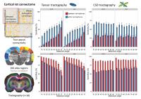
Evaluation of Diffusion MRI Based Feature Sets for the Classification of Primary Motor and Somatosensory Cortical Areas.
Tara Ganepola1,2, Jiaying Zhang2, Hui Zhang2, Martin I Sereno3, and Daniel C Alexander2
1Department of Cognitive, Perceptual and Brain Sciences, University College London, London, United Kingdom, 2Centre for Medical Image Computing, University College London, London, United Kingdom, 3Birkbeck-UCL Centre for Neuroimaging, University College London, London, United Kingdom
In the following work several diffusion based feature vectors (DTI, NODDI, spherical harmonic (SH) invariants and fourth order tensor invariants (T4)) are compared in order to validate their usability in grey matter investigations. It was found that using multi-shell data and non-biophysical models such as SH and T4 achieves the highest classification accuracy between the primary motor and somatosensory cortical areas, and thus is likely to characterise grey matter tissues domains more effectively.  
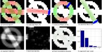

2003.   
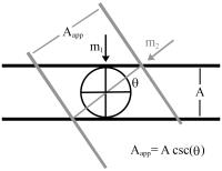
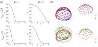
Inferring axon diameter from the apparent cylindrical geocentric diameter in the longitudinal plane
Farshid Sepehrband1 and Kristi A Clark1
1Laboratory of Neuro Imaging, USC Mark and Mary Stevens Neuroimaging and Informatics Institute, Keck School of Medicine of USC, Los Angeles, CA, United States
Recent diffusion-weighted imaging techniques have enabled the inference of axon diameter, a valuable neuroanatomical measure1,2. Current techniques fit a cylindrical model of axons to the acquired signal, primarily in the transverse direction. Despite many improvements, sensitivity to small axons is difficult to achieve, primarily due to the scanner’s physical limitations. Even with a strong gradient strength system such as the connectome scanner and high SNR, the minimum resolvable axon diameters are greater than 2μm, which accounts for only a small proportion of axons in the human brain. Here we utilize Neuman’s cylindrical model3, and generalize it to the geocentric direction in the longitudinal plane of axons (Figure 1) to decrease the minimum axon diameter resolvable with a given scanner.


2004.   
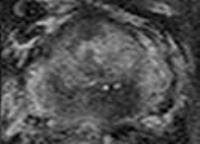
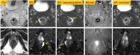
In vivo characterisation of mouse brain glioma using VERDICT MRI and validation with histology
Tom A Roberts1, Giulia Agliardi1, Andrada Ianus2, Ben Jordan1, James O Breen-Norris1, Rajiv Ramasawmy1, Angela D'Esposito1, Valerie Taylor1, Bernard Siow1, Eleftheria Panagiotaki2, Daniel C Alexander2, Mark F Lythgoe1, and Simon Walker-Samuel1
1Centre for Advanced Biomedical Imaging, London, United Kingdom, 2Centre for Medical Image Computing, London, United Kingdom
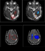
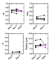
Vascular Extracellular and Restricted Diffusion for Cytometry in Tumours (VERDICT) is a diffusion MRI technique which uses a 3-compartment model to characterise the vascular (V), extracellular-extravascular (EES) and intracellular (IC) compartments in tumours. VERDICT allows for quantitation of tumour morphology including vascular fraction (fv), intracellular fraction (fic) and cellular radius, hence providing a non-invasive ‘biopsy’ that can be performed longitudinally. Previously, VERDICT has been applied to subcutaneous mouse tumours1 and human prostate cancer2. For the first time, we apply VERDICT in a mouse model of glioma, examine it in the context of other multi-compartment models and optimise it based on comparison with histological analysis.


2005.   
Generative statistical models of white matter microstructure for MRI simulations in virtual tissue blocks
Leandro Beltrachini1 and Alejandro Frangi1
1The University of Sheffield, Sheffield, United Kingdom
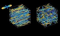
In silico studies of diffusion MRI are becoming a standard tool for testing the sensitivity of the technique to changes in white matter (WM) structures. To perform such simulations, realistic models of brain tissue microstructure are needed. However, most of the computational results are obtained considering straight and parallel cylinders models, which are known to be too simplistic for representing real-scenario situations. We present a statistical-driven approach for obtaining random models of WM tissue samples based on histomorphometric data available in the literature. We show the versatility of the method for characterising WM voxels representing bundles and disordered structures.


2006.   
Does Myelin Water Influence DWI?
Kevin D Harkins1 and Mark D Does1,2,3,4
1Institute of Image Science, Vanderbilt University, Nashville, TN, United States, 2Biomedical Engineering, Vanderbilt University, Nashville, TN, United States, 3Radiology and Radiological Sciences, Vanderbilt University, Nashville, TN, United States, 4Electrical Engineering, Vanderbilt University, Nashville, TN, United States
The presence and movement of myelin water is often neglected from models of DWI signal. This study presents a Monte Carlo simulation illustrating that myelin water diffusion can have a subtle but important impact on measured Dapp and Kapp values, and that incorporating myelin water diffusion can influence myelin-content dependent changes in Dapp and Kapp.


2007.   
Characterizing microstructural changes in Multiple Sclerosis lesions using advanced diffusion MRI at 3T and 7T
Silvia De Santis1,2, Matteo Bastiani2, Henk Jansma2, Amgad Droby3, Pierre Kolber3, Eberhard Pracht4, Tony Stoecker4, Frauke Zipp3, and Alard Roebroeck2
1Cardiff University, CUBRIC, Cardiff, United Kingdom, 2Dept. of Cognitive Neuroscience, Faculty of Psychology & Neuroscience, Maastricht University, Maastricht, Netherlands, 3Department of Neurology and Neuromaging Center, University Medical Center of the Johannes Gutenberg University, Mainz, Germany, 4German Center for Neurodegenerative diseases, Bonn, Germany
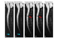
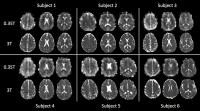
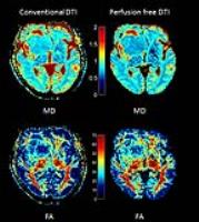
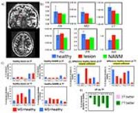
Aim of this work was to test the ability of conventional (i.e., DTI) and advanced (i.e., CHARMED, stretched exponential) diffusion methods to differentiate between Multiple Sclerosis lesions, normal appearing white matter and healthy controls, at both 3T and 7T. Advanced dMRI at 7T gives the best discriminating power between MS lesions and healthy tissue across WM; DTI is appropriate in areas of low fiber dispersion like the corpus callosum.


2008.   
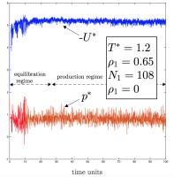
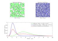
Exploring Structural, Diffusive and Thermodynamic Properties of Model Systems with Molecular Dynamics Simulations
Jonathan Phillips1
1Institute of Life Science, College of Medicine, Swansea University, Swansea, United Kingdom
This work aims at introducing methods of molecular dynamics (MD) simulation into diffusion MRI modelling. MD allows the study of transport properties (e.g. diffusion), structural properties (e.g. radial distribution functions) and thermodynamic properties (e.g. pressure). Access to all of these properties allows investigation into the links between them. We present the first steps into studying all of these properties (including the diffusion coefficient and kurtosis) in model systems for comparison with MRI data. The system is a binary mixture which includes a diffusing species (the solvent e.g. water) and a larger spatially-fixed species (modelling cellular-sized colloid particles).


2009.   
Axon diameter distribution influences diffusion-derived axonal density estimation in the human spinal cord: in silico and in vivo evidence
Francesco Grussu1, Torben Schneider1,2, Ferran Prados1,3, Carmen Tur1, Sébastien Ourselin3, Hui Zhang4, Daniel C. Alexander4, and Claudia Angela Michela Gandini Wheeler-Kingshott1,5
1NMR Research Unit, Queen Square MS Centre, Department of Neuroinflammation, UCL Institute of Neurology, University College London, London, United Kingdom, 2Philips Healthcare, Guildford, Surrey, England, United Kingdom, 3Translational Imaging Group, Department of Medical Physics and Biomedical Engineering, University College London, London, United Kingdom, 4Department of Computer Science and Centre for Medical Image Computing, University College London, London, United Kingdom, 5Brain Connectivity Center, C. Mondino National Neurological Institute, Pavia, Italy
Diffusion MRI-derived neurite density is a potential biomarker in neurological conditions. In the brain, neurites are commonly modelled as sticks for sufficiently long diffusion times and gradient durations. However, in the spinal cord, large axons are present and typical diffusion times (20-30 ms) may not be sufficiently long to support this model. We investigate via simulations and in vivo whether neurite density estimation is affected by the diffusion time in the spinal cord. Short diffusion times lead to bias, while long diffusion times improve accuracy but reduce precision. Therefore, a trade-off accuracy-precision needs to be evaluated depending on the application.


2010.   
A quantitative measurement of the cell membrane water permeability of expression-controlled AQP4 cells with diffusion weighted MRI
Takayuki Obata1, Jeff Kershaw2, Yasuhiko Tachibana1, Youichiro Abe3, Sayaka Shibata2, Yoko Ikoma2, Hiroshi Kawaguchi4, Ichio Aoki2, and Masato Yasui3
1Applied MRI Research, National Institute of Radiological Sciences, Chiba, Japan, 2Molecular Imaging Center, National Institute of Radiological Sciences, Chiba, Japan, 3Department of Pharmacology, Keio University, Tokyo, Japan, 4Human Informatics Research Institute, National Institute of Advanced Industrial Science and Technology (AIST), Tsukuba, Japan
We performed multi-b and multi-diffusion-time DWI on aquaporin-4-expressing and non-expressing cells, and demonstrated a clear difference between the signals from the two cell types. The data was interpreted with a two-compartment model including inter-compartmental exchange. It was also assumed that restricted diffusion of water molecules inside the cells leads to the intracellular diffusion coefficient being inversely proportional to the diffusion-time. Estimates of the water-exchange times with this model were comparable with those measured using an independent optical imaging technique, which suggests that this method might be used to characterize cell-membrane water permeability. As the technique can be applied in routine clinical examination, it has the potential to improve clinical diagnosis.


2011.   
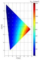
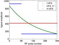
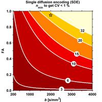
Acquisition Protocol Optimization for Axon Diameter Mapping at High-Performance Gradient Systems – A Simulation Study
Jonathan I Sperl1, Ek Tsoon Tan2, Miguel Molina Romero1,3, Marion I Menzel1, Chris J Hardy2, Luca Marinelli2, and Thomas K.F. Foo2
1GE Global Research, GARCHING, Germany, 2GE Global Research, NISKAYUNA, NY, United States, 3Institute of Medical Engineering, Technische Universität München, GARCHING, Germany
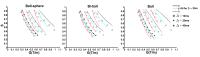
The measurement of axonal diameter by diffusion MRI techniques has assumed major interest in the research community. While most work has focused on developing and comparing various multi-compartment models, only minor efforts have been undertaken to optimize corresponding acquisition protocols. In this work we perform simulations using a rather simple two-compartment model, but study the effect of various choices of acquisition parameters on the precision and the bias of the fitted parameters. More precisely, we analyze potential sampling strategies in the 2D design space spanned by the two timing parameters (Δ, δ) of the diffusion encoding.


2012.   
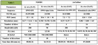
NODDI and AxCaliber diffusion-weighted imaging at ultrahigh 
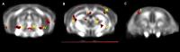
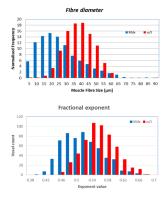
field for microstructural imaging of the mouse spinal cord
Ahmad Joman Alghamdi1,2, Hari K Ramachandran3, Ian M Brereton1, and Nyoman D Kurniawan1
1Centre for Advanced Imaging, The University of Queensland, Brisbane, Australia, 2College of Health Sciences, Taif University, Taif, Saudi Arabia, 3Computer Science and Engineering, SRM University, Kattankulathur, India
DTI has been used to measure changes in spinal cord WM, but lacks the specificity in measuring changes in GM and axonal diameter. This study aims to apply NODDI and AxCaliber techniques to measure characteristics of the lumbar spine in C57BL/6 mice, in-vivo at 9.4T and ex-vivo at 
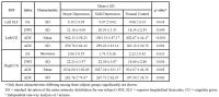
16.4T. The GM orientation distribution index is 3 times that of the WM, and the correlation of ODI to FA is r=–0.9, P<<0.01 for GM and r=–0.56, P<<0.01 for WM. AxCaliber analysis determined WM axon diameter populations with an average of 1.55±0.15mm (in-vivo); and 1.37±0.20 mm (ex-vivo). 


2013.   
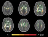
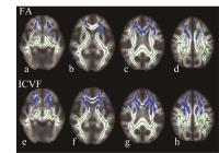
White matter alterations in young adults born extremely preterm: a microstructural point of view.
Zach Eaton-Rosen1, Andrew Melbourne1, Joanne Beckmann2, Eliza Orasanu1, Nicola Stevens3, David Atkinson4, Neil Marlow2, and Sebastien Ourselin1
1TIG, UCL, London, United Kingdom, 2UCL EGA Institute for Women's Health, London, United Kingdom, 3UCLH, London, United Kingdom, 4CMIC, UCL, London, United Kingdom
We used NODDI and DTI in order to investigate the differences in white matter between young adults born at term, and those born at fewer than 26 weeks completed gestation, using TBSS. The differences in FA were closely mirrored by the differences in orientation dispersion index (ODI) while the intra-axonal volume fraction (Vi) did not show significant differences in the same regions. This suggests that the ODI may be more sensitive to indicators of being born preterm than Vi in the white matter.
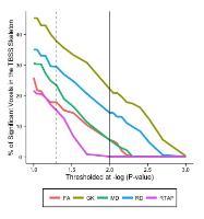

2014.   
Statistical assessment of a model combining IVIM and T2 decay for multi-b-value, multi-echo-time DW-MRI in abdominal organs
Matthew R Orton1, Neil P Jerome1, Thorsten Feiweier2, Dow-Mu Koh3, Martin O Leach4, and David J Collins4
1Radiotherapy and Imaging, Institute of Cancer Research, London, United Kingdom, 2Siemens Healthcare, Erlangen, Germany, 3Department of Radiology, Royal Marsden NHS Foundation Trust, London, United Kingdom, 4CRUK Cancer Imaging Centre, Division of Radiotherapy and Imaging, Institute of Cancer Research, London, United Kingdom
The IVIM model is essentially a two-compartment model, and it has previously been noted that the T2 relaxation times in each compartment may not be equal.  This work uses the Akaike Information Criterion to compare two combined IVIM-T2 models using data acquired in various abdominal organs with all combinations of five echo-times and six b-values.  The first model has the same T2 in each compartment, the second has different T2s, and we show that the second model has greater statistical support in the liver (but not spleen or kidney), implying that both T2 values can be measured in this organ.  


2015.   
Extensive White Matter Damage in Neuromyelitis Optica Assessed by Neurite Orientation Dispersion and Density Imaging: A Tact-Based Spatial Statistics study
Tomohiro Takamura1, Shou Murata2, Koji Kamagata3, Kouhei Tsuruta2, Masaaki Hori3, Michimasa Suzuki3, and Shigeki Aoki3
1University of Yamanashi, Yamanashi, Japan, 2Tokyo Metropolitan University, Tokyo, Japan, 3Juntendo University, Tokyo, Japan
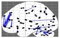
Recently, patients with neuromyelitis optica (NMO) have shown extensive white matter damage, which could be related not only to Wallerian degeneration resulting from lesions of spinal cord or optic tracts but also to demyelination by using diffusion-tensor (DT) MRI imaging. This study aimed to evaluate the expansion of white matter damage in NMO assessed using neurite orientation dispersion and density imaging (NODDI), as well as its relationship with disease severity by applying Tact Based Spatial Statistics (TBSS).


2016.   
Comparison of fast and conventional diffusion kurtosis imaging in an anisotropic synthetic phantom
Ganna Blazhenets1,2, Farida Grinberg1,3, Ezequiel Farrher1, Xiang Gao1, Mikheil Kelenjeridze4, Tamo Xechiashvili4, and N. Jon Shah1,3
1Institute of Neuroscience and Medicine - 4, Forschungszentrum Juelich, Juelich, Germany, 2Institute of Nuclear Physics, University of Cologne, Cologne, Germany, 3Department of Neurology, Faculty of Medicine, JARA, RWTH Aachen University, Aachen, Germany, 4Department of Physics, Georgian Technical University, Tbilisi, Georgia
We compare the sensitivity and applicability of two methods for the estimation of mean kurtosis in a multi-sectional, anisotropic diffusion phantom using conventional diffusion kurtosis imaging and a fast protocol for rapid mean kurtosis metric estimation suggested by Hansen et al. (2013). Both methods provide similar image quality and it can be concluded that fast estimation of mean kurtosis is a useful tool that can be used as a fast method for clinical applications. An interesting finding of this work is a stronger dependence of fast computed kurtosis metrics on the orientation of fibres with respect to the static magnetic field than of the conventional method.


2017.   
Evaluating mean diffusivity and mean kurtosis derived from different diffusion-encoding schemes and signal-to-noise ratio
Chia-Wen Chiang1, Shih-Yen Lin1,2, Yi-Ping Chao3, Yeun-Chung Chang4,5, Teh-Chen Wang6, and Li-Wei Kuo1
1Institute of Biomedical Engineering and Nanomedicine, National Health Research Institutes, Miaoli, Taiwan, 2Department of Computer Science, National Chiao Tung University, Hsinchu, Taiwan, 3Gradulate Institute of Medical Mechatronics, Chang Gang University, Taoyuan, Taiwan, 4Department of Medical Imaging, National Taiwan University Hospital, Taipei, Taiwan, 5Department of Radiology, National Taiwan University College of Medicine, Taipei, Taiwan, 6Department of Radiology, Taipei City Hospital Yang-Ming Branch, Taipei, Taiwan
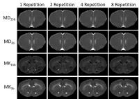
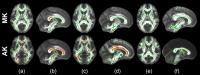
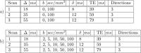
Diffusion kurtosis imaging (DKI), evaluating the non-Gaussianity of water diffusion, has been demonstrated to be sensitive biomarker in many neurological diseases. However, number of repetition is one of the factors, but people is trying less to investigate it. In this study, normal rats were performed using two different diffusion scheme protocols (15 b-values with six diffusion directions vs. 3 b-values with thirty directions) and with different repetitions. Our results suggesting the protocol with one repetition provides good image quality for DKI analysis in this case.


2018.   
Maximum b-value dependence of Diffusion kurtosis imaging sensitivity in detecting white matter microstructure
Miao Sha1, Yuanyuan Chen1, Xin Zhao1, Man Sun2, Weiwei Wang1, Hongyan Ni2, and Dong Ming1
1Tianjin University, Tianjin, China, People's Republic of, 2Tianjin First Center Hospital, Tianjin, China, People's Republic of
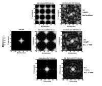
Diffusion kurtosis imaging is a powerful technique to measure the non-gaussion diffusion as well as the complicated microstructure. In this paper, we conducted a comparison between different acquisitions with different maximum b-value on normal volunteers. We found that the outcome of diffusion kurtosis imaging was influenced by the maximum b-value in the acquisition. And this influence was highly associated with the microstructure, including both radial profile and angular profile in the structure reconstruction, which indicated the mechanism of non-gaussion under high b-value.


2019.   
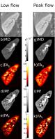
Determination of Microvascular Parameters from Diffusion-Weighted Images
Robert J Loughnan1,2, Damien McHugh1,3, Hamied A Haroon1, Douglas Garratt2, Rishma Vidyasagar1,4, Hojjatollah Azadbakht1, Penny H Cristinacce1, Geoff JM Parker1,5, and Laura M Parkes1
1Centre for Imaging Sciences, Faculty of Medical and Human Sciences, The University of Manchester, Manchester, United Kingdom, 2School of Physics and Astronomy, The University of Manchester, Manchester, United Kingdom, 3CRUK & EPSRC Cancer Imaging Centre in Cambridge & Manchester, Manchester, United Kingdom, 4Melbourne Brain Centre, The Florey Institute of Neuroscience and Mental Health, Melbourne, Australia, 5Bioxydyn Limited, Manchester, United Kingdom
Diffusion imaging has been used to probe microstructure and to investigate perfusion via the IVIM model. However, the contribution of microvasculature structure to the diffusion signal has largely been overlooked. Presented here is a novel method for imaging blood velocity and capillary segment length using diffusion-weighted images. We apply a model for extracting perfusion parameters from diffusion-weighted images from 23 people with a range of diffusion times (?=18, 35 and 55ms) and b-values (0-100s/mm2). Mean blood velocity was significantly slower (P<0.005) in white matter (0.92±0.03mm/s) compared to grey matter (0.95±0.04mm/s). Mean vessel segment length was significantly shorter (P<0.0001) in white matter (7.97±0.13µm) than in grey matter (10.35±0.20µm). 


2020.   
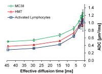
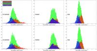
Detection of lymphocytes fractions using temporal diffusion spectroscopy
Johannes Riegler1, Maj Hedehus1, and Richard A. D. Carano1
1Biomedical Imaging, Genentech, South San Francisco, CA, United States
Inflammation and T-cell infiltration are important prognostic biomarkers for cancer immunotherapies.1 Current clinical practice relies on histological assessment of tissue biopsies which is invasive and prone to sampling errors. Temporal diffusion spectroscopy, particularly with short effective diffusion times can estimate cell sizes.2,3 Lymphocytes have small diameters compared to typical tumor cells. We therefore tested the ability of temporal diffusion spectroscopy to differentiate between pellets of tumor cells mixed with a varying amount of activated lymphocytes. We observed clearly separable diffusion characteristics for samples containing > 20% lymphocytes indicating that this approach may have potential to quantify inflammation in highly inflamed tissues.


2021.   
Estimation of Fiber Packing Correlation Length by Varying Diffusion Gradient Pulse Duration
Hong-Hsi Lee1, Gregory Lemberskiy1, Els Fieremans1, and Dmitry S. Novikov1
1New York University, Center for Biomedical Imaging, New York, NY, United States
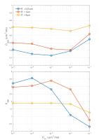
Finite pulse duration $$$\delta$$$ of diffusion gradient has typically been a source of bias for quantifying microstructure. Here, we suggest to use the diffusivity dependence on $$$\delta$$$ to reveal the correlation length of the fiber packing, an essential μm–level characteristic of microstructure, thereby turning the finite pulse duration to our advantage. We validate our method in a fiber phantom that mimics an axonal packing geometry, and the estimated correlation length matches the fiber radius. Future work will focus on the evaluation of its potential as biomarkers for in vivo brain scans, such as axonal density and outer axonal diameters.


2022.   
Detection of Early Emphysema by Quantifying Lung Terminal Airways with Hyperpolarized 129Xe Diffusion MRI
Weiwei Ruan1, Jianping Zhong1, Ke Wang2, Yeqing Han1, and Xin Zhou1
1Wuhan Institute of Physical and Mathematics,Chinese Academy of Sciences, Wuhan, China, People's Republic of, 2Department of MRI, zhongnan hospital of wuhan university, Wuhan, China, People's Republic of
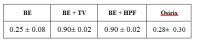
To detect the early emphysema, hyperpolarized xenon diffusion MRI with multi-b values was used to quantify the lung terminal airways in five initial stages of emphysematous rats and five control rats. The DL(longitudinal diffusion coefficient), r, h, LM and S/V in the emphysematous group showed significant differences compared to those in the control group (P<0.05) and also exhibited a strong linear correlation (|r|>0.8) to Lm from histology for all the rats. The results showed multi-b diffusion MRI of hyperpolarized xenon has potential for the diagnosis of emphysema at the early stage.


2023.   
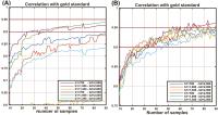
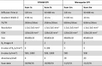
A Time-Efficient Acquisition Protocol For Multi-Purpose Diffusion-Weighted Microstructural Imaging At 7T
Farshid Sepehrband1,2, Kieran O’Brien1,3, and Markus Barth1
1Centre for Advanced Imaging, University of Queensland, Brisbane, Australia, 2Laboratory of Neuro Imaging, USC Mark and Mary Stevens Neuroimaging and Informatics Institute, Keck School of Medicine of USC, Los Angeles, CA, United States, 3Siemens Healthcare Pty Ltd, Brisbane, Australia
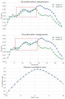
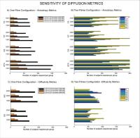
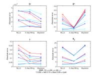
Several diffusion-weighted MRI techniques for modeling tissue microstructure have been developed and validated during the past two decades. While offering various neuroanatomical inferences, these techniques differ in their proposed optimal acquisition design, which impede clinicians and researchers to benefit from all potential inference methods, particularly when limited time is available. We examined the performance of the most common diffusion models with respect to acquisition parameters at 7T when limiting the acquisition time to about 10 minutes. The most balanced compromise among all combinations in terms of the robustness of the estimates was a two-shell scheme with b-values of 1,000 and 2,500 s/mm2 with 75 diffusion-encoding gradients, 25 and 50 samples for low and high b-values, respectively. 


2024.   
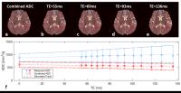
Extraction of Tissue-Specific ADC Based on Multi-Exponential T2 Analysis
Qiqi Tong1, Mu Lin1, Hongjian He1, Xu Yan2, Thorsten Feiweier3, Hui Liu2, and Jianhui Zhong1
1Center for Brain Imaging Science and Technology, Department of Biomedical Engineering, Zhejiang University, Hangzhou, China, People's Republic of, 2MR Collaboration NE Asia, Siemens Healthcare, Shanghai, China, People's Republic of, 3Siemens Healthcare, Erlangen, Germany
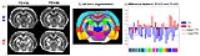
Multi-component diffusion models with each component of its own T2 value have been studied previously. When the diffusion signal is decomposed into three compartments (short, intermediate and long T2), the respective ADC values can be obtained. Our results from simulations and in vivo measurements show that the model successfully separates signal from different tissue types, allows extraction of tissue-specific ADC, and results are mostly free of partial volume problem. Moreover, an ADC without T2 effect can also be generated by combining the ADCs of all components.


2025.   
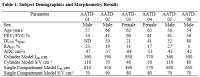
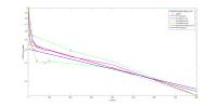
Single Compartment model estimates of acinar duct measurements from inhaled noble gas MRI: Proof of Concept in alpha-1 antitrypsin deficiency emphysema
Eric Lessard1, Alexei Ouriadov1, David G McCormack2, and Grace Parraga1
1Robarts Research Institute, The University of Western Ontario, London, ON, Canada, 2Department of Medicine, The University of Western Ontario, London, ON, Canada
Diffusion-weighted MRI provides a way to non-invasively estimate in vivo morphometry measurements of the alveolar ducts. Current modelling approaches may not be appropriate for cases of severe tissue destruction where the geometry of the acinar ducts may not be uniform, nor cylindrical.  Therefore, in this proof-of-concept evaluation, we used a single-compartment model and multiple b-value diffusion-weighted noble gas pulmonary MRI to generate estimates of acinar duct surface-to-volume ratio and mean-linear-intercept.  In cases of very severe emphysema that accompany alpha-one antitrypsin deficiency, this approach well-approximated the severity of lung disease, while the cylindrical model did not.


2026.   
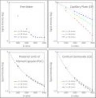
Use envelope bounding to improve the stability of intravoxel incoherent motion modeling
Cheng-Ping Chien1, Feng Mao Chiu2, and Queenie Chan3
1Institute of Biomedical Electronics and Bioinformatics, National Taiwan University, Taipei, Taiwan, 2Philips Healthcare, Taipei, Taiwan, 3Philips Healthcare, Hong Kong, China, People's Republic of
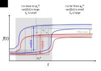
Intravoxel incoherent motion (IVIM) model is useful tool to observe the microcirculatory perfusion, but its stability still needs to be improved. We propose the envelope bounding technique to reduce the fluctuated signal at low b-value, and use this new signal profile to fit IVIM model. This improvement gives a more stable outcome with fast diffusion (D*) and perfusion fraction (PF).


2027.   
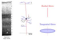
Modelling radial and tangential fibres in the neocortex
Luke J. Edwards1, Siawoosh Mohammadi1,2, Pierre-Louis Bazin3, Michiel Kleinnijenhuis4, Kerrin J. Pine1, Anne-Marie van Cappellen van Walsum5, Hui Zhang6, and Nikolaus Weiskopf1,3
1Wellcome Trust Centre for Neuroimaging, UCL Institute of Neurology, UCL, London, United Kingdom, 2Institut für Systemische Neurowissenschaften, Universitätsklinikum Hamburg-Eppendorf, Hamburg, Germany,3Department of Neurophysics, Max Planck Institute for Human Cognitive and Brain Sciences, Leipzig, Germany, 4FMRIB Centre, University of Oxford, Oxford, United Kingdom, 5Department of Anatomy, Donders Institute for Brain, Cognition and Behaviour, Radboud University, Nijmegen, Netherlands, 6Centre for Medical Image Computing, Department of Computer Science, UCL, London, United Kingdom
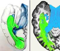
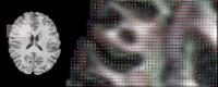
The structure of neocortical grey matter is complex due to the crossing intracortical neuronal connections involved in cortical processing. Herein we present a two-step method to capture radial and tangential fibre structure of neocortex from diffusion data: first the radial cortical orientation is extracted voxelwise using surface-based methods, and then a three-compartment diffusion model extracts radial and tangential fibre volume fractions. We demonstrate in a post mortem sample of human V1 tissue that this method captures structure known from histology and comparable diffusion models, implying potential future use as a probe of intracortical neuronal connectivity.


2028.   
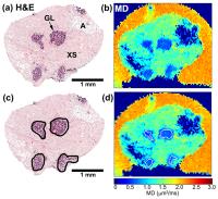
Correlation of diffusion-weighted MRI with cellularity in glandular breast tissue
Narina Norddin1,2, Nyoman Kurniawan3, Gary Cowin3, Carl Power4, Geoffrey Watson5, Esther Myint6, Laurence Gluch7, and Roger Bourne1
1University of Sydney, Sydney, Australia, 2International Islamic University Malaysia, Pahang, Malaysia, 3University of Queensland, Brisbane, Australia, 4University of New South Wales, Sydney, Australia, 5Royal Prince Alfred Hospital, Sydney, Australia, 6Douglass Hanly Moir Pathology, Sydney, Australia, 7The Strathfield Breast Centre, Sydney, Australia
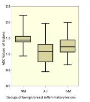
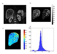
Although diffusivity (ADC) changes in tissue are commonly attributed to variations in ‘cellularity’, direct evidence from breast tissue studies is limited and inconsistent. Here we report a diffusion microimaging and histology investigation of the correlation of mean diffusivity (MD) with cellularity in the glandular component of breast tissue. Diffusion microimaging was performed at 16.4T on fixed normal and cancer tissue samples and matched with post MRI histology. There was a moderate correlation between MD and nuclear count, but only a weak correlation between MD and nuclear area.


2029.   
Time dependence of diffusion and kurtosis parameters in the rat spinal cord
Sune Nørhøj Jespersen1,2, Brian Hansen1, Daniel Nunes3, and Noam Shemesh3
1CFIN, Aarhus University, Aarhus, Denmark, 2Dep. Physics and Astronomy, Aarhus University, Aarhus, Denmark, 3Champalimaud Neuroscience Programme, Champalimaud Centre for the Unknown, Lisbon, Portugal
Non-vanishing diffusion kurtosis and time-dependent diffusion are both hallmarks of nongaussian diffusion in biological tissues. Here we combine measurements of time-dependent DTI parameters and time dependence of mean kurtosis using fast kurtosis imaging in rat spinal cord. We observe substantial time dependence of all parameters in both white and gray matter.


2030.   
Distinguishing between different microstructural changes using optimised diffusion-weighted acquisitions
Damien J. McHugh1,2 and Geoff J.M. Parker1,2,3
1Centre for Imaging Sciences, The University of Manchester, Manchester, United Kingdom, 2CRUK & EPSRC Cancer Imaging Centre in Cambridge & Manchester, United Kingdom, 3Bioxydyn Ltd., Manchester, United Kingdom
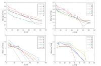
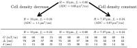
This work investigates the use of optimised diffusion-weighted acquisitions for distinguishing between different microstructural changes relevant to characterising tumour tissue. Optimised protocols are found for a 'baseline' microstructure, and for two distinct changes which would lead to an ADC increase: (1) volume fraction decrease with cell size constant (therefore a decrease in cell density), (2) cell size decrease and coupled volume fraction decrease (therefore a constant cell density). Model fitting simulations are performed with optimised and non-optimised protocols, demonstrating that the improved precision achieved with optimised protocols may be beneficial in terms of distinguishing between these microstructural changes.


2031.   
Oscillating Gradient Spin Echo Diffusion Tensor MRI of the Brain in Multiple Sclerosis Patients
Christian Beaulieu1, Corey Baron1, Penny Smyth2, Roxane Billey2, Leah White2, Fabrizio Giuliani1, Derek Emery3, and Robert Stobbe1
1Biomedical Engineering, University of Alberta, Edmonton, AB, Canada, 2Neurology, University of Alberta, Edmonton, AB, Canada, 3Radiology, University of Alberta, Edmonton, AB, Canada
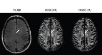
In diffusion tensor imaging, oscillating gradient spin echo (OGSE) gradient waveforms enable much shorter diffusion times (4 ms) than the typical pulsed gradient spin echo (PGSE, 40 ms) and OGSE was applied here for the first time in multiple sclerosis patients. A different dependence on diffusion time would suggest a change in micro-structural scale within the MS lesions. Compared to normal appearing white matter (NAWM), FLAIR-visible lesions showed reductions of fractional anisotropy (FA) on both PGSE and OGSE. The proportional FA decrease between NAWM and lesions was similar for OGSE and PGSE. 


2032.   
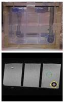
Longitudinal stability of astriction cotton as an anisotropic diffusion phantom
Koji Sakai1, Toshiaki Nakagawa1, and Kei Yamada1
1Kyoto Prefectural University of Medicine, Kyoto, Japan
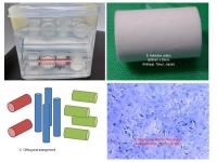
To obtain anisotropic diffusion phantom with ease, we evaluated the longitudinal stability of commercially available astriction cotton as an anisotropic diffusion phantom. DTI examinations were performed at 3 T using a whole-body scanner by 20ch head coil for 131 days intermittently (18 times). The DTI analysis was performed and diffusion metrics (ADC and FA) of the phantom were evaluated by comparing standard deviation in one day to the averaged change between two consequence days. The averaged changes of ADC and FA within the experimental term were 0.03 x 10⁻³sec/mm2  and 0.002, respectively. The commercially available astriction cotton showed stability on its diffusivity over four months.

2033.   
Brain white matter abnormalities in Alzheimer’s disease with and without cerebrovascular disease
Fang Ji1, Ofer Pasternak2, Yng Miin Loke1, Saima Hilal3,4, Mohammad Kamran Ikram1, Xin Xu3,4, Boon Yeow Tan5, Narayanaswamy Venketasubramanian6, Christopher Li-Hsian Chen3,4, and Juan Zhou1,4
1Center for Cognitive Neuroscience, Neuroscience and Behavioral Disorders Program, Duke-National University of Singapore Graduate Medical School, Singapore, Singapore, 2Department of Psychiatry, Brigham and Women's Hospital, Harvard Medical School, USA, Boston, MD, United States, 3Department of Pharmacology, National University Health System, Clinical Research Centre, Singapore, Singapore, 4Memory Aging & Cognition Centre, National University Health System, Singapore, Singapore, 5St. Luke’s Hospital, Singapore, Singapore, 6Raffles Neuroscience Centre, Raffles Hospital, Singapore, Singapore
Using a novel free-water method, we examined the white matter tissue deterioration and extracellular water content changes in Alzheimer’s disease with and without cerebrovascular disease and vascular dementia. We found that free-water and white matter hyperintensity (WMH) were highly correlated; both might reflect neuroinflammation in dementia. After correcting for increased extracellular water, the degree and extent of white matter integrity decreased in dementia subtypes; nevertheless, the cortical difference between groups remained. Intriguingly, free water compartment (but not WMH volume) was associated with symptom severity. Our findings suggested the potential of free-water method in differential diagnosis and disease progression monitoring. 


2034.   
In vivo exploration of the human brainstem complex pathways at 3 Tesla with track-density imaging: a digital three-dimensional microscope for anatomists
Sophie Sébille1, Romain Valabregue1, Anne-Sophie Rolland1, Chantal François1, and Eric Bardinet1
1Brain and Spine Institute, CNRS UMR 7225 - INSERM U 1127 - UPMC-P6 UMR S 1127, Paris, France
We applied super-resolution TDI, as a tool to gain spatial resolution using post-processing methods, to one healthy individual to highlight the fine details of the anatomical fibers tracts in the brainstem. A 1.25 mm isotropic diffusion data acquired in vivo at 3T was used to calculate a 0.2 mm isotropic TDI map. We demonstrated that the super-resolution TDI clearly improved the spatial resolution, as well as the emphasis on different contrast information. These maps can be of help to anatomists to explore the brainstem complex organization by identifying subject-specific tracts.


2035.   
Optimization of acquisition parameters for diffusion MRI using chemical tracing
Giorgia Grisot1,2, Julia Lehman3, Suzanne N Haber3, and Anastasia Yendiki2
1Harvard-MIT Health Sciences and Technology, Massachusetts Institute of Technology, Cambridge, MA, United States, 2Athinoula A. Martinos Center for Biomedical Imaging, MGH, Charlestown, MA, United States,3University of Rochester School of Medicine, Rochester, NY, United States
Determining the optimal diffusion MRI (dMRI) acquisition scheme for reconstructing a brain network of interest with tractography is an open problem, and the lack of ground truth on brain connections makes it challenging to resolve. We use chemical tracing and ex vivo dMRI in macaques to optimize dMRI acquisition with respect to tractography accuracy. We present preliminary results illustrating that 1. There is an upper bound to the angular resolution of dMRI, beyond which tractography accuracy does not improve, and 2. That this finding likely generalizes to in vivo human dMRI.


2036.   
The best of both worlds: Combining the strengths of TBSS and tract-specific measurements for group-wise comparison of white matter microstructure
Greg D Parker1, Dafydd LLoyd2, and Derek K Jones1,3
1CUBRIC, School of Psychology, Cardiff University, Cardiff, United Kingdom, 2Ysgol Gyfun Gwyr, Swansea, United Kingdom, 3Neuroscience and Mental Health Research Institute (NMHRI), School of Medicine, Cardiff University, Cardiff, United Kingdom
Tract-specific microstructural measurements are key to many white matter studies. Common tract-specific measurement strategies average measurements along tracts of interest, but are insensitive to localised changes. Alternatively, by searching radially to a co-registered tract skeleton, tract based spatial statistics1 provides desirable localised comparisons. However, considering one value at each point (the highest value found by radial search), increases susceptibility to outliers, and misses the SNR benefit of averaging multiple estimates within a locale. We propose a hybrid method using tract skeletons to divide streamlines into localised sections, comparing averages within each section. Example results in remitted depression are presented.


2037.   
High-resolution DTI-based cortical connectome reconstructions match incompletely with true axonal projections in rat brain
Michel R.T. Sinke1, Willem M. Otte1,2, Annette van der Toorn1, R. Angela Sarabdjitsingh3, Marian Joëls3, and Rick M. Dijkhuizen1
1Biomedical MR Imaging and Spectroscopy Group, Center for Image Sciences, University Medical Center Utrecht, Utrecht, Netherlands, 2Department of Pediatric Neurology, Brain Center Rudolf Magnus, University Medical Center Utrecht, Utrecht, Netherlands, 3Department of Translational Neuroscience, Brain Center Rudolf Magnus, University Medical Center Utrecht, Utrecht, Netherlands
The exact relationship between DTI-based tract representations and true axonal projections remains uncertain. We compared the accuracy of tensor-based and constrained spherical deconvolution (CSD)-based tractography, against neuroanatomical tracer data in rat brain. Our study with high spatial and angular resolution postmortem DTI data revealed low tractography accuracy, characterized by significant amount of false positive and false negative streamline connections. Accounting for crossing fibers by CSD did not significantly improve sensitivity and specificity. Because DTI-based tract reconstructions correlate incompletely with true axonal projections in rat brain, even when using an advanced algorithm like CSD, DTI-based connectomics should be interpreted with care. 


2039.   
Application of a combined IVIM-DTI model in ECG-triggered imaging of the human kidney
Fabian Hilbert1, Simon Veldhoen1, Tobias Wech1, Henning Neubauer1, Thorsten Alexander Bley1, and Herbert Köstler1
1Department of Diagnostic and Interventional Radiology, University of Würzburg, Würzburg, Germany
Diffusion tensor imaging (DTI) accounts for anisotropy of diffusion, while the intravoxel incoherent motion (IVIM) model considers a fast moving pseudo-diffusion compartment. In the kidney DTI and IVIM parameters vary significantly depending on the time they are acquired within the cardiac cycle. A combined IVIM-DTI model incorporates anisotropic diffusion and anisotropic pseudo-diffusion parameters. The purpose of this study was to investigate the impact of the cardiac cycle on the combined IVIM-DTI model. While in DTI the fractional anisotropy of the diffusion tensor (FAD) varies within the cardiac cycle, FAD does not change in the IVIM-DTI model.


2040.   
Diffusion-weighted imaging with multiple diffusion time to assess water-exchange between restricted and hindered diffusion components in vivo
Yasuhiko Tachibana1,2, Takayuki Obata1,2, Hiroki Tsuchiya1, Tokuhiko Omatsu1,2, Riwa Kishimoto1,2, Thorsten Feiweier3, and Hiroshi Tsuji1
1Research Center of Charged Particle Therapy, National Institute of Radiological Science, Chiba, Japan, 2Applied MRI Research, National Institute of Radiological Science, Chiba, Japan, 3Siemens Healthcare GmbH, Erlangen, Germany
We performed multi-b and multi-diffusion-time DWI (MbMdt-DWI) on human brain to visualize the mixture of restricted and hindered diffusion components, and also the water exchange between them. The diffusion parameters including the exchange time were calculated. The observed signal patterns clearly indicated the existence of the inter-compartmental water exchange. The calculated exchange time was within the appropriate range assumed from a previous cell-based study in vitro. MbMdt-DWI may be useful for assessing micro-diffusion in human brain.


2038.   
Model-free Global Tractography
Henrik Skibbe1, Elias Kellner2, Valerij G Kiselev2, and Marco Reisert2
1Faculty of Informatics, Ishii Lab, Kyoto University, Kyoto, Japan, 2Medical Physics, University Medical Center Freiburg, Freiburg, Germany
Tractography based on diffusion-weighted MRI investigates the large scale arrangement of neurite fibers in brain white matter. It is usually assumed that the signal is a convolution of a fiber response function (FRF)  with a fiber orientation distribution (FOD). The FOD is the focus of tractography. While in the past the FRF was estimated beforehand and was usually assumed to be fix, more recent approaches estimate the FRF during tractography. This work proposes a novel objective function independent of the FRF, just aiming for FOD reconstruction. The objective is integrated into global tractography showing promising results. 


2041.   
Thinking Outside the Voxel: A Joint Spatial-Angular Basis for Sparse Whole Brain HARDI Reconstruction
Evan Schwab1, Rene Vidal2, and Nicolas Charon3
1Electrical and Computer Engineering, Johns Hopkins University, Baltimore, MD, United States, 2Biomedical Engineering, Johns Hopkins University, Baltimore, MD, United States, 3Applied Mathematics and Statistics, Johns Hopkins University, Baltimore, MD, United States
Sparse modeling of dMRI signals has become of major interest for advanced protocols such as HARDI which require a large number of q-space measurements.  With few exceptions, prior work have developed bases to sparsely represent q-space signals per voxel with additional constraints of spatial regularity.  In this work, we propose a single basis to represent an entire HARDI dataset by modeling spatial and angular domains jointly, achieving an unprecedented level of sparsity. With a globally compressed representation we can then redefine HARDI processing, diffusion estimation, feature extraction and segmentation, and drastically reduce acquisition time and data storage.


2042.   
Assessment of Early Renal Fibrosis Induced in a Murine Model of Streptozotocin Induced Diabetes
Yet Yen Yan1, Tiffany Hennedige1, Tong San Koh1, Lei Zhou2, Septian Hartono3, Helmut Rumpel3, Laurent Martarello4, James Boon Kheng Khoo1, Dow-Mu Koh5, Kai Hsiang Chuang6, Tony Kiat Hon Lim3, Yock Young Dan2, and Choon Hua Thng1
1National Cancer Centre Singapore, Singapore, Singapore, 2National University Hospital, Singapore, Singapore, 3Singapore General Hospital, Singapore, Singapore, 4Roche Translational Medicine Hub, Singapore, Singapore, 5Royal Marsden Hospital, London, United Kingdom, 6Singapore BioImaging Consortium, Singapore, Singapore
Streptozotocin induced diabetes was created in twenty mice while eighteen mice served as control. DTI & IVIM were performed at 0, 12 and 24 weeks after injection of streptozotocin. Histopathological analysis confirmed fibrosis in all diabetic mice. Increase in ADC & tissue diffusivity were found in the diabetes group at week 12, which might reflect an increased tubule volume that outweighed the effects of early fibrosis. FA was significantly reduced in the diabetes group at week 12 and represented tubular damage of renal fibrosis. This study showed the potential of FA as a biomarker of early diabetic nephropathy.


2043.   
Can Cramer-Ráo Lower Bound be used to find optimal b-values for IVIM?
Oscar Gustafsson1,2, Maria Ljungberg1,2, and Göran Starck1,2
1Department of Radiation Physics, Institute of Clinical Sciences, The Sahlgrenska Academy, University of Gothenburg, Gothenburg, Sweden, 2Department of Medical Physics and Biomedical Engineering, Sahlgrenska University Hospital, Gothenburg, Sweden
Cramer-Ráo Lower Bound is commonly used in experiment design optimization. Here we use it to find optimal b-value schemes for IVIM imaging. The optimization was generalized with regard to averaging and the characteristics of the results, given the input and the constraints, were studied. The resulting schemes never included more than the minimum number of four unique b-values, even though multiple sets of tissue parameters were included in the optimization. The optimized b-value schemes were compared to a typical one using simulations.


2045.   
White matter microstructural deficits in schizophrenia using generalized kurtosis
Arash Nazeri1, Lipeng Ning2, Jon Pipitone1, David J. Rotenberg1, Yogesh Rathi2, and Aristotle N. Voineskos1
1Research Imaging Centre, Centre for Addiction and Mental Health, University of Toronto, Toronto, ON, Canada, 2Brigham and Women's Hospital, Harvard Medical School, Boston, MA, United States
Numerous studies have used diffusion tensor imaging to investigate schizophrenia-related white matter microstructural deficits. Diffusion tensor imaging assumes a Gaussian distribution for the water molecule displacement. However, this assumption may not be valid in the complex biological tissues such as white matter. Using directional radial basis function it is possible to estimate ensemble average diffusion propagator and generalized kurtosis of water diffusion (a measure of non-Gaussianity). Our results suggest that white matter generalized kurtosis is more sensitive to differences between persons with schizophrenia and healthy controls than diffusion tensor model parameters (particularly in frontotemporal superficial white matter areas).


2046.   
What is the best method for robust statistical inference on connectomic graph metrics?
Mark Drakesmith1,2, David Linden2, Anthony S David3, and Derek K Jones1,2
1CUBRIC, Cardiff University, Cardiff, United Kingdom, 2Neuroscience and Mental health Research Institute, Cardiff University, Cardiff, United Kingdom, 3Institute of Psychiatry, Psychology and Neurosceince, Kings College London, London, United Kingdom
Connectomic network analyses, while powerful, suffer from high instability, which is problematic for robust statistical inference. The area under the curve (AUC) across thresholds is a common approach, but lacks robustness to this instability. A superior approach is multi-threshold permutation correction (MTPC), but this is computationally expensive. Smoothed AUCs (smAUCs) are less costly and theoretically can achieve the same level of sensitivity as MTPC. smAUC was tested and compared with MTPC in a virtual patient-control comparison. Results show that smAUC sensitivity is not consistently comparable to MTPC and that exhaustive searching across the threshold space is required for robust inference. 


2047.   
Evaluation of IVIM Perfusion Parameters as Biomarkers for Paediatric Brain Tumours
Emma Meeus1,2,3, Jan Novak2,3, Stephanie Withey2,3,4, Lesley MacPherson3, and Andrew Peet2,3
1Physical Sciences of Imaging in Biomedical Sciences (PSIBS) Doctoral Training Centre, University of Birmingham, Birmingham, United Kingdom, 2Institute of Cancer and Genomic Sciences, University of Birmingham, Birmingham, United Kingdom, 3Department of Oncology, Birmingham Children's Hospital, Birmingham, United Kingdom, 4RRPPS, University Hospitals Birmingham NHS Foundation Trust, Birmingham, United Kingdom
This study investigated the bi-exponential IVIM fitting methods and their robustness for applications in the brain. Data simulations relevant to normal brain and tumour were computed to assess the accuracy and precision of the IVIM perfusion parameters.  The paediatric patient cohort evaluated the correlation between the IVIM and DSC-MRI derived parameters. The simulation results showed that the perfusion fraction (IVIM-f) was robust enough to provide reliable values using the constrained 1-parameter fit. The robustness was further confirmed with the significant correlation observed between the IVIM-f and DSC-CBV. Therefore, IVIM-f could provide an alternative non-invasive perfusion measure for paediatrics.


2048.   
Correcting spatial misalignment between fiber bundles segments for along-tract group analysis
Samuel St-Jean1, Max Viergever1, Geert Jan Biessels2, and Alexander Leemans1
1Image Sciences Institute, University Medical Center Utrecht, Utrecht, Netherlands, 2Department of Neurology, Rudolf Magnus Institute of Neuroscience, University Medical Center Utrecht, Utrecht, Netherlands
For diffusion MRI studies relying on statistics computed along fiber trajectories, the extracted values might not be optimally aligned between subjects in the metric space, which could lead to subsequent erroneous statistical analysis. We thus propose a 1D fast Fourier transform based correction algorithm for a fast realignment (< 1 second) directly in the metric space. Our experiments with a) synthetic signals and b) FA values along the uncinate fasciculus from real data show that our fiber-tract realignment algorithm improves the overlap of extracted metrics. This could help researchers uncover relationships of interest which were hidden by residual misalignment at first.


2049.   
A Theoretical Framework for Sampling and Reconstructing Ensemble Average Propagators in Diffusion MRI
Divya Varadarajan1 and Justin P Haldar2
1University of Southern California, 90089, CA, United States, 2University of Southern California, Los Angeles, CA, United States
Diffusion MRI can be modeled as sampling the Fourier transform of the Ensemble Average Propagator (EAP). This is potentially advantageous because of extensive theory that has been developed to characterize sampling requirements, accuracy, and stability for Fourier reconstruction.  However, previous work has not taken advantage of this characterization.  This work presents a novel theoretical framework that precisely describes the relationship between the estimated EAP and the true original EAP.  The framework is applicable to arbitrary linear EAP estimation methods, and for example, provides new insights into the design of q-space sampling patterns and the selection of EAP estimation methods.


2050.   
Phase-correcting Non-local Means Denoising for Diffusion-Weighted Imaging
Sevgi Gokce Kafali1,2, Tolga Çukur1,2, and Emine Ulku Saritas1,2
1Electrical and Electronics Engineering, Bilkent University, Ankara, Turkey, 2National Magnetic Resonance Research Center (UMRAM), Bilkent University, Ankara, Turkey
Diffusion-weighted imaging (DWI) intrinsically suffers from low SNR due to diffusion-induced signal losses. Multiple acquisitions have to be averaged to attain reasonable SNR level in high-spatial-resolution DWI images. However, subject motion during diffusion-sensitizing gradients creates varying phase offsets between repeated acquisitions, prohibiting a direct complex averaging of the image repetitions. Here, we propose a phase-correcting non-local means denoising filter that combines multiple DWI acquisitions while effectively reducing noise and phase cancellations. Results are demonstrated in vivo in the cervical spinal cord at 3T, using a reduced field-of-view DWI with 0.9 x 0.9 mm2 in-plane resolution. 


2051.   
Rapid Estimation of IVIM Pseudo-Diffusion Fraction with Correction of TE Dependence
Neil Peter Jerome1, Matthew R Orton1, Thorsten Feiweier2, Dow-Mu Koh3, Martin O Leach1, and David J Collins1
1CRUK Cancer Imaging Centre, Division of Radiotherapy & Imaging, Institute of Cancer Research, London, United Kingdom, 2Siemens Healthcare GmbH, Erlangen, Germany, 3Department of Radiology, Royal Marsden Hospital, London, United Kingdom
The biexponential IVIM model of diffusion does not account for distinct T2 values for the two components, commonly interpreted as blood and tissue, leading to a TE dependence of the pseudo-diffusion volume fraction parameter f. In this volunteer study, the addition of a small number of DWI scans at different TEs allows for fitting of an extended T2-IVIM model, returning TE-independent estimations of liver f (18.26±7.3 % compared to 27.88±6.0 % from conventional IVIM fitting), and T2s of 77.6 ± 30.2 and 42.1 ± 6.8 ms for pseudo- and true diffusion compartments, respectively. 


2052.   
Validation of MD map calculation from DWI acquired on a 0.35T MRI scanner in Malawi for acute cerebral malaria
Yuchuan Zhuang1, Samuel D. Kampondeni2,3, Madalina Tivarus2, Michael J. Potchen2, Gretchen L. Birbeck4, and Jianhui Zhong2
1Electrical and Computer Engineering, University of Rochester, Rochester, NY, United States, 2Department of Imaging Sciences, University of Rochester Medical Center, Rochester, NY, United States, 3MRI Center, Queen Elizabeth Central Hospital, Blantyre, Malawi, 4Department of Neurology, University of Rochester Medical Center, Rochester, NY, United States
Cerebral malaria (CM) is an often fatal disease that still devastates children in Africa. In Malawi, MRIs at 0.35T are obtained on pediatric CM patients, but quantitative analysis remains challenging. This report validates the 0.35T DWI measurements by comparing diffusion scans of normal adult subjects on both 0.35T and 3T MRI scanners. We used ROI analysis, regression analysis and histogram for quantitative validation. Strong consistency between the two data sets indicates that the DWI findings obtained on the 0.35T in Malawi can be used despite its inherent limitations.


2053.   
Real valued diffusion weighted imaging using decorrelated phase filtering
Tim Sprenger Sprenger1,2, Jonathan I. Sperl2, Brice Fernandez3, Axel Haase1, and Marion Menzel2
1Technische Universität München, Munich, Germany, 2GE Global Research, Munich, Germany, 3GE Healthcare, Munich, Germany
Due to the intrinsic low signal to noise ratio in diffusion weighted imaging (DWI), magnitude processing often results in an overestimation of the signal’s amplitude. This results in low estimation accuracy of diffusion models and reduced contrast because of a superposition of the image signal and the noise floor. We adopt a new phase correction (PC) technique yielding real valued data and maintaining a Gaussian noise distribution. The advantage of PC is shown in a DSI experiment where the Ensemble average propagator is better delineated in the real valued data and delineation improves as the noise floor is lowered.


2054.   
Non-linear Distortion Correction in Human Optic Nerve Diffusion Imaging
Joo-won Kim1,2, Jesper LR Andersson3, Peng Sun4, Sheng-Kwei Song4, Robert Naismith5, and Junqian Xu1,2,6
1Department of Radiology, Icahn School of Medicine at Mount Sinai, New York, NY, United States, 2Translational and Molecular Imaging Institute, Icahn School of Medicine at Mount Sinai, New York, NY, United States,3Oxford Centre for Functional MRI of the Brain, University of Oxford, Oxford, United Kingdom, 4Department of Radiology, Washington University, St. Louis, MO, United States, 5Department of Neurology, Washington University, St. Louis, MO, United States, 6Department of Neuroscience, Icahn School of Medicine at Mount Sinai, New York, NY, United States
A major challenge in optic nerve diffusion MRI is the non-linear optic nerve distortion induced by eye-ball movement. In this work, we developed and evaluated a non-linear registration scheme to improve optic nerve edge alignment over conventional diffusion imaging distortion correction methods. Optic nerve edge plots (both 1D and 2D) were used to evaluate the optic nerve edge alignment for different non-linear registration methods (FSL/fnirt and ANTs) after FSL/topup and FSL/eddy correction of unprocessed diffusion images. Overall, the additional non-linear registration step, regardless of the non-linear registration method used, substantially improved optic nerve edge alignment along all diffusion measurement frames.


2055.   
Fast implementations of contextual PDE’s for HARDI data processing in DIPY
Stephan Meesters1, Gonzalo Sanguinetti1, Eleftherios Garyfallidis2, Jorg Portegies1, and Remco Duits1
1Department of Mathematics and Computer Science, Eindhoven University of Technology, Eindhoven, Netherlands, 2Computer Science Department, University of Sherbrooke, Sherbrooke, QC, Canada
We present a novel open-source module that implements a contextual PDE framework for processing HARDI data. It’s potential in enhancement of ODF/FOD fields is demonstrated where the aim is to enhance the alignment of elongated structures while preserving crossings. The method for contextual enhancement is based on a hypo-elliptic PDE defined in the domain of coupled positions and orientations and can be solved with a shift-twist convolution. The module is available in the DIPY (Diffusion Imaging in Python) software library, which makes it widely available for the neuroimaging community.


2056.   
Flow-based White Matter Supervoxel Parcellation using Functional Bregman Divergence between Orientation Distribution Functions
Teng Zhang1, Kai Liu1, Lin Shi2,3, and Defeng Wang4,5
1Department of Imaging and Interventional Radiology, The Chinese University of Hong Kong, Hong Kong, Hong Kong, 2Department of Medicine and Therapeutics, The Chinese University of Hong Kong, Hong Kong, Hong Kong, 3Chow Yuk Ho Technology Centre for Innovative Medicine, The Chinese University of Hong Kong, Hong Kong, Hong Kong, 4Research Center for Medical Image Computing, Department of Imaging and Interventional Radiology, The Chinese University of Hong Kong, Hong Kong, Hong Kong, 5Shenzhen Research Institute, The Chinese University of Hong Kong, Shenzhen, China, People's Republic of
We propose a flow-based supervoxel parcellation method to split white matter into supervoxels with homogeneous diffusion property. In particular, we defined a new similarity metric between orientation distribution functions derived from q-ball imaging according to functional Bregman divergence. The proposed method was applied to high quality data from Human Connectome Project. Our work demonstrated a methodological feasibility to generate supervoxel approach tractography, construction of WM connectivity network, etc.


2057.   
Generalisability of Image Quality Transfer: Can we approximate in-vivo human brains from dead monkey brains?
Aurobrata Ghosh1, Viktor Wottschel2, Enrico Kaden1, Jiaying Zhang1, Hui Zhang1, Stamatios N. Sotiropoulos3, Darko Zikic4, Tim B. Dyrby5, Antonio Criminisi4, and Daniel C Alexander1
1Centre for Medical Image Computing, University College London, London, United Kingdom, 2Institute of Neurology, University College London, London, United Kingdom, 3Oxford Centre for Functional Magnetic Resonance Imaging of the Brain (FMRIB), University of Oxford, Oxford, United Kingdom, 4Microsoft Research, Cambridge, United Kingdom, 5Danish Research Centre for Magnetic Resonance, Centre for Functional and Diagnostic Imaging and Research, Copenhagen University Hospital Hvidovre, Hvidovre, Denmark
The Image-Quality Transfer (IQT) framework enhances low quality images by transferring information from high quality images acquired on expensive bespoke scanners. Although IQT has major potential in medical imaging, one key question is its dependence on training data. We demonstrate the generalisability of IQT used for super-resolution by showing that reconstruction of in-vivo human images degrades minimally from training on human data from the same study, to data from a different demographic and imaging protocol, to data from fixed monkey brains. Remarkably, a patchwork of fixed monkey brain image-pieces is hardly distinguishable from a reconstruction using pieces of human data.


2044.   
Abnormal brain white matter skeleton in patients with disorders of consciousness
Huan Wang1, Youqiu Xie2, Ling Weng1, Qing Ma2, Ling Zhao1, Ronghao Yu2, Miao Zhong1, Xiaoyan Wu1, and Ruiwang Huang1
1Center for the Study of Applied Psychology, Guangdong Key Laboratory of Mental Health and Cognitive Science, School of Psychology, South China Normal University, GuangZhou, China, People's Republic of, 2Coma research group and coma recovery unit, neuroscience institute, guangzhou general hospital of Guangzhou command, GuangZhou, China, People's Republic of
What we did was to use the tract-based spatial statistics (TBSS) approach to examine the changes of diffusion parameters in whole brain white matter of DOC patients relative to healthy controls, and to detect the correlation between the diffusion parameters of white matters and clinical variables.


2058.   
Anisotropic fractional-motion-based diffusion MRI in the human brain
Yun Liu1, Yang Fan1,2, and Jia-Hong Gao11Peking University, Beijing, China, People's Republic of, 2MR Research China, GE Healthcare, Beijing, China, People's Republic of
Several anomalous diffusion models, both empirical and theoretical, were proposed to explain the departure from purely mono-exponential decay of DWI signal in biological tissues. Recently, a fractional motion (FM) based diffusion MRI theory was proposed, which was claimed to be a proper model in the description of diffusion processes in biological systems. However, the tensorial properties of FM related parameters is still unknown. In this work, diffusion magnetic field gradients were applied in several non-linear directions to acquire DWI images. Then, the FM-based parameters were obtained in each diffusion direction and found direction dependent. 


2059.   
Perfusion-free Diffusion Tensor Imaging of Brain Tumors
Zhongwei Zhang1, Zhuhao Li2, Yu-Chien Wu3, Dawen Zhao4, and Mark E Schweitzer5
1Radiology, UT Southwestern Medical Center, Dallas, TX, United States, 2Radiology, The 1st Affiliated Hospital, Sun Yat-Sen University, Guangzhou, China, People's Republic of, 3Radiology and Imaging Sciences, Indiana University, Indianapolis, IN, United States, 4Biomedical Engineering and Cancer Biology, Wake Forest School of Medicine, Winston-Salem, NC, United States, 5Radiology, Stony Brook University, Stony Brook, NY, United States
In conventional DTI, the quantitation of various DTI indices was strongly influenced by b-value. In this study, we proposed a new approach that perfusion-free DTI can be fulfilled using IVIM and DTI models.


2060.   
The value of DTI and DTT in evaluating the protective effect of neuregulin-1 on spinal cord transection models of Sprague–Dawley rats.
Tao Gong1, Guangbin Wang2, and Weibo Chen3
1Shandong University, Jinan, China, People's Republic of, 2Jinan, China, People's Republic of, 3Shanghai, China, People's Republic of
DTI can noninvasive evaluate the injury and prognosis of spinal cord, and NRG-1 has the function of protecting and repairing of injury spinal cord in rats.


2061.   
Is voxel-based apparent diffusion coefficient reproducible?
Masamitsu Hatakenaka1, Koichi Onodera1, Naomi Koyama1, and Mitsuhiro Nakanishi2
1Diagnostic Radiology, Sapporo Medical University, Sapporo, Japan, 2Division of Radiology, Sapporo Medical University Hospital, Sapporo, Japan
To evaluate reproducibility of voxel-based ADC, voxel-based ADC of  the phantom was measured using clinical 3T MRI system. The direction of motion probing gradient affected the voxel-based ADC significantly in both echo planar imaging and turbo spin echo diffusion-weighted imaging. Also voxel-based ADC differed both between identical positioning examinations and between slightly different positioning examinations. Voxel-based ADC could not be reproduced sufficiently even in a phantom study. It would be recommended to pay enough attention when performing voxel-based ADC study like histogram analysis for tumor ADC.

2062.   
Magnetic ROIs enable improved tractography accuracy through oriented prior
Maxime Chamberland1,2,3, Benoit Scherrer3, Sanjay Prabhu3, Joseph Madsen3, David Fortin4, Kevin Whittingstall2,5, Maxime Descoteaux1, and Simon K Warfield3
1Computer science, Université de Sherbrooke, Sherbrooke, QC, Canada, 2Nuclear Medicine and Radiobiology, Université de Sherbrooke, Sherbrooke, QC, Canada, 3Boston Children's Hospital, Harvard Medical School, Boston, MA, United States, 4Division of Neurosurgery and Neuro-Oncology, Université de Sherbrooke, Sherbrooke, QC, Canada, 5Department of Diagnostic Radiology, Université de Sherbrooke, Sherbrooke, QC, Canada
Streamline tractography algorithms infer connectivity by following directions which are maximally aligned at every voxel. This rule has even been the definition of the probability of connectivity, with the difference in current and next orientation being defined as uncertainty in connectivity. However, our experiments demonstrate that in regions where multiple fiber pathways interdigitate (e.g. temporal lobe), this heuristic is inadequate and does not necessarily reflect the underlying human brain architecture. Furthermore, we demonstrate that inference of connectivity can be improved by incorporating anatomical knowledge of the expected fiber orientation in regions where this information is known. We applied this heuristic through a new tractography region of interest (ROI) and demonstrate that it provides improved delineation of the expected anatomy. 


2063.   
Effects of cortical regions of interests on tractography and brain connectivity quantification
Tina Jeon1, Virendra Mishra2, and Hao Huang1,3
1Radiology Research, Children's Hospital of Philadelphia, Philadelphia, PA, United States, 2Lou Ruvo Center for Brain Health, Cleveland Clinic, Las Vegas, NV, United States, 3Perelman School of Medicine, University of Pennsylvania, Philadelphia, PA, United States
Dense white matter (WM) zones just beneath cerebral cortex impede tracking from a cortical region of interest with diffusion MRI data. To address this tracing problem, we can either dilate the parcellated cortex into the adjacent WM to initiate tracing or trace directly from WM interior to these dense WM zones. Here we evaluated with diffusion MRI data from three developmental age groups 1) how much dilation from the segmented cortical gyrus would be sufficient for appropriate WM tracing; and 2) if tracing directly from the WM immediately beneath the dense WM zones will yield the same tractography results. 


2064.   
LOCAL ANALYSIS OF TRACK DENSITY IMAGING FOR THE DETECTION OF WHITE MATTER ALTERATIONS
Rodrigo de Luis-Garcia1, Angel Luis Guerrero2, Miguel Angel Tola-Arribas3, and Santiago Aja-Fernandez1
1Universidad de Valladolid, Valladolid, Spain, 2Hospital Clinico Universitario, Valladolid, Spain, 3Hospital Universitario Rio Hortega, Valladolid, Spain
Track-Density Imaging (TDI) can provide super resolution images of the white matter of the brain. As it is based on the results of a whole brain tractography process, it comprises information from different features of the white matter diffusion. We exploit this information by proposing a local analysis approach for TDI, and test it on two different datasets where conventional TBSS analysis using FA did not yield any significant differences. Results revealed the proposed method to be extremely sensitive in the detection of white matter abnormalities, making it a promising tool for white matter group studies.


2065.   
Minimum number of diffusion encoding directions required to yield a rotationally invariant powder average signal in single and double diffusion encoding
Filip Szczepankiewicz1, Carl-Fredrik Westin2, Freddy Ståhlberg1, Jimmy Lätt3, and Markus Nilsson4
1Dept. of Medical Radiation Physics, Lund University, Lund, Sweden, 2Dept. of Radiology, Brigham and Women’s Hospital, Harvard Medical School, Boston, MA, United States, 3Center for Medical Imaging and Physiology, Skåne University Hospital, Lund, Sweden, 4Lund University Bioimaging Center, Lund University, Lund, Sweden
Several analysis techniques of diffusion-weighted data make use of the powder average to yield signal that is invariant to rotation. However, rotational invariance is achieved only at a sufficient directional resolution, which depends on the tissue anisotropy and diffusion encoding strength. In this work we present the minimum number of diffusion directions necessary to yield a rotationally invariant powder average, at arbitrary anisotropy and encoding strength, for single and double diffusion encoding.


2066.   
Structural connectivity analysis at the voxel level
Jan-Gerd Tenberge1 and Patrick Schiffler1
1University of Münster, Münster, Germany
We present a GPU-accelerated method to compute a structural connectome of the human brain with voxel-level resolution from diffusion weighted images.


2067.   
On the feasibility of data-driven estimation of Markov random field parameters for IVIM modelling of abdominal DW-MRI: insights into which parameters can be reliably estimated from clinical data
Matthew R Orton1, Neil P Jerome1, Mihaela Rata1, David J Collins1, Khurum Khan2, Nina Tunariu3, David Cunningham2, Thorsten Feiweier4, Dow-Mu Koh3, and Martin O Leach1
1CRUK Cancer Imaging Centre, Division of Radiotherapy and Imaging, Institute of Cancer Research, London, United Kingdom, 2Department of Medical Oncology, Royal Marsden NHS Foundation Trust, London, United Kingdom, 3Department of Radiology, Royal Marsden NHS Foundation Trust, London, United Kingdom, 4Siemens Healthcare, Erlangen, Germany
The intravoxel incoherent motion model is of great interest as it gives a more complete characterization of DWI signals. However, estimates of the pseudo-diffusion coefficient D* are noisy, which can be mitigated using Markov random field (MRF) models.  The MRF smoothing weights are usually subjectively chosen; by removing this requirement, we show that while the smoothing weights for the pseudo-diffusion volume fraction and diffusion coefficient can be estimated from the data, smoothing weights for D* cannot.  This suggests that with currently available data, D* estimates require stabilization by imposing subjective constraints of some kind, such as the MRF used here.


2068.   
A T1 and DTI fused 3D Corpus Callosum analysis in MCI subjects with high and low cardiovascular risk profile
Yi Lao1,2, Binh Nguyen2, Sinchai Tsao2, Niharika Gajawelli1,2, Meng Law1,3, Helena Chui1,3, Yalin Wang4, and Natasha Lepore1,2
1Biomedical Engineering, University of Southern California, Los Angeles, CA, United States, 2Radiology, Children's Hospital Los Angeles, Los Angeles, CA, United States, 3Radiology, Keck School of Medicine, University of Southern California, Los Angeles, CA, United States, 4School of Computing, Informatics, and Decision Systems Engineering, Arizona State University, Temple, AZ, United States
Understanding how vascular disease and its risk factors influence Alzheimer's disease (AD) progression may enhance predictive accuracy as well as guide early interventions. Here, we apply a novel T1 and DTI fusion analysis on the 3D corpus callosum (CC) of mild cognitive impairment (MCI) populations with different levels of cardiovascular risk, with the aim of decoupling vascular factors in the prodromal AD stage. Our new fusion method detected significant differences in the anterior CC between MCI subjects with high and low vascular risk profiles.  These findings may help to elucidate the interdependent relationship between MCI and vascular risk factors.


2069.   
Neuroimaging biomarkers for predicting treatment response in schizophrenia based on alteration patterns of the whole brain white matter tracts
Jing-Ying Huang1, Yu-Jen Chen1, Chih-Min Liu2, Tzung-Jeng Hwang2,3, Yun-Chin Hsu1, Yu-Chun Lo1, Hai-Gwo Hwu2,3, and Wen-Yih Isaac Tseng1,3,4,5,6
1Institute of Medical Device and Imaging, National Taiwan University College of Medicine, Taipei, Taiwan, 2Department of Psychiatry, National Taiwan University Hospital, Taipei, Taiwan, 3Graduate Institute of Brain and Mind Sciences, National Taiwan University College of Medicine, Taipei, Taiwan, 4Institute of Biomedical Engineering, National Taiwan University, Taipei, Taiwan, 5Department of Medical Imaging, National Taiwan University Hospital, Taipei, Taiwan, 6Molecular Imaging Center, National Taiwan University, Taipei, Taiwan
This study aims to identify image biomarkers for schizophrenia patients in order to predict treatment responses on individual subject basis. We develop algorithm that can discriminate remission or non-remission in patients with schizophrenia based on the difference in microstructural integrity of the white matter tracts. The ROC analysis shows that the accuracy of the prediction is 76%.


2070.   
Individualized prediction of schizophrenia based on patterns of altered tract integrity over the whole brain using diffusion spectrum imaging
Yu-Jen Chen1, Chih-Ming Liu2, Tzung-Jeng Huang2, Yun-Chin Hsu1, Yu-Chun Lo1, Hai-Gwo Hwu2, and Wen-Yih Isaac Tseng1,3
1National Taiwan University, Institute of Medical Device and Imaging, Taipei, Taiwan, 2National Taiwan University Hospital, Department of Psychiatry, Taipei, Taiwan, 3National Taiwan University, Molecular Imaging Center, Taipei, Taiwan
In this study, we examined the performance of predicting patients with schizophrenia based on the patterns of altered tract integrity over the whole brain. The whole-brain tract information was compared with predefined differences between schizophrenia patients and healthy participants to calculate an index of SLI indicating the similarity to schizophrenia. Our results showed that the prediction performance was high (AUC = 0.86 for males; AUC = 0.77 for females) when we compared the white matter integrities at specific segments on fiber pathways. 


2071.   
De-noising of diffusion-weighted MRI data by averaging of inconsistent input data in wavelet space
Henrik Marschner1, Cornelius Eichner1, Alfred Anwander1, André Pampel1, and Harald E. Möller1
1Max Planck Institute for Human Cognitive and Brain Sciences, Leipzig, Germany
Diffusion Weighted Images datasets with high spatial resolution and strong diffusion weighting are often deteriorated with low SNR. Here, we demonstrate the feasibility of a recently presented repetition-free averaging based de-noising (AWESOME). That technique reduces noise by averaging over a series of N images with varying contrast in wavelet space and regains intensities and object features initially covered by noise. We show that high resolution DWIs are achievable in a quality that almost equals to that obtained from 6fold complex averaging.


2072.   
Linear Acceleration of SADD Method for Three Compartments
Ana Karen Loya1 and Mariano Rivera1
1Computer Science, Centro de Investigacion en Matematicas AC, Guanajuato, Mexico
The proposal attempts to model more properly the intra-voxel information from DW-MRI signals in order to obtain tissue diffusion properties in white matter. The method is based on Sparse and Adaptive Diffusion Dictionary (SADD) strategy that dynamically adapts a dictionary of diffusion functions by changing size and orientation of the diffusion tensors. In ISMRM2015, we demonstrated that our accelerated version (LASADD) reduces complexity and computational cost wrt SADD with similar quality results. This work extends the idea of LASADD to three compartments (intracellular, extracellular and cerebrospinal fluid) and presents experimental results depicting the computed properties about the diffusion structure.


2073.   
Robust assessment of the brain's sheet structure using normalized convolution
Chantal M.W. Tax1,2, Carl-Fredrik Westin2, Tom Dela Haije3, Andrea Fuster3, Max A. Viergever1, Luc Florack3, and Alexander Leemans1
1Image Sciences Institute, University Medical Center Utrecht, Utrecht, Netherlands, 2Department of Radiology, Brigham and Women's Hospital, Harvard Medical School, Boston, MA, United States, 3Department of Mathematics and Computer Science, Eindhoven University of Technology, Eindhoven, Netherlands
The theory that brain fiber pathways cross in sheet-like structures has been a topic of debate. This theory is mainly supported by qualitative findings using diffusion MRI tractography, and a comprehensive quantitative analysis is necessary. To this end, an approach was developed to quantify the degree of “sheetness” based on constructing a large amount of loops with tractography. This approach, however, is computationally expensive, cannot cope well with missing peaks, and is only an approximation when the loops are not infinitesimally small. Here we present an alternative, fast, robust, and elegant approach for the computation of the degree of sheetness.


2074.   
Introducing a structural similarity index and map for quality control in tractography performed using multi-band EPI
Yuichi Suzuki1, Akira Kunimatsu1, Kouhei Kamiya1, Masaki Katsura1, Harushi Mori1, Katsuya Maruyama2, Thorsten Feiweier3, Kenji Ino1, Yasushi Watanabe1, Jiro Sato1, Keiichi Yano1, and Kuni Ohtomo1
1The Department of Radiology, The University of Tokyo Hospital, Bunkyo-ku, Japan, 2Siemens Japan K.K., Shinagawa-ku, Japan, 3Siemens AG, Erlangen, Germany
We quantitatively evaluated the quality of tractography images captured using the multi-band EPI (MBEPI) compared with those obtained without using MBEPI. We also demonstrated the potential weakness of classic Dice similarity coefficients (DSCs) and introduced a structural similarity (SSIM) index and map as a new method for evaluating the quality of tractography images. A numerical evaluation was enabled by the SSIM index and that the SSIM map was advantageous in that it allows visual confirmation of the structural similarity ratio; in contrast, the DSCs only offered a numerical evaluation.


2075.   
Single-shot diffusion mapping through overlapping-echo detachment planar imaging technique
Lingceng Ma1, Congbo Cai1, Shuhui Cai1, and Zhong Chen1
1Electronic Science Department, Xiamen University, Xiamen, China, People's Republic of
   Conventional diffusion MRI tends to be of limited use in real-time imaging, because motion can distort the images from multiple scans. In this study, we propose a new imaging method, single-shot diffusion mapping through overlapping-echo detachment planar (DM-OLED) method, together with corresponding signal separation algorithm, to achieve reliable single-shot diffusion mapping in the order of milliseconds. Numerical simulations were performed to verify the proposed method. The results show that the method is accurate and efficient.


2076.   
"Noise" in diffusion tractography connectomes is not additive
Michael Paquette1, Gabriel Girard1,2,3, Maxime Chamberland1,2,3, and Maxime Descoteaux1,2
1Sherbrooke Connectivity Imaging Lab, Computer Science, Université de Sherbrooke, Sherbrooke, QC, Canada, 2Centre de Recherche CHUS, Université de Sherbrooke, Sherbrooke, QC, Canada, 3Sherbrooke Neuro-Analysis Imaging Lab, Department of Nuclear Medicine and Radiobiology, Université de Sherbrooke, Sherbrooke, QC, Canada
With the increasing popularity of diffusion MRI tractography-based connectomes in the literature, a better analysis of their reproducibility is crucial. Studying connectome differences across a test-retest dataset allows us to investigate their variance. In this work we highlight the non-additive nature of tractography based connectome “noise”. This observation holds even when accounting for some of the biggest tractography biases such as seeding density, seeding region, tract volume and fiber length.


2077.   
Correcting diffusion weighted MR images for signal pile-up and distortions near gas pockets
Laurens D. van Buuren1, Daniel Polders1, Maaike T. Milder1, Floris J. Pos1, Stijn W. Heijmink1, Baukelien van Triest1, and Uulke A. van der Heide1
1Department of Radiation Oncology, The Netherlands Cancer Institute, Amsterdam, Netherlands
Echo-planar imaging is widely used to obtain diffusion images within acceptable time limits. These images suffer from geometric distortions. Additionally, the diffusion signal intensity can be obscured by signal pile-up, when strong variations of the magnetic field occur, for example near gas pockets. We demonstrate in a water phantom that both the signal pile-up and geometric distortions can be corrected by combining the information from EPI images obtained with opposite gradients and a magnetic field map. We applied this method in two patients and show a reduction in signal pile-up and geometric distortions near gas pockets in the rectum.


2078.   
FRACTAL DIMENSION AS A GLOBAL DESCRIPTOR OF THE WHITE MATTER IN DIFFUSION MRI GROUP STUDIES
Rodrigo de Luis-Garcia1, Miguel Angel Tola-Arribas2, Claudio Delrieux3, and Carlos Alberola-Lopez1
1Universidad de Valladolid, Valladolid, Spain, 2Hospital Universitario Rio Hortega, Valladolid, Spain, 3Universidad Nacional del Sur, Bahia Blanca, Argentina
Simple global measures describing the complexity of the white matter architecture can provide useful information when analyzing diffusion MRI data, and can be even capable of finding statistical differences between groups. We propose the use of the fractal dimension of the FA maps for that purpose, and illustrate its potential on a dataset composed of elderly subjects and patients from three different stages of Alzheimer’s disease.


2079.   
Sensitivity of diffusion metrics in complex white matter configurations
Pedro Angel Luque Laguna1,2, Luis Lacerda1,2, Steve C.R. Williams1, and Flavio Dell'Acqua1,2
1Neuroimaging, King's College London, LONDON, United Kingdom, 2Natbrainlab, LONDON, United Kingdom
In the context of studies using diffusion MRI, an important criterion to choose between the available diffusion metrics is the sensitivity to detect pathological changes. Sensitivity of diffusion metrics has been shown to vary widely across brain regions although the biological factors behind such variability remain undetermined. In this work we use computational simulations to evaluate the effect that different white matter configurations have on the sensitivity of existing metrics of diffusion and anisotropy. We show that for the same biological change, features of microstructural organisation like the angle of crossing fibres have a significant and characterising effect in the sensitivity of each particular metric.


2080.   
An assessment of Bayesian IVIM model fitting
Oscar Gustafsson1,2, Mikael Montelius1, Göran Starck1,2, and Maria Ljungberg1,2
1Department of Radiation Physics, Institute of Clinical Sciences, The Sahlgrenska Academy, University of Gothenburg, Gothenburg, Sweden, 2Department of Medical Physics and Biomedical Engineering, Sahlgrenska University Hospital, Gothenburg, Sweden
Bayesian model fitting has been proposed as an alternative to the commonly used least squares fitting of the IVIM model. In this work we used Monte Carlo simulations to study the convergence of a Markov Chain Monte Carlo implementation of Bayesian model fitting and compared the resulting model parameters to two least squares model fitting methods.  We saw that the convergence of the Bayesian model fitting procedure was affected by noise and compartment sizes. Bayesian model fitting was beneficial for the diffusion coefficient and the perfusion fraction, especially at low SNR


2081.   
A Fast and Effective Strategy for Artifact Identification and Signal Restoring with HARDI data
Elisa Scaccianoce1,2, Francesca Baglio2, Giuseppe Baselli1, and Flavio Dell'Acqua3
1Department of Electrinics, Informations and Bioengineering, Politecnico di Milano, Milano, Italy, 2RM Lab, Don Carlo Gnocchi Foundation ONLUS, IRCCS S. Maria Nascente, Milano, Milano, Italy, 3NATBRAINLAB, Department of Neuroimaging, Institute of Psychiatry, Psychology and Neuroscience, King’s College, London, United Kingdom, London, United Kingdom
HARDI datasets are often prone to different type of artifacts, difficult to detect even by expert users. In this work we propose a fast and effective pipeline for outlier identification and correction of  HARDI datasets. Here corrupted data is first identified as outlier and then regenerated using a framework based on signal decomposition using spherical harmonics.  This approach was tested on healthy controls and validated with simulated dataset. Our study confirms the efficacy of using SH for artifacts identification and correction. 


2082.   
Novel Strategy for Quantitative Analysis of IVIM Diffusion MRI in Ewing’s Sarcoma Family of Tumours
Esha Baidya Kayal1, Devasenathipathy K2, Kedar Khare3, Jayendra Tiru Alampally2, Sameer Bakhshi4, Raju Sharma2, and Amit Mehndiratta1,5
1Centre for Biomedical Engineering, Indian Institute of Technology Delhi, New Delhi, India, 2Department of Radiology, All India Institute of Medical Sciences, New Delhi, India, 3Department of Physics, Indian Institute of Technology Delhi, New Delhi, India, 4BRA IRCH, All India Institute of Medical Sciences, New Delhi, India, 5Department of Biomedical Engineering, All India Institute of Medical Sciences, New Delhi, India
Quantitative analysis of IVIM effect revels both diffusion and perfusion component of tissue. As widely used bi-exponential model is not very reliable, we propose two penalty function: a) Total Variation and b) Huber Penalty function with bi-exponential model for IVIM parametric analysis of soft tissue tumours. Results show better fit to IVIM dataset by our two methods compared to standard BE model and freeware Osirix. IVIM analysis using Total Variation Reduction methodology showed qualitatively and quantitatively better estimation of both perfusion and diffusion component in soft tissue tumours. 


2083.   
Spatial Heterogeneity Mapping of Brain Tumors from 3.0 T Diffusion MR: Quantitative Results Versus Histological Tumour Grade
Lalit Gupta1, Sundararaman VK1, and Rakesh K Gupta2
1Philips India Ltd., Bangalore, India, 2Department of Radiology, Fortis Memorial Research Institute, Gurgaon, India
In a previous study a method based on “texture analysis” of apparent diffusion coefficient maps was proposed for tumor grading, with validation on limited 1.5T data. In this study, we use a modified method and show additional results (46 patients’ data) on 3.0 T data. There was significant difference between high and low grade tumors using heterogeneity measure (p<0.05). 39 out of 46 patients were found to be correctly classified using a threshold in-between mean values of high and low grade tumors. In the other seven patients, the tumors were either very small or had undergone surgical interventions.


2084.   
The Apparent Range of Spin Movement in Diffusion MRI Data
Tom Dela Haije1, Andrea Fuster1, and Luc Florack1
1Mathematics and Computer Science, Eindhoven University of Technology, Eindhoven, Netherlands
In this work we investigate the potential of diffusion MRI to measure the maximum range of motion due to diffusion within spatially homogeneous voxels. We show that it is possible to characterize this range even in clinical scanners, and show in data of the human brain how this leads to interesting new ways to extract information from diffusion MRI.


2085.   
Comparison of Image Quality and Apparent Diffusion Coef?cient Reproducibility with Water Exitation Using Binominal Scheme 11, 121, 1331, 14641 in iShim Sequence
Xiaolu Li1, Mengchao Zhang1, Hong Zeng1, and Lin Liu1
1Jilin University Sino-Japan Hospital, Chang Chun, China, People's Republic of
Our study is to compare the homogeneity and degree of fat saturaton when using water excitation at different binominal scheme (11, 121, 1331, 14641) in iShim (sequence integrated shimming) DWI of liver.We found that the binominal scheme 1331 and 14641 in water excitation can improve the homogeneity of fat saturation in abdominal iShim DWI without effecting ADC values.We believe that it will be good for the clinical image quality.


2086.   
The Influence of Parallel Imaging in Diffusion Tensor Imaging Using Slice Accelerated Multiband Sequence
Yuanyuan Chen1, Miao Sha1, Xin Zhao1, Xu Yan2,3, Weiwei Wang1, Xiong Zhang1, Hongyan Ni3, and Dong Ming1
1Tianjin University, Tianjin, China, People's Republic of, 2MR Collaboration NE Asia, Siemens Healthcare, Shanghai, China, People's Republic of, 3Tianjin First Central Hospital, Tianjin, China, People's Republic of
The use of simultaneous multiband radiofrequency (RF) pulses to accelerate volume coverage along the slice direction is becoming increasingly popular. In this work, we attempt to evaluate the impact on parameter calculations of parallel imaging in combination with multiband excitation for DTI applications. The image quality as well as the indexes was compared. This experiment shows that the accelerated multiband sequence are highly reproducible in voxel-based analysis for different parallel imaging factors, with no significant differences (p < 0.001). In addition, the parallel imaging factor may have an influence on SNR and distortion of the diffusion images.


2087.   
A Novel and Cost-effective IVIM MRI Quality Assurance Method
LEI JIANG1 and CHEN LIN1
1Radiology and Imaging Sciences, Indiana University School of Medicine, Indianapolis, IN, United States
Intravoxel incoherent motion (IVIM) MRI measures the combined effect of perfusion in the capillaries and water diffusion in the extracellular extravascular space. However, verification of the accuracy of IVIM is not performed routinely as it requires a flow phantom and a pump with accurate and constant output. Our goal is to develop a practical IVIM quality assurance method based on a simple and compact flow phantom driven by a power injector. We have demonstrated that using a simple phantom and a power injector as standard is feasible and can be easily implemented on many clinical/research scanners


2088.   
Combining TBSS and atlas-based analysis may reveal white matter abnormalities in Early Tourette Syndrome Children
Yue Liu1, Jishui Zhang1, Yue Zhang1, Hongwei Tian1, and Yun Peng1
1Beijing Children's hospital, Beijing, China, People's Republic of
Tourette syndrome (TS) is a childhood-onset neurobehavioral disorder. The present study investigate the microstructural changes of the white matter involved in children with TS by Diffusion tensor imaging (DTI). This is the first study that used both Tract-Based Spatial Statistics (TBSS) and Atlas-based approach to analyze DTI data of TS children.We found that FA/AD decrease and RD/MD increase in white matter tracts in cortico-striato-thalamo-cortical(CSTC) as well as basalganglia and thalamus. The positive relation between higher RD, MD and more tics and the negative correlation between higher regional FA values and fewer tics, suggests that these alterations of white matter microstructure represent adaptive reorganization of somatosensory and motor processing in TS.


2089.   
Brain Structural Connectome using PROPELLER Echo-planar Diffusion Tensor Imaging and Probabilistic Tractography
Ya-Ling Lin1,2, Tsyh-Jyi Hsieh3, and Ming-Chung Chou1
1Department of Medical Imaging and Radiological Sciences, Kaohsiung Medical University, Kaohsiung, Taiwan, 2Department of Radiation Oncology, Kaohsiung Municipal Ta-Tung Hospital, Kaohsiung, Taiwan, 3Department of Radiology, Chi-Mei Medical Center, Tainan, Taiwan
Diffusion tensor imaging (DTI) was demonstrated to successfully trace three-dimensional trajectory of neuronal fiber tracts in vivo and has been widely utilized in many clinical applications. However, there are two major disadvantages when using conventional single-shot DTI, including the problems of intra-voxel fiber crossings and susceptibility distortions. Therefore, the purpose of this study was to utilize PROPELLER echo-planar DTI technique and probabilistic tractography to construct brain connectivity networks. The results showed that susceptibility distortions significantly deteriorated the results of brain connectivity networks and might erroneously enhance the network difference in clinical applications.


2090.   
Denoised diffusion spectrum imaging of white matter tracts in the brainstem
Cristina Granziera1,2,3,4, Samuel St-Jean5, Alessandro Daducci3, Gunnar Krueger6, and Maxime Descoteaux7
1Radiology, A.A. Martinos Center for Biomedical Imaging, Massachussetts General Hospital and Harvard Medical School, Charlestown, MA, United States, 22. Neuroimmunology Unit, Neurology, Department of Clinical Neurosciences, Centre Hospitalier Universitaire Vaudois (CHUV) and University of Lausanne (UNIL), Lausanne, Switzerland, 3Signal Processing Laboratory 5 (LTS5), Ecole Polytechnique Féderale Lausanne (EPFL), Lausanne, Switzerland, 4Advanced Clinical Imaging Technology (HC CMEA SUI DI BM PI), Siemens Healthcare AG, Lausanne, Switzerland, 56. Image Sciences Institute, University Medical Center Utrecht, Utrecht, Netherlands, 6Siemens Medical Solutions USA, Inc., Boston, MA, United States, 7Sherbrooke Connectivity Imaging Laboratory (SCIL), University of Sheerbrooke, Sherbrooke, Canada

High-angular resolution diffusion (HARDI) MRI, like diffusion spectrum imaging-DSI, provides an attractive tool to investigate the complex white matter structure in the brainstem. However, due to the application of high b-values in the HARDI acquisition, the raw images are SNR limited (SNR<10). In this study, we applied a novel  denoising alogorithm to low-SNR  DSI data. Our results showed that Generalized Anisotropy maps and tractography seeding the periacqueductal grey matter, a small structure in the mesencephalon, match more   accurately the underlying anatomy when applying the denoising algorithm.


The International Society for Magnetic Resonance in Medicine is accredited by the Accreditation Council for
Continuing Medical Education to provide continuing medical education for physicians.