|
MR Safety
|
2209.
 |
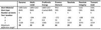
Magnetically Induced Force Measurements per ASTM F2052 of Active
Implantable Medical Device Lead Materials 
Michael Childers1, Roya Hashemi Rad1,
Richard Williamson1, and Shiloh Sison1
1St. Jude Medical, Sylmar, CA, United States
This abstract presents magnetically induced force
measurements per ASTM F2052 of materials commonly used in
implantable leads. Implantable leads which are constructed
solely from tested materials which pass the magnetically
induced force testing acceptance criteria (i.e. gravity
force), may not require magnetically induced force testing
per ASTM F2052 for MR conditionality with 3 T MR scanners.
|
 |
2210.
 |
Detailing the MR Safety of Intraocular Tantalum Markers Used for
Treatment Planning of Proton Beam Therapy of Uveal Melanoma: A
7.0T Study
Eva Oberacker1, Katharina Paul1, Lukas
Winter1, Celal Oezerdem1, Antje Els1,
Andreas Pohlmann1, Laura Boehmert1,
Stefanie Kox1, Min-Chi Ku1, Till
Huelnhagen1, Oliver Stachs2, Jens
Heufelder3,4, Andreas Weber3,4, and
Thoralf Niendorf1,5
1Berlin Ultrahigh Field Facility (B.U.F.F.), Max
Delbrück Center for Molecular Medicine in the Helmholtz
Association, Berlin, Germany, 2Department
of Ophthalmology, University of Rostock, Rostock, Germany,3Department
of Ophthalmology, Charité University Medicine, Berlin,
Germany, 4BerlinProtonen,
Helmholtz Zentrum Berlin, Berlin, Germany, 5Experimental
and Clinical Research Center (ECRC), a joint cooperation
between the Charité Medical Faculty and the Max Delbrück
Center for Molecular Medicine in the Helmholtz Association,
Berlin, Germany
This work examines the MR safety of intraocular tantalum
markers used in proton beam therapy of uveal melanoma. RF
power deposition induced heating was studied using
electromagnetic field and temperature simulations. Magnetic
force acting on the marker was investigated and image
artifacts were assessed. Minor local increase of RF power
deposition was observed for SAR0.075g but
not detectable for SAR1g. Measurements showed no
detectable magnetic attraction of the implant. FSE based
imaging showed only small artifacts barely exceeding the
thickness of the sclera. Our studies indicate that
intraocular tantalum markers do not constitute a per se
contraindication for 7.0T MRI.
|
|
2211.
 |
SAR/B1+ calibration workflow for safe, high duty-cycle parallel
transmission imaging at ultra-high field
Filiz Yetisir1, Bastien Guerin2,
Lawrence Wald2,3, and Elfar Adalsteinsson1,3
1Electrical Engineering and Computer Science,
Massachusetts Institute of Technology, Cambridge, MA, United
States, 2Dept.of
Radiology, Martinos Center for Biomedical Imaging,
Charlestown, MA, United States,3Harvard-MIT
Division of Health Sciences Technology, Institute of Medical
Engineering and Science, Cambridge, MA, United States
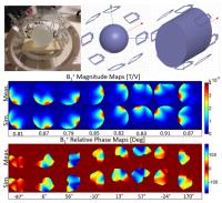
In this work, we propose a pTX safety workflow that will
enable high duty cycle imaging at high field systems.
Several SAR and B1+ calibration
steps are suggested for a complete analysis including
modeling the TX array, testing it over time and different
loads and finding a safety margin to account for RF system
imperfections. Good qualitative agreement was achieved
between the simulated and measured B1+ maps
for the TX array. 11% and 6° standard deviation was observed
in the magnitude and the relative phase maps over time. A
maximum difference of 16% was observed between offline and
online calculated local SAR values due to RF system
imperfections.
|
|
2212.
 |
Direct optical measurement of the RF electrical field for MRI
Isabelle Saniour1, Anne-Laure Perrier2,
Gwenaël Gaborit2,3, Jean Dahdah3,
Lionel Duvillaret3, and Olivier Beuf1
1CREATIS, Université de Lyon ; CNRS UMR5220 ;
Inserm U1044 ; INSA-Lyon ; Université Claude Bernard Lyon 1,
Villeurbanne, France, 2IMEP-LAHC,
UMR 5130 ; Université de Savoie, Le Bourget-du-Lac, France,3Kapteos,
Sainte-Hélène du Lac, France
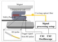
In MRI, a real time monitoring of the magnitude of the
electric field prevents the patient from safety hazards due
to heating phenomenon. A sub-cm electro-optical probe was
used to localize and measure the E-field in 4.7-T MRI. This
probe is formed from an electro-optic crystal that changes
its refractive indexes according to the applied E-field. The
results show that the probe is non-perturbative regarding
the E-field and does not affect the quality of MR images.
Six clear E-field concentrations were localized at proximal
and distal sides of the transceiver coil. Their magnitudes
vary between 10000V/m and 20000V/m.
|
|
2213.
 |
On Peripheral Nerve Stimulation of a Compact, Asymmetric
Head-Only Gradient Coil: Head Orientation Dependence
Seung-Kyun Lee1, Kishore V. Mogatadakala2,
Dominic Graziani1, Jean-Baptiste Mathieu2,
Thomas K.-F. Foo1, and Matt A. Bernstein3
1GE Global Research, Niskayuna, NY, United
States, 2GE
Healthcare, Florence, SC, United States, 3Mayo
Clinic, Rochester, MN, United States
Head orientation dependence of the peripheral nerve
stimulation (PNS) thresholds and the induced electric fields
of a high-performance, asymmetric head-only gradient coil
were studied experimentally and by numerical simulation. In
the experiment, the gradient field direction was fixed and
the subject head was rotated in the transverse plane. The
subject-reported PNS thresholds nearly doubled when the
head's anterior-posterior direction was parallel to the
gradient compared to when the head was approximately
perpendicular to the gradient. Human-body-model simulation
suggested that the orientation dependence may be primarily
due to locally concentrated electric fields in the
corrugated regions of the face.
|
|
2214.
 |
Positioning to decrease hot spots caused by an intramedullary
rod implanted in a forearm
Yu Kikuchi1, Minghui Tang1, and Toru
Yamamoto2
1Graduate School of Health Sciences, Hokkaido
university, sapporo, Japan, 2Faculty
of Health Sciences, Hokkaido university, sapporo, Japan
RF heating causes most of incidents during MRI examinations.
There still are patients who were implanted metallic
products before the advent of MRI and MR compatibility of
most such products is unknown. It was reported that an MRI
examination of a patient implanted an intramedullary rod in
his forearm was aborted due to a heating claim from the
patient. In this study, we confirm RF heating of such
patient by using an electromagnetic analysis software
dedicated for MRI, and shows that positioning of an
implanted arm can decrease SAR sufficiently enough to take
MRI examinations.
|
|
2215.
 |
Assessment of Radio Frequency Induced Heating On or Near
Implants during MRI – some open issues
Mikhail Kozlov1,2 and
Gregor Schaefers1,3
1MR:comp GmbH, Gelsenkirchen, Germany, 2Max
Planck Institute for Human Cognitive and Brain Sciences,
Leipzig, Germany, 3Magnetic
Resonance Institute for Safety, Technology and Research
GmbH, Gelsenkirchen, Germany
We evaluated locations of maximum temperature rise (max(ΔT))
and the dependence of max(ΔT)
on RF-induced power deposition (Ptotal) for
some generic implants. ΔT spatial
and temporal variations were investigated. To fulfill ASTM
F2182-11a setup requirements, the temperature probe should
be placed with submillimetre precision at location that
cannot be predicted by a full wave electromagnetic
simulation alone. It is a challenge to validate with small
uncertainty Ptotal calculated
using EM simulation by only measuring SAR or VLD value at
some points in space, if the field probe sensor size is
larger than one tenth of the wire diameter.
|
|
2216.
 |
Reduction of the E field at the tip of implanted wires generated
by pTx coils using RF current measurements
Gerd Weidemann1, Frank Seifert1, and
Bernd Ittermann1
1Physikalisch-Technische Bundesanstalt (PTB),
Braunschweig and Berlin, Germany
The possibility to reduce implant heating is an added value
option of parallel transmission. An orthogonal-projection
method (OPM) is presented to reduce the E fields at the tip
of wire type implants by using voltage vectors orthogonal to
the vector inducing the worst case RF current at the
protruding end of the implant. Experiments confirm that the
minimization of RF current at the protruding end leads to a
distinct reduction of the electric field at the tip of the
wire. Low-hazard steering conditions for n-element
pTx coils can be determined in real time during an MR
investigation from the measurement of only n complex
valued RF currents at the protruding end of the implant.
|
|
2217.
 |
Visualization and Localization of Implanted Devices with
Parallel Transmit Array Using Reversed RF Polarization
Parnian Zarghamravanbakhsh1, John M Pauly1,
and Greig Scott1
1Electrical Engineering, Stanford University,
Stanford, CA, United States
The radiofrequency (RF) transmit field can induce current in
implanted devices; therefore, it is essential to detect and
minimize coupling to stimulator leads and guide-wire
structures. Reverse polarization has been proposed as
low-RF-power method to safely detect current in the
implanted devices using birdcage coil. The purpose of this
study is to demonstrate feasibility of combining knowledge
of coil current and location with reverse polarization
method using parallel transmit array to detect and localize
implanted wires.
|
|
2218.
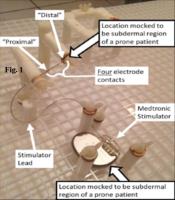 |
Heating of lead electrodes disconnected from sacral stimulator
during routine lumbar MRI at 3T with receive-only coil
Pallab K Bhattacharyya1, Howard Goldman2,
Mark J Lowe1, Adrienne Quirouet2, and
Stephen E Jones1
1Imaging Institute, Cleveland Clnic, Cleveland,
OH, United States, 2Glickman
Urological Institute, Cleveland Clnic, Cleveland, OH, United
States
RF heating testing during lumbar scans of Medtronic
Interstim II (Model 3058) implantable pulse generator (IPG)
connected to Medtronic Quadipolar Nerve Stimulator Lead
(Model 3889) at 3T whole body Siemens TIM Trio scanner with
receive-only cervical-lumbar-thoracic coil was performed.
Temperatures of the electrodes were measured by using fiber
optic sensors with fluoroptic monitoring with the IPG and
lead placed inside an ASTM gel phantom. No electrode heating
was observed when the lead was connected with the IPG in any
of the scans, while considerable heating was observed when
the IPG was disconnected and taken out of the phantom.
|
|
2219.
 |
Comparing RF heating simulations and experimental results in pTx
coils: an evaluation of three simulation methods
Hongbae Jeong1, Peter Jezzard1, and
Aaron Hess2
1FMRIB Centre, University of Oxford, Oxford,
United Kingdom, 2Department
of Cardiovascular Medicine, University of Oxford, Oxford,
United Kingdom
In this study, we conducted thermal simulations using EM
simulation software and compared these to proton resonance
frequency (PRF) thermometry using an ultra-high-field MR
phantom. RF heating was measured in the magnet environment
using a PRF-based 3D GRE on a 8-channel pTx coil. Three
types of simulation method were assessed and compared with
experimental data. Amongst the three simulation methods the
realistic capacitance simulation was closest to the
experimental measurement. In conclusion, PRF RF heating
measurements with real fiber optic temperature changes can
be used to assess and validate different types of RF
simulation.
|
|
2220.
 |
Statistical Equivalence Test Protocol for RF Performance of AIMD
Systems
Li-Yin Lee1, Shiloh Sison2, Shi Feng3,
Kishore Kondabatni4, and Richard Williamson5
1BioStatistics, St. Jude Medical, Sylmar, CA,
United States, 2Electrical
Engineering, St. Jude Medical, Sunnyvale, CA, United States, 3Electrical
Engineering, St. Jude Medical, Sylmar, CA, United States, 4St.
Jude Medical, Sylmar, CA, United States, 5Program
Management, St. Jude Medical, Sylmar, CA, United States
Test methods for MRI safety and RF safety of AIMD systems
has been defined through ISO/TS 10974 are cumbersome to
perform on every device and lead combination. A clear
method for determination that two likely equivalent systems
has not been described. The Concordance Correlation
Coefficient has been described for this purpose in assay
comparison. This paper evaluates the CCC method for RF
equivalence in presence of measurement uncertainty, and
confirms that the CCC method is simple and robust for this
purpose.
|
|
2221.
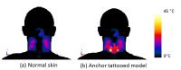 |
Modelling the RF safety of tattoo pigment ink for subjects
undergoing 7 Tesla MRI
Hongbae Jeong1, Aaron Hess2, and Peter
Jezzard1
1FMRIB Centre, University of Oxford, Oxford,
United Kingdom, 2Department
of Cardiovascular Medicine, University of Oxford, Oxford,
United Kingdom
Despite many reports of skin burns in the region of tattoos,
there are few safety studies concerning RF heating caused by
tattoos. Manufacturers of tattoo ink are numerous and use a
range of dye ingredients, making it difficult to assess the
electromagnetic properties of each ink pigment. An
anchor-shaped tattoo was modelled 1mm under the skin layer
in the region of the cervical spine to predict a possible
skin burn generated by RF coil. A simulation model of RF
heating in tattoo pigment is proposed, which shows that
certain tattoo pigments may lead to severe skin burns when
performing high field MRI.
|
|
2222.
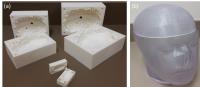 |
Heterogeneous gelatin-based head phantom for evaluating DBS
heating
Clare McElcheran1, Benson Yang2, Fred
Tam2, Laleh Golenstani-Rad3, and Simon
Graham2
1University of Toronto, Toronto, ON, Canada, 2Sunnybrook
Health Sciences Centre, Toronto, ON, Canada, 3Massachusetts
General Hospital, Charlestown, MA, United States
A method to create a heterogeneous head phantom with long
implanted wires to improve the evaluation of tissue heating
surrounding deep brain stimulation (DBS) leads is
presented. The phantom consists of three different
oil-in-gelatin dispersions with electrical properties that
mimic grey matter, white matter and cerebral spinal fluid
(CSF) as well as a human skull. 3D printing technology was
used to create gelatin moulds and an acrylic casing. A CT
scan of the human skull was obtained to create a mesh-based
digital representation. Thus, the physical phantom has an
associated mesh-based digital model which can be used in
electromagnetic simulation.
|
|
2223.
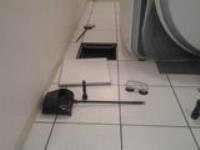 |
Potentially hazardous materials left behind after an MRI
installation
Ken E Sakaie1, Wanyong Shin1, and Lowe
J Mark1
1Imaging Institute, The Cleveland Clinic,
Cleveland, OH, United States
We share our experience discovering and removing metallic
objects left behind after a routine MRI hardware upgrade.
The results suggest that vigilance is necessary despite the
routine nature of such an upgrade.
|
|
2224.
 |
Increased Signal Intensity of brain structures on unenhanced
T1-weighted images following 35 or more GBCA administrations
Yang Zhang1,2, Yan Cao1, George Shih1,
Elizabeth Hecht3, and Martin R Prince1,4
1Radiology, Weill Cornell Medical Center, New
York, NY, United States, 2Radiology,
Qilu Hospital, Shandong University, Jinan, China, People's
Republic of, 3Columbia
University, New York, NY, United States, 4Radiology,
Columbia University, New York, NY, United States
In 16 patients with 35 or more linear GBCA administrations
increased T1 signal on unenhanced images was observed in
dentate nucleus (100%), globus pallidus (100%), cerebral
peduncles (100%), substantial nigra (88%), red nucleus
(88%), colliculi (81%), posterior thalamus (75%), superior
cerebellar peduncle (56%), internal capsule (50%), head of
caudate nucleus (31%), body of caudate nucleus (25%) , whole
thalamus (25%), pons (13%), anterior commissure (13%),
posterior brain stem (6%), pituitary gland (6%), mammillary
body (6%) and putamen (6%). The source of T1 signal
increase is unknown but may relate to GBCA administration.
No clinical significance was identified.
|
|
2225.
 |
Power deposition into a metallic hip prosthesis exposed to
switched gradient fields
Luca Zilberti1, Oriano Bottauscio1,
Mario Chiampi2, Jeffrey Hand3, Hector
Sanchez Lopez4, Rüdiger Brühl5, and
Stuart Crozier6
1Istituto Nazionale di Ricerca Metrologica,
Torino, Italy, 2Dipartimento
Energia, Politecnico di Torino, Torino, Italy, 3Division
of Imaging Sciences and Biomedical Engineering, King’s
College London, London, United Kingdom, 4Department
of Engineering, Universitas Dian Nuswantoro, Semarang,
Indonesia, 5Physikalisch-Technische
Bundesanstalt, Berlin, Germany, 6School
of Information Technology and Electrical Engineering,
University of Queensland, St. Lucia, Australia
Concern has been recently raised about the possible heating
of massive metallic implants, in particular hip prostheses,
due to the gradient fields used in MRI. Thus, this
contribution discusses the computation of the power density
deposited by the magnetic field into the implant, which
represents the first step to estimate the thermal heating.
The analysis is based on numerical simulations, performed
through a computational formulation applied to an anatomical
model of the body. The results provide evidence of the role
of the three gradient coil axes and of the different
harmonic components of the signals in this power deposition
process.
|
|
2226.
 |
Testing of a compact ultrasound scanner for use inside clinical
interventional MRI suite
Chi Ma1, Zaiyang Long1, Diana M
Lanners1, Donald J Tradup1, Joel P
Felmlee1, David A Woodrum1, Nicholas J
Hangiandreou1, and Krzysztof R Gorny1
1Department of Radiology, Mayo Clinic, Rochester,
MN, United States
The suitability of a compact Samsung ultrasound (US) system
for real-time imaging guidance of treatment device
positioning inside 1.5T interventional magnetic resonance
imaging (iMRI) suite was assessed. The US system was tested
in a proposed site-specific configuration. Magnetic
displacement forces exerted by the static magnetic field on
each of the US system components were estimated at the
proposed locations. Image quality of both MRI and US systems
with the US system set to different operating modes were
evaluated. Results demonstrate that this particular US
system is suitable for use in the site-specific
configuration at our 1.5T iMRI suite.
|
|
2227.
 |
An Evaluation of Radio Frequency Induced Power Deposition of
Coaxial Leads with an Implant Model
Mikhail Kozlov1,2 and
Gregor Schaefers1,3
1MR:comp GmbH, Gelsenkirchen, Germany, 2Max
Planck Institute for Human Cognitive and Brain Sciences,
Leipzig, Germany, 3Magnetic
Resonance Institute for Safety, Technology and Research
GmbH, Gelsenkirchen, Germany
We performed 3-D electromagnetic simulations of coaxial
leads and numerically obtained the lead models to evaluate
power deposition and the voltage induced at the lead
proximal end with the lead models. No correlation between
peak volume loss density and deposited powers at the tip and
the ring was observed. In some cases deposited power at the
ring exceeded deposited power at the tip. However further
extensive simulations of induced heating behavior should be
done before final conclusions regarding coax lead design
preferences are made.
|
|
2228.
 |
Influence of electrical properties of lead insulation on radio
frequency induced heating during MRI
Mikhail Kozlov1,2 and
Gregor Schaefers1,3
1MR:comp GmbH, Gelsenkirchen, Germany, 2Max
Planck Institute for Human Cognitive and Brain Sciences,
Leipzig, Germany, 3Magnetic
Resonance Institute for Safety, Technology and Research
GmbH, Gelsenkirchen, Germany
We evaluated the dependence of RF-induced power deposited at
a hot spot (p) on insulating
electrical properties for insulated stainless steel wires of
1.5 mm in diameter with insulation thickness of 0.5 mm. Lead
transfer functions (TF) were obtained by 3-D electromagnetic
simulations. TF and p depended
significantly on electrical properties of insulation.
Increased insulator conductivity resulted in decreased p.
For all insulated wires investigated non-uniform RF
excitation resulted in higher power deposition than uniform
RF excitation.
|
|
2229.
 |
Design and simulation of a nested 4 channel 1H and 3 channel 13C
coil for glycogen NMR experiments in the calf muscle at 7 T
Sigrun Goluch1,2,3, Roberta Kriegl2,3,
Elmar Laistler2,3, Martin Gajdošík 4,5,
and Martin Krššák 1,4,5
1Division of Endocrinology and Metabolism,
Department of Internal Medicine III, Medical University of
Vienna, Vienna, Austria, 2MR
Center of Excellence, Medical University of Vienna, Vienna,
Austria, 3Center
for Medical Physics and Biomedical Engineering, Medical
University of Vienna, Vienna, Austria, 4High-Field
MR Center, Department of Biomedical Imaging and Image-Guided
Therapy, Medical University of Vienna, Vienna, Austria, 5Christian
Doppler Laboratory for Clinical Molecular MR Imaging,
Medical University of Vienna, Vienna, Austria
Due to the inherently low sensitivity of carbon-13 NMR, 13C
spectroscopic experiments at 7T require specifically
optimized double tuned local RF transceive arrays for high
SNR, exhibiting sufficient electrical isolation between the
arrays to enable 1H decoupling and high SAR efficiency as to
not invoke SAR limits during proton decoupling. In this work
we present the simulation and optimization of a 7 channel
nested 1H/13C
RF transceive coil array for 13C
metabolic studies in the human calf muscle at 7 T.
|
|
2230.
 |
Assessment of RF induced heating of intracranial Micro-depth
electrodes during MRI
Anastasia Papadaki1,2, David Carmichael3,
Andrew McEvoy4,5, Anna Miserocchi4,5,
Tarek Yousry1,2, Beate Diehl4,6, Louis
Lemieux4, and John S Thornton1,2
1Lysholm Department of Neuroradiology, National
Hospital for Neurology and Neurosurgery, UCLH, London,
United Kingdom, 2Department
of Brain Repair and Rehabilitation, UCL Institute of
Neurology, London, United Kingdom, 3Imaging
and Biophysics Unit, UCL Institute of Child Health, London,
United Kingdom, 4Department
of Clinical and Experimental Epilepsy, UCL Institute of
Neurology, London, United Kingdom,5Department of
Neurosurgery, National Hospital for Neurology and
Neurosurgery, London, United Kingdom, 6Department
of Neurophysiology, National Hospital for Neurology and
Neurosurgery, London, United Kingdom
In this study we assessed temperature changes (?T) during
MRI in the vicinity of microwires EEG electrodes in a
phantom. Measurements were performed at 1.5T during a high
SAR TSE sequence for two different depth electrode
arrangements with and without microwires. Although we
observed a small temperature rise due to the presence of
microwires the maximum temperature change ?T did not exceed
1°C at 1.5T.
|
|
2231.
 |
SAR and patient orientation for 3 T 2-channel parallel transmit
pelvis imaging
Mariya Lazebnik1
1GE Healthcare, Waukesha, WI, United States
This work investigates the impact of patient orientation on
SAR for 3 Tesla two-channel parallel transmit (pTx) pelvis
imaging. SAR simulations were performed on two human body
models in a supine position in a 70 cm-diameter 3 T body
coil in a pelvis landmark, in both “head first” and “feet
first” patient entry orientations. Whole body SAR, peak
spatial SAR, and SAR ratio (= peak SAR / whole body SAR)
were computed for quadrature and pTx excitations. Patient
position and orientation can cause peak SAR and SAR ratio to
vary significantly and must be considered when evaluating
pTx excitation.
|
|
2232.
 |
Subject-specific SAR prediction in adults and children at 7.0T
Gianluigi Tiberi1,2, Mauro Costagli1,2,
Laura Biagi2, Alessio De Ciantis3,
Nunzia Fontana4, Riccardo Stara5,6,
Mark R Symms7, Mirco Cosottini8, Renzo
Guerrini3, and Michela Tosetti1,2
1Imago7, Pisa, Italy, 2IRCCS
Stella Maris Foundation, Pisa, Italy, 3Meyer
Children’s Hospital, Firenze, Italy, 4Dipartimento
di Ingegneria dell'Informazione, Pisa, Italy, 5National
Institute of Nuclear Physics (INFN), Pisa, Italy, 6Stanford
University, Stanford, CA, United States, 7General
Electric ASL Scientist (EMEA), Pisa, Italy, 8Department
of Translational Research and New Surgical and Medical
Technologies, Pisa, Italy
In this study we propose a procedure which allows the
prediction of global and local subject-specific SAR exposure
for commonly used 7.0T sequences. Prerequisites for such
prediction are: sequences’ SAR exposure simulated on the
generic anatomic models; subject-specific measured B1+ maps.
Validation has been provided through phantom experiment. We
observed that: SILENT and FLAIR can be safely used in all
subjects, both adults and children; FLAIR is more SAR
demanding than SILENT; predicted SAR exposure does not show
a significant variation with subject weight.
|
|
2233.
 |
Safety of MR Imaging of Patients with Cardiac Implanted Devices
El-Sayed H. Ibrahim1, Laura Horwood1,
Jadranka Stojanovska1, Luba Frank1,
Anil Attili1, Hakan Oral1, and Frank
Bogun1
1University of Michigan, Ann Arbor, MI, United
States
This study examines whether MRI is safe in patients with
cardiac implantable electronic device (CIED) excluded from
published protocols, e.g. patients with abandoned leads or
pacemaker dependency. A total of 162 MRI scans were obtained
in 142 consecutive patients with CIED’s. Cardiac scans were
performed in 94 patients and spinal/brain scans were
performed in 47 patients. Only one patient developed
ventricular tachycardia during a spine scan and was removed
from the scanner for device reactivation without
consequences. No other adverse events were noted. The
devices interrogated parameters essentially remained the
same immediately, 1-week after, and 3-months after the
scans.
|
|
2234.
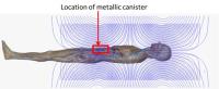 |
The Potential for Eddy Current Induced Peripheral Nerve
Stimulation from an Active Implanted Device Canister
Xin Chen1, Jonathan Edmonson2, and
Michael Steckner1
1Toshiba Medical Research Institute USA, Inc.,
Mayfield Village, OH, United States, 2Medtronic
CRHF, Mounds View, MN, United States
We used numerical simulations to investigate the potential
for increased PNS likelihood with implanted device. Modeling
of a gradient coil loaded with a human subject with a
metallic implanted canister showed that the electric field
around the device can increase by up to 3 fold, suggesting
increased PNS likelihood.
|
|
2235.
|
Prospective Observational Post-marketing Study on the Safety of
Gadoterate Meglumine - Final Results in the pediatric cohort of
over 1,600 children
Yun Peng1
1Beijing Children's Hospital, Beijing, China,
People's Republic of
An observational post-marketing study was conducted in 10
countries to prospectively collect safety data in adults and
children who were scheduled to undergo routine Magnetic
Resonance Imaging (MRI) with administration of gadoterate
meglumine (Dotarem®). The incidence of Nephrogenic Systemic
Fibrosis (NSF) in routine practice was assessed through
specific follow-up of patients with moderate to severe renal
impairment. Final results in a large pediatric
sub-population of over 1,600 children showed a very good
safety profile of gadoterate meglumine with only one adverse
event reported in a child and no suspicion of NSF reported.
|
|
2236.
 |
Scanner-specific verification of Transmit RF Body Coil B1-field
to inform clinical triage of patients with implanted devices
Chi Ma1, Krzysztof R Gorny1,
Christopher P Favazza1, Robert E Watson1,
and Heidi A Edmonson1
1Radiology, Mayo Clinic, Rochester, MN, United
States
Exclusion of scanning with transmit RF body coil may
prohibit access to life-saving diagnoses for patients with
MR-conditional implantable devices. Manufacturer provided
plots of RF B1-field indicate that RF energy over the
implant may be significantly reduced if the implant is kept
outside of the 50-55cm long transmit RF body coil.
Scanner-specific B1-field measurements and RF-induced
heating measurements confirm reduction in heating as
conductive material moves away from scanner isocenter.
B1-field measurements lateral to the central scanner axis
demonstrate local peaks in the B1-field that would not be
identified from the IEC-required manufacturer plots.
|
|
2237.
 |
Magnetic Displacement Force and Safety of Coronary Artery Stents
at 7 Tesla.
Christian Hamilton-Craig1,2, Jess Cameron1,
Gregory Brown1, and Graham Galloway1,3
1Centre for Advanced Imaging, University of
Queensland, Brisbane, Australia, 2Richard
Slaughter Centre of Excellence in CVMRI, The Prince Charles
Hospital, Brisbane, Australia, 3Translational
Research Institute, Brisbane, Australia
Currently, there are minimal data regarding the magnetically
induced displacement force of coronary artery stents, in 7.0
T MR. We tested a range of commonly implanted coronary
artery stents for maximal displacement force at 7T. CoCr
stents appear to have safe deflection properties at 7T.
However 316L-SS and PtCr stents exhibit increased
magnetically induced displacement forces, and may be not be
considered conditionally safe at 7.0T
|
|
2238.
 |
Electrocorticography grids might cause excessive heating during
MR imaging
Emad Ahmadi1, Reza Atefi1, Emad
Eskandar2, Alexandra J. Golby3,
Michael H. Lev1, Rajiv Gupta1, and
Giorgio Bonmassar1
1Radiology, Massachusetts General Hospital,
Boston, MA, United States, 2Neurosurgery,
Massachusetts General Hospital, Boston, MA, United States, 3Neurosurgery,
Brigham and Women's Hospital, Boston, MA, United States
Electrocorticography grids are routinely implanted over the
cortex for pre-surgical planning in epilepsy surgery. We
propose that MR imaging at 3T might cause heating injury in
patients with implanted electrocorticography grids.
|
|
2239.
 |
Sugar free tissue-mimicking MRI phantoms for improved
signal-to-noise ratio
Carlotta Ianniello1,2, Ryan Brown1,
Martijn Cloos3, Qi Duan4, Jerzy
Walczyk3, Graham Wiggins3, Daniel K
Sodickson2,3, and Riccardo Lattanzi2,3
1Radiology, Center for Advanced Imaging
Innovation and Research (CAI2R) and Center for Biomedical
Imaging, Department of Radiology, New York University School
of Medicine, New York, NY, United States, 2The
Sackler Institute of Graduate Biomedical Science, New York
University School of Medicine, New York, NY, United States, 3Center
for Advanced Imaging Innovation and Research (CAI2R) and
Center for Biomedical Imaging, Department of Radiology, New
York University School of Medicine, New York, NY, United
States, 4Laboratory
of Functional and Molecular Imaging, NINDS, National
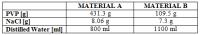
Institutes of Health, Bethesda, MD, United States
We investigated Polyvinylpyrrolidone (PVP) as an alternative
to sugar to control relative permittivity in
tissue-mimicking MR phantoms. We constructed a
two-compartment phantom filled with water solutions of PVP
and NaCl, the latter used to control conductivity. A lower
amount of PVP than sugar is required, allowing low
permittivity materials to be realized. While signal
decreases rapidly in sugar-based phantoms, PVP materials
have long T2*/T2, making
PVP-based phantoms suitable for the validation of MR-based
electrical properties mapping techniques that rely on high
SNR of signal and B1+ maps.
PVP solutions are relatively inexpensive, easy to mix and do
not require preservatives.
|
|
2240.
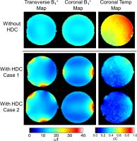 |
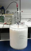
Experimental evaluation of heating and SAR reduction with a
dielectric insert at 3T
Christopher Sica1, Sebastian Rupprecht1,
and Qing X Yang1
1Radiology, Penn State College of Medicine,
Hershey, PA, United States
Prior work has suggested that a dielectric insert can reduce
the SAR in the brain at 3T. These previous results were
obtained via electromagnetic simulations. Here, we present
an experimental evaluation of SAR reduction in a phantom
with a dielectric insert.
|
|