ISMRM 24th Annual Meeting & Exhibition • 07-13 May 2016 • Singapore

Traditional Poster Session: Molecular Imaging

2304 -2327 Molecular Imaging & Contrast Agents
2328 -2350 Hyperpolarised C-13 & Other Nuclei


2304.   
Fluorescence-based Quantification of Gadolinium-bound Liposomes using Carbostyril 124-sensitized DTPA
Lindsay Kathleen Hill1,2, Stewart Russell3,4, Dung Minh Hoang1, and Youssef Zaim Wadghiri1
1Radiology, NYU School of Medicine, New York, NY, United States, 2Biomedical Engineering, SUNY Downstate Medical Center, Brooklyn, NY, United States, 3Thayer School of Engineering, Dartmouth College, Hanover, NH, United States, 4Department of Mechanical Engineering, The City College of New York, New York, NY, United States
Advancement in the field of Gadolinium-bound contrast agent discovery is reliant on the development and characterization of novel constructs made in-house. However, the assessment of physicochemical properties and in vivo pharmacokinetics, requiring highly sensitive measurements, is often impeded by the lack of analytical techniques that are simultaneously sensitive, affordable, and accessible. Here we demonstrate that Carbostyril 124-sensitized DTPA can be incorporated into a lipid-based microparticle, allowing for rapid quantification of Gadolinium concentration with nanomolar sensitivity using a readily available fluorescence plate reader. This sensitive and convenient technique could rapidly propel the characterization of novel MR contrast agents. 


2305.   
Nanocrystal Loaded Polymeric Microbubbles for Multimodal Imaging
Nutte Teraphongphom1, Peter Chhour2, John Eisenbrey3, Pratap Chandra Naha2, Walter Witschey2, Borirak Opasanont4, Lauren Jablonowski1, David Peter Cormode2, and Margaret Wheatley1
1Biomedical Engineering, Drexel University, Philadelphia, PA, United States, 2Radiology, University of Pennsylvania, Philadelphia, PA, United States, 3Radiology, Thomas Jefferson University, Philadelphia, PA, United States, 4Chemical and Biological Engineering, Drexel University, Philadelphia, PA, United States
To create multimodal contrast agents, we hypothesized that the shell of polymeric microbubbles could accommodate additional payloads. We therefore modified microbubbles by encapsulating nanoparticles including quantum dots, magnetic iron oxide nanoparticles, or gold nanoparticles to create bi-modality platforms in a manner that minimally compromised the performance for each individual imaging technique (ultrasound, fluorescence imaging, computed tomography and MRI). 


2308.   
A Targeted Host-Guest MRI Contrast Agent for Breast Cancer Molecular Imaging
Zhuxian Zhou1,2, Zheng Han1, and Zheng-Rong Lu1
1Biomedical Engineering, Case Western Reserve University, Cleveland, OH, United States, 2Chemical and Biological Engineering, Zhejiang University, Hangzhou, China, People's Republic of
To produce MRI detectable signal enhancement for the biomarker on cancer cell surface, we developed a targeted host-guest nanosized contrast agent cRGD-POSS-βCD-(DOTA-Gd)-Cy5. The nanosized host-guest systems are preferable for facile synthesis of customized and functionalized imaging agent. Here, a cyclic peptide cRGD targeting to cancer cell αvβ3-integrin, a macrocyclic Gd(III) chelate and Cy5 fluorescent probes were loaded on the nanosized carrier by host-guest interaction. cRGD-POSS-βCD-(DOTA-Gd)-Cy5 can provide strong contrast enhancement to delineate malignant tumors during molecular MR and fluorescent imaging in a mouse 4T1 breast cancer model.


2306.   
Development of facile protocols for stable nanoparticle formulations of 19F MR molecular imaging probes
Eric A Tanifum1,2, Chandresh Patel1, Robia Pautler2, and Ananth Annapragada1,2
1Texas Children's Hospital, Houston, TX, United States, 2Baylor College of Medicine, Houston, TX, United States
Perfluorocarbons and perfluoropolyethers are currently the major molecules of choice in 19F MRI contrast agents. These molecules generally have magnetically diverse 19F atoms and are very hydrophobic. The later characteristic greatly hinders easy access to stable formulations for broad usage and the former generates chemical shift artifacts which result in blurred images. We have synthesized several hydrophilic organofluorine molecules all bearing magnetically equivalent 19F atoms and demonstrated that they are amenable to facile liposome nanoparticle formulation protocols. The resulting particles are highly stable and present a great potential for diverse applications as 19F MRI molecular imaging probes. 


2307.   
A Ratiometric Bioresponsive MRI Contrast Agent for Rapid Monitoring of Biological Processes
Tanja Savic1, Serhat Gündüz2, Rolf Pohmann3, Nikos Logothetis4, Klaus Scheffler3, and Goran Angelovski1
1Research group "MR Neuroimaging agents", Max Planck Institute for Biological Cybernetics, Tübingen, Germany, 2Research Group "MR Neuroimaging agents", Max Planck Institute for Biological Cybernetics, Tübingen, Germany, 3High-Field Magnetic Resonance, Max Planck Institute for Biological Cybernetics, Tübingen, Germany, 4Physiology of Cognitive Processes, Max Planck Institute for Biological Cybernetics, Tübingen, Germany
A number of bioresponsive MRI contrast agents have been developed, with the aim of producing the maximal signal difference for a given biological event. This paper introduces an approach which substantially improves the detection of physiological events with fast kinetics. A nanosized, calcium-sensitive dendrimeric probe was developed and characterized by means of a balanced steady-state free precession imaging protocol. Results show an almost four times greater contrast gain per unit of time as compared to conventional T1-weighted imaging with small sized contrast agents. Consequently, this ratiometric methodology has a profound significance for future studies of biological dynamic processes by means of MRI.


2309.   
Biodegradable glycoged-based nanoprobe as a multimodal tumor-targeting contrast agent
Andrea Galisova1, Daniel Jirak1, Marketa Jiratova1, Martin Hruby2, Maria Rabyk2, Aneta Pospisilova2, and Milan Hajek1
1MR Unit, Institute for Clinical and Experimental Medicine, Prague, Czech Republic, 2Academy of Sciences, Institute of Macromolecular Chemistry, Prague, Czech Republic
An effective cancer diagnostic and therapeutic contrast agent with suitable properties including high specificity and safety is on high demand. In this study, accumulation of a biocompatible and biodegradable glycogen-based nanoprobe (GG-Gd-DOTA-Dy) was tested and compared to a commercially available contrast agent (gadoterate meglumine). Relaxivity and MR imaging of the probe was performed on the phantoms. The uptake of the agents was measured on the tumor-bearing rats at several time points after the contrast agent administration by MRI and fluorescence imaging. We found out that the novel probe is superior to a commercially available contrast agent regarding the relaxivity and accumulation in the tumor tissue.


2310.   
In-vivo quantification of focal vessel wall changes following vascular injury in a murine model of atherosclerosis
Begona Lavin Plaza1, Alkystis Phinikaridou1, Marcelo Andia2, Silvia Lorrio Gonzalez1, and Rene Botnar1
1Imaging Sciences and Biomedical Engineering, King's College London, London, United Kingdom, 2Pontificia Universidad Catolica de Chile, Santiago de Chile, Chile
Despite the beneficial effect of percutaneous transluminal coronary angioplasty and stent implantation, negative vascular remodeling remains as one of the most important complications of interventional cardiology. These procedures may damage the vessel wall, particularly the endothelium, leading to a dysfunctional state characterized by impaired vasodilation, increased leukocyte adhesion and permeability that constitute the initial steps of atherosclerosis. The arterial tree can be divided in either “athero-susceptible” areas, e.g. arterial branches and curvatures, where blood flow is turbulent and shear stress is multidirectional or “athero-resistant” areas, e.g. abdominal aorta, where blood flow is laminar and shear stress is low. In this study, we investigated (1) whether an “atherosclerotic-resistant” segment of the vascular tree, like the aorta, can be switched into an “atherosclerotic-susceptible” area following endothelial injury and (2) whether such a switch in vessel wall remodeling is associated with changes in vascular permeability that can be assessed in-vivo using the albumin binding MR contrast agent, gadofosveset.


2311.   
Vascular-targeted Magnetic Nanoparticles for Image-guided Cancer Therapy
Sudath Hapuarachchige1, Robert Ivkov2, and Dmitri Artemov1
1Department of Radiology and Radiological Science, Johns Hopkins University School of Medicine, Baltimore, MD, United States, 2Department of Radiation Oncology and Molecular Radiation Sciences, Johns Hopkins University School of Medicine, Baltimore, MD, United States
Bionized nanoferrites are magnetic nanoparticles, which can be used as contrast agents and therapeutic platforms for alternating magnetic field (AMF) induced hyperthermia. One important application is enhancing of vascular permeability in tumors for delivery of nanodrugs. We studied BNF nanoparticles specifically targeted to the tumor vasculature via VEGF receptors ligands. Targeted BNF particles were visualized by intravital multiphoton and MR imaging, and increased accumulation of targeted BNF was detected in breast cancer models.


2312.   
BBN-assembled Gadolinium oxide nanoprobe for targeted bimodal imaging in vitro and in vivo
Danting Cui1, Xiaodan Lu1, Chenggong Yan1, Xiang Liu1, Yingjie Mei2, Meirong Hou1, Yikai Xu1, and Ruiying Liu3
1Department of Medical Imaging Center, Nanfang Hospital, Southern Medical University, Guangzhou, China, People's Republic of, 2Philips Healthcare, Guangzhou, China, People's Republic of, 3School of Pharmaceutical Sciences, Southern Medical University, Guangzhou, China, People's Republic of
Bombesin (BBN) is a peptide exhibiting high affinity for the gastrin-releasing peptide receptor (GRPr), To develop a BBN-assembled nanoprobe based on Gd2O3 was efficent for earlier tumor detection. Gd2O3 was conjugated with 5(6)-carboxyfluorescein and bombesin for MR/optical bimodal imaging of GRPr positive tumor. Gd2O3-FI-PEG NPs without bombesin-modified NPs were tested as non-targeted control.  MRI and opitical  imaging in vitro and vivo confirmed BBN-assembled Gd2O3 nanoprobe exhibited better binding affinity to GRPr positive tumor than the control group. The nanoprobe may provide opportunities to further biomedical application.


2313.   
Combining Multi-therapy in Singlet Vehicle
Yuqi Yang1, Shizhen Chen1, Sha Li1, Lianhua Liu1, and Xin Zhou1
1Wuhan Institute of Physics and Mathematics, Chinese Academy of Sciences, Wuhan, China, People's Republic of
Two types of water-insoluble texphyrins (TP), anticancer drug Gd-TP and photosensitizer Lu-TP, were synthesized and loaded onto RGD-functionalized graphene quantum dots (GQDs) via π-π stacking. The obtained complex could be used as a MRI-fluorecent imaging multi-model probe for cancer therapy. Compared with conventional photodynamic therapy (PDT), our method demonstrated better therapy efficiency for deeper tissue, because a laser with longer wavelength was applied to active the photosensitizer Lu-TP. Furthermore, reactive oxygen species resulted from the reaction between redox active drug Gd-TP and cellular reducing metabolites and photothermal effect from GQDs led cancer cells more impressionable to PDT from Lu-TP. 


2314.   
Short-lived mesenchymal stem cells accelerate healing of acid skin burns – an MRI cell tracking study using iron oxide, fluorine and bioluminescence imaging
Ghulam Muhammad1,2, Jiadi Xu3, Jeff W.M. Bulte1, Anna Jablonska1, Piotr Walczak1,4, and Miroslaw Janowski5
1The Russell H. Morgan Department of Radiology and Radiological Science, Johns Hopkins University, Baltimore, MD, United States, 2Stem Cell Laboratory, University of the Punjab, Lahore, Pakistan, 3F.M. Kirby Research Center, Kennedy Krieger Institute, Baltimore, MD, United States, 4Department of Radiology, University of Warmia and Mazury, Olsztyn, Poland, 5Johns Hopkins University, Baltimore, MD, United States
Incidence of acid burns due to accidents and attacks is on the rise and mesenchymal stem cell transplantation is a promising therapeutic strategy. Cell tracking makes treatment more precise. We have compared two MRI tracking strategies: SPIO nanoparticles-based 1H MRI and 19F nanoemulsion “hot-spot” MRI. Bioluminescence imaging was used as a reference standard for monitoring cell survival. Susceptibility artifacts due to skin injury compromised the interpretation of 1H imaging, while 19F MRI was capable of providing information on cell location and survival. The SPIO nanoparticles and fluorine nanoemulsion had no detrimental effect on the therapeutic activity and survival of MSCs.


2315.   
Absolute Quantification of Stem Cell Transplant in MRI
Muhammad Jamal Afridi1, Arun Ross2, and Erik M Shapiro3
1Department of Radiology, Department of Computer Science, Michigan State University, East Lansing, MI, United States, 2Department of Computer Science, Michigan State University, East Lansing, MI, United States,3Department of Radiology, Michigan State University, East Lansing, MI, United States
We describe an image analysis strategy for quantifying the location and number of transplanted stem cells from MRI images. MRI-based single cell detection facilitates the use of machine learning algorithms for spot detection. Using convolutional neural networks, automatic and intelligent cell enumeration was first developed on in vitro agarose samples containing a known number of labeled cell mimics. Then, the validated image analysis approach was used to quantify stem cell transplants in rodent brains. An accuracy of 99.8% was achieved on in vitro samples and 94.6% on in vivo examples.


2316.   
Automatic in vivo detection of transplanted cells in MRI using transfer learning paradigm
Muhammad Jamal Afridi1, Arun Ross2, Steven Hoffman2, and Erik M Shapiro3
1Department of Radiology and Department of Computer Science, Michigan State University, East Lansing, MI, United States, 2Department of Computer Science, Michigan State University, East Lansing, MI, United States, 3Department of Radiology, Michigan State University, East Lansing, MI, United States
Despite advances in machine learning and computer-vision, many MRI studies rely on tedious manual procedures for quantifying imaging features, i.e. cell numbers, contrast area etc. Development of intelligent, automatic tools for quantifying imaging data requires large scale data for their training and tuning, which in the clinical arena is challenging to obtain.  Here, we present an approach that obviates the need for large scale data collection to develop an intelligent and automatic tool for single cell detection in MRI. Our strategy achieves 91.3% accuracy for in vivo cell detection in MRI despite using only 40% of the data for training.


2317.   
Cell tracking with fluorine-19 MRI and zirconium-89 PET - a multi-modal approach
Kai D. Ludwig1, Benjamin L. Cox1,2,3, Myriam N. Bouchlaka4,5, Stephen A. Graves1, Justin J. Jeffery5, R. Jerry Nickles1, Bryan P. Bednarz1,6, Christian M. Capitini4,5, and Sean B. Fain1,6,7
1Medical Physics, University of Wisconsin-Madison, Madison, WI, United States, 2Morgridge Institute for Research, Madison, WI, United States, 3Laboratory for Optical and Computational Instrumentation, University of Wisconsin-Madison, Madison, WI, United States, 4Pediatrics, University of Wisconsin-Madison, Madison, WI, United States, 5Carbone Cancer Center, University of Wisconsin-Madison, Madison, WI, United States,6Radiology, University of Wisconsin-Madison, Madison, WI, United States, 7Biomedical Engineering, University of Wisconsin-Madison, Madison, WI, United States
Methods for non-invasive cell tracking may greatly enhance the ability to assess efficacy of cellular-based therapies. A dual-labeled (89Zr and 19F) cell labeling approach could inform and potentially improve in vivo cell tracking sensitivity and clinical adoption. Here, we show longitudinal detection of localized cell injections with 19F MRI and the ability to quantify the number of cells within a voxel. Additionally, 89Zr cell tracking results shows a high sensitivity for intravenous delivery of cells with longitudinal signal detection. Future work aims to combine both cell tracking approaches utilizing the dual-modality imaging platform on a PET/MRI system.


2318.   
Interaction of Manganese and Iron in R1 mapping in a Low Concentration Setting
Chien-Lin Yeh1, Carlos J. Perez-Torres1, and Ulrike Dydak1,2
1School of Health Sciences, Purdue University, West lafayette, IN, United States, 2Radiology and Imaging Sciences, Indiana University School of Medicine, Indianapolis, IN, United States
Being able to use MRI to estimate brain Mn accumulation is of high interest in occupational Mn exposure settings. As a first step towards absolute quantification of brain Mn concentration in exposed humans using MRI, the interaction between Mn and Fe and their combined effect to R1 need to be explored. Our results suggest that a model only assuming independent linear contributions of Mn and Fe already explains the R1 data well. However introducing a cross term of Mn and Fe in the equation improves the fits, suggesting a Mn-Fe interaction.   


2319.   
Glutamate-sensitive CEST and MEMRI as novel biomarkers for studying ALS pathophysiology
Amit Kumar Srivastava1,2, Jiadi Xu3, Peter C.M. van Zijl2,3, Nicholas J Maragakis4, and Jeff W.M. Bulte1,2,3
1Cellular Imaging Section, Institute for Cell Engineering, Johns Hopkins University, Baltimore, MD, United States, 2Russel H. Morgan Department of Radiology and Radiological Science, Johns Hopkins University, Baltimore, MD, United States, 3F.M. Kirby Research Center for Functional Brain Imaging, Kennedy Krieger Institute, Baltimore, MD, United States, 4Neurology, Johns Hopkins University, Baltimore, MD, United States
Amyotrophic lateral sclerosis (ALS) is characterized by selective loss of motor neurons. ALS treatment is very difficult because disease manifestation and diagnosis often happen much later than when ALS pathology occurs in the patient. In this study, we developed two non-invasive MRI biomarkers for the early detection of disease pathology and its progression thereafter. A newly developed Glutamate-sensitive CEST showed higher signal intensity in the spinal cord level of ALS at pre-symptomatic stage, an indicator of initiation of ALS pathology. Manganese-enhanced MRI showed higher T1-weighted signal in the ALS spinal cord at post-symptomatic stage suggesting activation of astrocytes.          


2320.   
Longitudinal MEMRI Characterization of a Novel Mouse Medulloblastoma Model
Harikrishna Rallapalli1, Eugenia Rafaela Volkova1, I-Li Tan2, Alexandre Wojcinski2, Alexandra L Joyner2, and Daniel H Turnbull1
1Skirball Institute and Radiology, New York University School of Medicine, New York, NY, United States, 2Developmental Biology, Sloan Kettering Institute, New York, NY, United States
In this work, we describe a powerful longitudinal Manganese-enhanced magnetic resonance imaging (MEMRI) strategy to characterize a novel mouse medulloblastoma model. An activated Smoothened mutation was engineered to induce proliferative growth in the cerebellum. Lesions were monitored using MEMRI up to postnatal day P100, and 3D tumors were segmented for quantitative volumetric analysis. Qualitative analysis has shown a ~50% chance of regression overall (n=21), and preliminary quantitation has suggested a combined progression/regression growth model. With this model, we aim to guide diagnostic decisions from early timepoints and quantify therapeutic efficacy.


2321.   
Mn cell uptake mechanisms in organotypic rat hippocampal slice cultures
Alexia Daoust1, Stephen Dodd1, and Alan Koretsky1
1NINDS, LFMI, NIH, Bethesda, MD, United States
MEMRI can be used for different applications such as tracing neuronal connections or functional imaging. However, Mn cellular uptake is still unclear. We studied this mechanism by the use of MEMRI in a hippocampal organotypic slice culture. After added Mn to the medium for 2h, we obtained an optimal MR contrast that was affected by Ca channel manipulation. Mn cellular uptake was also affected by the presence of other metals that use divalent metal transporters (DMT-1). Our results suggest a strong capacity of our technique to study the cellular mechanisms related to MEMRI.


2322.   
Distributed T2 relaxation model for polydisperse nanoparticle systems
Bashar Issa1
1Physics, UAE University, Al-Ain, United Arab Emirates
Theories describing 1/T2 enhancement due to the presence of superparamagnetic particles agree well with experimental and Monte Carlo (MC) simulation data under the condition that the particles are monodisperse both in size and magnetization. We present a 1/T2 distributed model that takes into account the particle size and magnetization distributions. We average the individual 1/T2 components exhibited by each group of particle with a uniform particle size. MC simulations of the model successfully predict 1/T2 within the MAR regime confirming the implicit assumption that the spins are able to sample all the particles’ radii and magnetizations within the echo time.


2323.   
Quantifying exchange in host-guest systems for hyperpolarized xenon
Sergey Korchak1, Wolfgang Kilian1, Leif Schröder2, and Lorenz Mitschang1
1Physikalisch-Technische Bundesanstalt (PTB), Braunschweig and Berlin, Germany, 2Leibniz-Institut für Molekulare Pharmakologie (FMP), Berlin, Germany
The reversible binding of xenon to host structures is fundamental to the development of novel contrast agents employing hyperpolarized xenon and chemical exchange saturation transfer (HyperCEST) for molecular imaging. The rates for entering and leaving the host depend on atomic details and affect the obtainable contrast rendering them pivotal for the selection of hosts and optimization of imaging methods. However, different exchange processes may apply whose contributions are difficult to assign. Exchange spectroscopy experiments are proposed which enable straightforward disentanglement of the exchange kinetics and quantification of individual contributions. The approaches are exemplified for the cryptophane-xenon host-guest system.


2324.   
CEST Nuclear Overhauser Enhancement imaging of protein misfolding in mice at different stages of prion disease
Eleni Demetriou1, Mohamed Tachrount1, Marilena Rega2, Franscisco Torrealdea1, Karin Shmueli3, Mark Farrow4, and Xavier Golay1
1Brain Repair and Rehabilitation, Institute of Neurology, London, United Kingdom, 2Institute of Neurology, London, United Kingdom, 3Medical Physics and Biomedical Engineering, University College of London, London, United Kingdom, 4MRC prion unit, Institute of Neurology, London, United Kingdom
Prion diseases are fatal neurodegenerative disorders which are caused by abnormal conformational changes of cellular prion protein. Chemical Exchange Saturation Transfer (CEST) imaging of NOE effects has been proposed as a new imaging mechanism to monitor protein folding by MRI. In this study, prion-infected mice were imaged at three stages of prion disease (asymptomatic, early-stage and late-stage) to investigate whether prion propagation could be detected in their brains. We concluded that NOE values at different stages of prion disease provide additional evidence of prion protein misfolding occurring in the brains of diseased mice. 


2325.   
SUV-quantification in physiological lung tissue in an integrated PET/MR-System: Impact of lung density and bone tissue.
Ferdinand Seith1, Holger Schmidt1, Sergios Gatidis1, Ilja Bezrukov2, Christina Schraml1, Christina Pfannenberg1, Christian la Fougère3, Konstanin Nikolaou1, and Nina Schwenzer1
1Radiology, Universitätsklinikum Tübingen, Tübingen, Germany, 2Max-Planck-Institut, Tübingen, Germany, 3Nuclear Medicine, Universitätsklinikum Tübingen, Tübingen, Germany
Attenuation correction (AC) plays a key role in the quantification of tracer uptake in positron emission tomography (PET), expressed as standardized uptake value (SUV). The segmentation method is the standard approach for AC in whole-body PET/magnetic resonance imaging (MRI) that has been implemented into the software of most vendors. However, this method is neglecting bone and applies only one single patient-independent attenuation coefficient for the whole lung. Our study could demonstrate that both, differences lung density and surrounding bone tissue can have significant influence on SUV measurement of physiological lung tissue, mostly affecting the posterior regions.


2326.   
IN VIVO 19F MRI QUANTIFICATION USING B1+/B1-CORRECTION
Ina Vernikouskaya1,2, Alexander Pochert3, Mika Lindén3, and Volker Rasche1,2
1Internal Medicine II, University Hospital of Ulm, Ulm, Germany, 2Small Animal MRI, University of Ulm, Ulm, Germany, 3Inorganic Chemistry II, University of Ulm, Ulm, Germany
Quantification of 1H MR contrast agents (CA) is limited by the only indirect visualization of the changes of the relaxation properties of the surrounding tissue. Using alternative nuclei such as fluorine (19F) as CA enables direct and quantifiable readout of local CA aggregations, since the 19F signal linearly correlates with its local concentration. However non-uniformity of the transmit/receive radiofrequency fields impact the resulting absolute signal, leading to wrong quantification results. Application of an easy-to-use time-efficient B1+/B1--mapping technique for correction of the 19F signal in vivo is presented in this work.


2327.   
Temporally flexible artifact suppression high field  SSFP images using Golden Angle incremented linear combination Steady-state Free Precession (LCSSFP) in DESPOT1/2 images
H. Douglas Morris1 and J. Andrew Derbyshire2
1NIH Mouse Imaging Facility, National Institutes of Health, Bethesda, MD, United States, 2Functional MRI Facility, National Institutes of Health, Bethesda, MD, United States
Rapid imaging of endogenously labeled neuroprogenitor cells in the rat brain is shown using high-field MRI and efficient Steady-State Free Precession sequences.  A RF-phase cycle progression based on the Golden Angle is used to produce Linear Combination SSFP (LCSSFP) images without banding artifacts.  The method yields high resolution images with few global distortions suitable for cell tracking and calculating relaxation images.

2328.   
Development of Calibrationless Parallel Imaging Methods for Clinical Hyperpolarized Carbon-13 MRI Studies
Yesu Feng1, Jeremy Gordon1, Peter Shin1, Cornelius von Morze1, Michael Lustig2, Peder E.Z. Larson1, Michael A. Ohliger1, Lucas Carvajal1, James Tropp3, John M Pauly4, and Daniel B. Vigneron1
1Radiology and Biomedical Imaging, UCSF, San Francisco, CA, United States, 2EECS, UC Berkeley, Berkeley, CA, United States, 3GE Healthcare, Fremont, CA, United States, 4Electrical Engineering, Stanford, Stanford, CA, United States
Hyperpolarized (HP) 13C imaging requires fast data acquisition due to the fast T1 relaxation. Parallel imaging methods are well suited for acceleration of data acquisition, yet conventional parallel imaging schemes require explicit calibration of coil sensitivity which presents significant challenge to HP 13C imaging. In this study, a calibrationless parallel imaging method was tested and applied to HP 13C MRI. A 2-fold acceleration was achieved when this technique was applied together with a 2D EPI readout. This strategy is being extended for 3D HP 13C EPI for improved volumetric coverage and better temporal resolution for future clinical studies. 


2329.   
Using a Low Rank plus Sparse Reconstruction Approach to Accelerate 3D Dynamic bSSFP Hyperpolarized Carbon-13 MR Imaging
Eugene Milshteyn1, Cornelius von Morze1, Galen D Reed2, Hong Shang1, Peter J Shin1, Peder EZ Larson1, and Daniel B Vigneron1
1Radiology and Biomedical Imaging, UCSF, San Francisco, CA, United States, 2HeartVista, Menlo Park, CA, United States
Hyperpolarized 13C MR can provide unique imaging assessments of metabolism and perfusion in various disease conditions in vivo. High spatiotemporal resolution is needed to best characterize these processes. This project used a low rank plus sparse reconstruction with the bSSFP acquisition to achieve high isotropic resolution dynamic 3D imaging with multiple hyperpolarized substrates.


2330.   
Direct arterial injection of hyperpolarized compounds into tumor tissue enables rapid detection of metabolism with minimal dilution
Steven Reynolds1, Stephen Metcalf2, Rebecca Collins3, Edward Cochrane3, Simon Jones3, Martyn Paley1, and Gillian Tozer2
1Academic unit of radiology, University of Sheffield, Sheffield, United Kingdom, 2Department of Oncology and Metabolism, University of Sheffield, Sheffield, United Kingdom, 3Department of Chemistry, University of Sheffield, Sheffield, United Kingdom
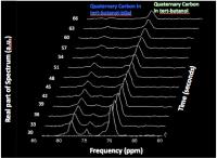
Hyperpolarizing drug candidates could allow insights into their mode of action and metabolic fate. However, administering drug molecules at high concentrations can lead to adverse effects in animals. We have developed a method for directly administering substrates to tumor tissue by infusion through a single supplying artery, thus maximizing tumor drug delivery and minimizing T1 relaxation and systemic toxicity. The net signal gain for arterially injected 13C-pyruvate was x54, compared with the systemically administered venous route. Hyperpolarized custom 13C-labeled CA1 was arterially administered and its parent peak observed, in vivo, at its expected chemical shift (58ppm).


2331.   
Characterisation of adipose tissue-derived mesenchymal stem cell using hyperpolarized MRS
Anja Bille Bohn1, Nathalie Nielsen2, Christoffer Laustsen2, Hans Stødkilde-Jørgensen2, and Lotte Bonde Bertelsen2
1The department of Clinical Immunology, Aarhus University Hospital, Aarhus, Denmark, 2MR Research Centre, Aarhus University, Aarhus University Hospital, Aarhus, Denmark
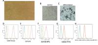
Synopsis: Studies of metabolism in stem cells have revealed a shift in the balance between glycolysis, mitochondrial oxidative phosphorylation and oxidative stress during the maturation of stem cells. In the stem cells, pyruvate from glycolysis will mainly be metabolized to lactate as a result of an uncoupling of the citric acid cycle and the oxidative phosphorylation pathway, thus the application of a novel metabolic cell culture tool could add valuable information to the studies of stem cell characterisation during development. In the present study we use hyperpolarised [1-13C] pyruvate to characterise mesenchymal stem cells harvested from adipose tissue.


2332.   
The formulation of hyperpolarized 13C pyruvate solutions influences the labeling of myocardial metabolites in vivo
Hikari A. I. Yoshihara1, Jessica A. M. Bastiaansen2, Corinne Berthonneche3, Arnaud Comment1, and Juerg Schwitter4
1Institute of Physics of Biological Systems, Swiss Federal Institute of Technology (EPFL), Lausanne, Switzerland, 2Department of Radiology, University Hospital Lausanne (CHUV) and University of Lausanne (UNIL), Lausanne, Switzerland, 3Cardiovascular Assessment Facility, University Hospital Lausanne (CHUV), Lausanne, Switzerland, 4Division of Cardiology and Cardiac MR Center, University Hospital Lausanne (CHUV), Lausanne, Switzerland
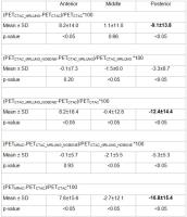
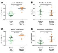
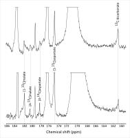
In developing an intact rat model for myocardial ischemia using hyperpolarized 13C pyruvate, different compound formulations were evaluated. Infusion of 4-hydroxy-TEMPO-polarized sodium [1-13C]pyruvate was compared to an equivalent dose of buffered trityl radical-polarized [1-13C]pyruvic acid. Whereas higher levels of polarization and MRS signal were obtained with trityl radical, the metabolite signals normalized to total signal were lower. In particular, [1-13C]lactate signal relative to total signal was markedly higher using TEMPO-polarized pyruvate. [13C]bicarbonate and [1-13C]alanine signals were affected to a lesser degree. This study demonstrates the composition of the infused hyperpolarized pyruvate solution can significantly affect its metabolism in vivo.


2333.   
Rapid decarboxylation of hyperpolarized [13C]ketobutyrate in mouse liver in vivo
Cornelius von Morze1, Irene Marco-Rius1, Celine Baligand1, Robert Bok1, John Kurhanewicz1, Daniel Vigneron1, and Michael Abram Ohliger1,2
1Radiology and Biomedial Imaging, University of California San Francisco, San Francisco, CA, United States, 2UCSF Liver Center, San Francisco, CA, United States
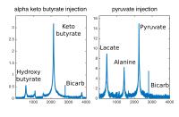
We investigate the rapid metabolic conversion of hyperpolarized (HP) [1-13C]α-ketobutyrate, a molecular analog of pyruvate, in mouse liver in vivo as compared to [1-13C]pyruvate.  Previously, it has been noted that in liver, there is relatively less conversion of [1-13C]α-ketobutyrate to its reduction product, [1-13C]hydroxybutyrate when compared to the conversion of [1-13C]pyruvate to [1-13C]lactate. This difference in conversion likely represents a different LDH activity in liver1. In this study, we examine the decarboxylation of ketobyrate into bicarbonate, which we have found to be unexpectedly elevated when compared to pyruvate, presumably also via PDH and/or a related enzyme.


2334.   
Intraperitoneal substrate administration for ¹³C metabolic imaging in a mouse model of abdominal metastasis
Justin Y.C. Lau1,2, Aws Abdul-Wahid3, Albert P. Chen4, Jean Gariépy1,3, and Charles H. Cunningham1,2
1Medical Biophysics, University of Toronto, Toronto, ON, Canada, 2Physical Sciences, Sunnybrook Research Institute, Toronto, ON, Canada, 3Biological Sciences, Sunnybrook Research Institute, Toronto, ON, Canada,4GE Healthcare, Toronto, ON, Canada
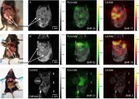
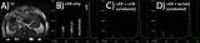
Conventionally, hyperpolarized 13C substrates are administered via intravenous injection. In this abstract, a novel route of hyperpolarized substrate delivery via intraperitoneal injection is demonstrated for observing metabolism in a mouse model of abdominal metastasis. 2D CSI revealed lactate signal in tumour-bearing mice, but only pyruvate signal in a control mouse. An extended time window of dynamic metabolic imaging may be possible with intraperitoneal administration due to the longer in vivo pyruvate T1 of 54 s as measured by dynamic 3D EPI. Intraperitoneal administration of hyperpolarized 13C substrates is a promising complementary technique well suited for observing poorly vascularized metastatic nodules.


2335.   
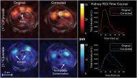
In vivo Assessment of Metabolic Derangements in Renal Ischemia-Reperfusion Injury using Carbon-13 HP-MRI
Mehrdad Pourfathi1,2, David D. Aufhauser3, Douglas R. Murken3, Zhonglin Wang3, Stephen J. Kadlecek1, Heather Gatens1, Ali Naji3, Matthew H. Levine3,4, and Rahim R. Rizi1
1Radiology, University of Pennsylvania, Philadelphia, PA, United States, 2Electrical and Systems Engineering, University of Pennsylvania, Philadelphia, PA, United States, 3Department of Surgery, Division of Transplant Surgery, University of Pennsylvania, Perelman School of Medicine, Philadelphia, PA, United States, 4Department of Surgery, Children's Hospital of Philadelphia, Philadelphia, PA, United States
Renal ischemia repercussion injury (IRI) and its manifestation of acute kidney injury (AKI) is a significant source of morbidity in diverse medical and surgical scenarios, for which for which there is no current therapeutic modality. AKI contributes significantly to hospital stay, morbidity, and mortality. Despite the extensive metabolic derangements that accompany renal IRI, there is an absence of clinically useful markers to predict the clinical course following AKI in an expedient manner.  
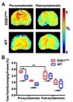
Here, we demonstrate the feasibility of using hyperpolarized carbon-13 MRI to image metabolic activity in the mice recovering from renal IRI.


2336.   
[13C]-tert-butanol-2-ß-D-galactose: A potential new hyperpolarized imaging agent for in vivo imaging of senescent cells
Keshav Datta1,2, Shie-Chau Liu1, Stephen R Lynch3, Zixin Chen1, Ralph Hurd4, Jianghong Rao1, and Daniel Mark Spielman1,2
1Dept. of Radiology, Stanford University, Stanford, CA, United States, 2Dept. of Electrical Engineering, Stanford University, Stanford, CA, United States, 3Dept. of Chemistry, Stanford University, Stanford, CA, United States, 4Applied Sciences Lab, GE Healthcare, Menlo Park, CA, United States
We evaluated the potential for the use of ([13C]-tert-butanol-bGal as hyperpolarizeable agent for in vivo imaging of senescent cells.   The chemical shift between [13C]-tert-butanol-bGal and bGal-cleaved [13C]-tert-butanol was found to be 7.4ppm, more than adequate for in vivo detection.  [13C]-tert-butanol-bGal was also found to polarize well(~30%) with [13C]-tert-butanol-bGal and [13C]-tert-butanol yielding T1 relaxation times of 22s and 34s respectively, very promising for in vivo studies.


2337.   
Concentration-dependent hepatic metabolism in vivo using a near physiological dose range of hyperpolarized [1-13C]pyruvate
Emine Can1, Jessica A. M. Bastiaansen2,3, Hikari A. I. Yoshihara4,5, Rolf Gruetter3,5,6, and Arnaud Comment1
1Institute of Physics of Biological Systems, École Polytechnique Fédérale de Lausanne (EPFL), Lausanne, Switzerland, 2Department of Radiology, University Hospital Lausanne (CHUV), Lausanne, Switzerland,3Department of Radiology, University of Lausanne (UNIL), Lausanne, Switzerland, 4Institute of Physics of Biological Systems, EPFL, Lausanne, Switzerland, 5Laboratory for Functional and Metabolic Imaging, EPFL, Lausanne, Switzerland, 6Department of Radiology, University of Geneva, Geneva, Switzerland
Hyperpolarized 13C-labeled pyruvate provides assessment of real-time liver mitochondrial enzymatic activities directly by labeling TCA cycle intermediates. However the technique is limited by the requirement of supraphysiological concentrations due to the low basal concentrations of metabolic intermediates. In this study we showed the feasibility of detecting liver metabolism in vivo with HP 13C pyruvate administered at plasma concentrations of at most 7-fold of the basal levels. Different metabolic response to the concentration change shows that the adaptation to supraphysiological levels can obscure feeding state-depending metabolic differences in liver.


2338.   
Pool size effects in experiments with hyperpolarized [13C]ketobutyrate
Cornelius von Morze1, Peder E Larson1, Michael A Ohliger1, Ralph E Hurd2, John Kurhanewicz1, and Daniel B Vigneron1
1Department of Radiology and Biomedical Imaging, University of California, San Francisco, San Francisco, CA, United States, 2GE Healthcare, Menlo Park, CA, United States
The purpose of this abstract was to investigate pool size effects in experiments with hyperpolarized [13C]α-ketobutyrate (αKB), a molecular analog of pyruvate which also has substantial activity with LDH. In contrast to pyruvate, formation of the reduction product [13C]α-hydroxybutyrate (αHB) necessarily reflects net metabolic flux as opposed to label exchange. We observed little change when co-injecting αHB but a large increase in the αHB-to-αKB ratio when co-injecting lactate. This suggests that the observed conversion of αKB to αHB only reflects net metabolic flux even in the presence of a large pool of reduction product.


2339.   
Feasibility of sensing small molecule thiols using hyperpolarized [13C]cyanate
Cornelius von Morze1, Chloe Najac1, Robert R Flavell1, David E Korenchan1, Pavithra Viswanath1, Lucas Carvajal1, John Kurhanewicz1, Sabrina M Ronen1, Daniel B Vigneron1, and David M Wilson1
1Department of Radiology and Biomedical Imaging, University of California, San Francisco, San Francisco, CA, United States
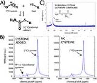
The purpose of this study was to show basic feasibility of non-invasively detecting small molecule thiols using hyperpolarized [13C]cyanate. We detected rapid formation of the expected hyperpolarized S-[13C]carbamyl thiol adduct after adding cysteine to liquid hyperpolarized [13C]cyanate samples. This work demonstrates a new non-enzymatic approach for detecting small molecule thiols such as reduced glutathione, which could be very useful for research on oxidative stress.


2340.   
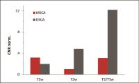
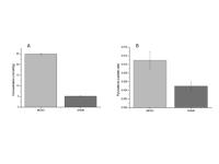
13C dynamic nuclear polarization NMR for quantification of metabolic flux of endothelial progenitor cells
Nathalie Nielsen1, Christoffer Laustsen1, Hans Stødkilde-Jørgensen1, and Lotte Bonde Bertelsen1
1MR Research Centre, Department of Clinical Medicine, Aarhus University Hospital, Aarhus University, Aarhus, Denmark
This study aims to quantify the metabolic flux in EPCs in order to characterize the metabolic changes occurring during in-vitro culturing utilized for cell expansion, 3D scaffolds and suspension. [1-13C] hyperpolarized pyruvate is injected to a NMR compatible bioreactor system and the conversion is detected and measured as the lactate/pyruvate ratio. Activation assays and qPCR is performed to support the results. The lactate/pyruvate (6±1,07 fold) and LDH activity is increased in cell suspension culturing. Together with an elevated PDH expression in suspension cultures our conclusion is that adherent cells metabolically compensate in the suspension culture due to the environmental conditions.  


2341.   
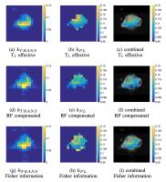
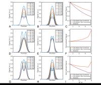
Optimizing flip angles for metabolic rate estimation in hyperpolarized carbon-13 MRI
John Maidens1, Jeremy W. Gordon2, Murat Arcak1, and Peder E. Z. Larson2
1Electrical Engineering & Computer Sciences, University of California, Berkeley, Berkeley, CA, United States, 2Radiology & Biomedical Imaging, University of California, San Francisco, San Francisco, CA, United States
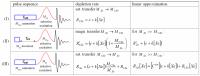
Hyperpolarized carbon-13 MRI experiments typically aim to distinguish between healthy and diseased tissues based on the rate at which they metabolize an injected substrate. Existing approaches to determine flip angle sequences for kinetic measurements have used metrics such as signal variation and signal-to-noise ratio, but are not optimized to provide the most reliable metabolic rate estimates. Here we present a flip angle sequence that maximizes the Fisher information about the metabolic rate. We demonstrate through numerical simulation that flip angle sequences optimized using the Fisher information lead to lower variance in metabolic rate estimates than existing sequences. We then validate this optimized sequence in vivo with experiments in a prostate cancer mouse model.


2342.   
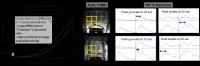
Dual-Echo EPI Sequence for Integrated Distortion Correction in 3D Time-Resolved Hyperpolarized 13C MRI
Benjamin J. Geraghty1,2, Albert P. Chen3, and Charles H. Cunningham1,2
1Physical Sciences, Sunnybrook Research Institute, Toronto, ON, Canada, 2Dept. of Medical Biophysics, University of Toronto, Toronto, ON, Canada, 3GE Healthcare, Toronto, ON, Canada
A novel dual echo EPI sequence is proposed for providing a built-in correction for off-resonance in time resolved, volumetric hyperpolarized 13C metabolic mapping with [1-13C]pyruvate. The phase evolution between two echoes was used to correct EPI distortion and improve spatial registration with the underlying anatomy. A correction term obtained from a fully phase encoded dual echo EPI proton reference scan was required to account for odd/even echo asymmetry in the 13C phase maps. Proof-of-concept dual echo EPI in vivo rat data was acquired on a clinical 3T MR scanner and corrected images are presented.


2343.   
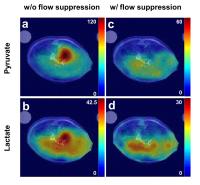
Optimization and application of bipolar gradient for flow-suppressed hyperpolarized 13C CSI in mouse liver at 9.4T
Hansol Lee1, Joonsung Lee2, Eunhae Joe1, Seungwook Yang1, Jae Eun Song1, Young-suk Choi3, Eunkyung Wang3, Ho-Taek Song3, and Dong-Hyun Kim1
1Department of Electrical & Electronic Engineering, Yonsei university, Seoul, Korea, Republic of, 2Center for Neuroscience Imaging Research, Institute for Basic Science, Sungkyunkwan University, Suwon, Korea, Republic of, 3Department of Radiology, Yonsei University College of Medicine, Seoul, Korea, Republic of
In hyperpolarized 13C MRI, high signal intensity of vasculature can cause errors in quantification of metabolites or conversion rate constants. The bipolar gradient was used to suppress vascular signal for accurate quantification. However, the velocity of vessel can vary depending on anesthetic level and pulsation. Furthermore, additional T2* relaxation signal loss can be induced by delayed data acquisition in ultra-high field (9.4T) due to short T2*. In this study, the bipolar gradient was optimized to minimize additional signal loss and mitigate variable velocity, then the optimized bipolar gradient was implemented for hyperpolarized 13C CSI and applied to mouse liver experiment.


2344.   
Mis-Estimation and Bias of Hyperpolarized ADC Measurements Due to Slice Profile Effects
Jeremy W Gordon1, Eugene Milshteyn1, Irene Marco-Rius1, Michael Ohliger1, Daniel B Vigneron1, and Peder EZ Larson1
1Radiology & Biomedical Imaging, University of California - San Francisco, San Francisco, CA, United States
Hyperpolarized diffusion weighted imaging has the potential to noninvasively assess transporter expression and probe specific metabolite microenvironments. However, the imperfect RF excitation profile and the transient, non-recoverable hyperpolarization lead to non-uniform depletion of Mz. After multiple RF pulses, this results in excess signal at later excitations, potentially biasing ADC estimation. Scaling the slice-select gradient can correct for this deviation, minimizing bias and providing more precise ADC measurements of hyperpolarized substrates.


2345.   
Design and test of a double-nuclear RF coil array for 1H MRI and 13C MRS at 7T
Omar Rutledge1, Tiffany Kwak1, Peng Cao1, and Xiaoliang Zhang1
1Radiology and Biomedical Imaging, University of California, San Francisco, San Francisco, CA, United States

RF coil operation at 7T is fraught with technical challenges, making expansion of 7T into clinical imaging difficult. In this work, a microstrip transmission line and a wire loop coil were combined to form a double-nuclear RF coil array for proton magnetic resonance imaging and carbon magnetic resonance spectroscopy at the ultrahigh magnetic field strength of 7T. Network analysis revealed a high Q-factor and excellent decoupling between the coils. Proton images and carbon spectra were acquired with high sensitivity. The successful testing of this novel double-coil array demonstrates the feasibility of this design for multi-nuclear studies at 7T.


2346.   
Feasibility of probing lactate metabolism and neuroprotection in a mouse model of stroke using hyperpolarized 13C-lactate
Mor Mishkovsky1, Lara Buscemi2, Ximena Castillo2, Mario Lepore3, Arnaud Comment4, Lorenz Hirt2, and Jean-Noël Hyacinthe5,6
1Laboratory of Functional and Metabolic Imaging, Ecole Polytechnique Fédérale de Lausanne (EPFL), Lausanne, Switzerland, 2Department of Clinical Neurosciences, Centre Hospitalier Universitaire Vaudois, Lausanne, Switzerland, 3Centre d'Imagerie Biomédicale (CIBM), Ecole Polytechnique Fédérale de Lausanne (EPFL), Lausanne, Switzerland, 4Institute of Physics of Biological Systems, Ecole Polytechnique Fédérale de Lausanne (EPFL), Lausanne, Switzerland, 5School of Health Sciences - Geneva, University of Applied Sciences and Arts Western Switzerland, Geneva, Switzerland, 6Image Guided Intervention Laboratory, University of Geneva, Geneva, Switzerland
Stroke is a major public health challenge in the context of the current demographic changes. Among a wide range of applications, hyperpolarized magnetic resonance enables in vivo real-time measurement of biochemical transformations of hyperpolarized 13C-labeled precursors, including lactate, a known neuroprotectant in stroke at the preclinical level. This study shows the feasibility of measuring lactate metabolism in vivo in a mouse model of stroke (MCAO) following intravenous injection of hyperpolarized L-[1-13C]lactate. Calculated pyruvate-to-lactate ratio shows an increased labeling of the pyruvate pool in MCAO when compared to sham. This feasibility study suggests new perspectives to understand lactate biodistribution and its neuroprotective effect in stroke.


2347.   
Robust, Quantitative Methods Applied to Clinical Hyperpolarized C-13 MR of Prostate Cancer Patients
Peder Eric Zufall Larson1, Jeremy Gordon1, John Maidens2, Murat Arcak2, Hsin-Yu Chen1, Galen Reed1, Ilwoo Park1, Rahul Aggarwal3, Robert Bok1, Sarah J Nelson1, John Kurhanewicz1, and Daniel B Vigneron1
1Radiology and Biomedical Imaging, University of California - San Francisco, San Francisco, CA, United States, 2Electrical Engineering & Computer Sciences, University of California - Berkeley, Berkeley, CA, United States, 3Medicine, University of California - San Francisco, San Francisco, CA, United States
Clinical evaluation of metabolic MRI using hyperpolarized C-13 agents has begun in earnest at multiple sites with the availability of the SpinLab commercial polarizer.  For this technology to succeed, robust imaging and analysis methods for quantification of metabolic activity are required.  We have developed and are applying efficient dynamic imaging methods, robust kinetic models, and specialized calibration schemes to enable accurate and reproducible quantification in clinical hyperpolarized MR studies.


2348.   
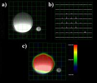
A Molecular Imaging Approach to Mercury Sensing Based on Hyperpolarized 129Xe Molecular Clamp Probe
Qianni Guo1, Qingbin Zeng1, Weiping Jiang1, Xiaoxiao Zhang1, Qing Luo1, and Xin Zhou1
1Wuhan Institute of Physics and Mathematics, Chinese Academy of Sciences, Wuhan, China, People's Republic of
Mercury contamination is widespread and arises from a variety of natural sources.We propose the use of hyperpolarized 129Xe nuclear magnetic resonance (NMR) spectroscopy for the sensitive detection of Hg2+ions in aqueous solution.We develop a biosensor whose molecular structure is like a clamp. When interact with Hg2+ in aqueous solution, the molecular structure of the biosensor could be changed as a clamp from “open” to “closed”. This molecular structure change causes the distance between the two cryptophane cages of the biosensor become closer, and the electron cloud of them overlapped. As a result, comparing with normal downfield chemical shifts of the reported xenon biosensors formetallic ions, the Xe caged in the cryptophane moiety shows a upfield chemical shift change from 66.5 ppm to 66.1 ppm. Images were obtained using a CSI method preciously used for clinical MRI.


2349.   
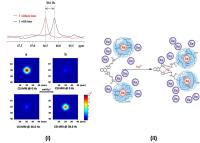
Investigating Spectral Selectivity of the bSSFP Sequence for High Resolution 3D Dynamic Hyperpolarized 13C MRI at 3T Using C2-Pyruvate and Urea
Eugene Milshteyn1, Cornelius von Morze1, Hong Shang1, Galen D Reed2, and Daniel B Vigneron1
1Radiology and Biomedical Imaging, UCSF, San Francisco, CA, United States, 2HeartVista, Menlo Park, CA, United States
Hyperpolarized 13C MR imaging can provide simultaneous assessments of metabolism and perfusion to study disease processes. High resolution dynamic imaging is needed to fully understand these processes, but is challenging, especially on clinically relevant systems. This project investigated new methods for spectral selectivity with SNR-efficient bSSFP sequences to provide improved high resolution 3D dynamic in vivo HP 13C MR imaging at 3T. 


2350.   
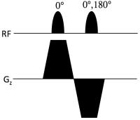
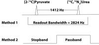
Toward Spectroscopically Selective Imaging of Hyperpolarized Pyruvate and its Metabolites Using Binomial Pulses In Balanced Steady-State Free Precession
Gopal Varma1, Patricia Coutinho de Souza1, Leo Tsai1, Rupal Bhatt2, and Aaron Grant1
1Radiology, Beth Israel Deaconess Medical Center and Harvard Medical School, Boston, MA, United States, 2Medicine, Beth Israel Deaconess Medical Center and Harvard Medical School, Boston, MA, United States
Balanced steady-state free-precession (bSSFP) offers high sensitivity and good temporal resolution, and makes efficient use of hyperpolarized magnetization.  Several strategies for spectroscopically selective imaging with bSSFP have been proposed [1-5].  Here we investigate the use of simple binomial excitation pulses to selectively null the signals from either pyruvate or lactate, the two dominant metabolites in tumors, thereby obtaining images that are dominated by either lactate or pyruvate, respectively.  The method is robust to off-resonance effects, and can be used to augment existing spectroscopic bSSFP techniques.
 

The International Society for Magnetic Resonance in Medicine is accredited by the Accreditation Council for
Continuing Medical Education to provide continuing medical education for physicians.