|
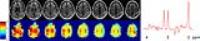
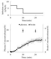
2365.
 |
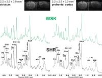
Accounting for GABA editing efficiency and macromolecule
co-editing to allow inter-vendor comparisons of GABA+
measurements
Ashley D Harris1,2,3,4,5, Nicolaas AJ Puts1,5,
Laura Rowland6, S. Andrea Wijtenburg6,
Mark Mikkelsen7, Peter B Barker1,5, C.
John Evans7, and Richard AE Edden1,5
1FM Kirby Center for Functional Brain Imaging,
Kennedy Krieger Institute, Baltimore, MD, United States, 2CAIR
Program, Alberta Children's Hospital Research Institute,
University of Calgary, Calgary, AB, Canada,3Radiology,
University of Calgary, Calgary, AB, Canada, 4Hotchkiss
Brain Institute and Alberta Children's Hospital Research
Institute, Calgary, AB, Canada, 5Russell
H Morgan Department of Radiology and Radiological Science,
The Johns Hopkins University, Baltimore, MD, United States, 6Maryland
Psychiatric Research Center, Department of Psychiatry,
University of Maryland School of Medicine, Baltimore, MD,
United States,7CUBRIC, School of Psychology,
Cardiff University, Cardiff, United Kingdom
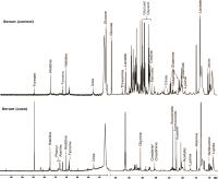
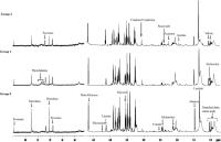
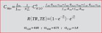
Differences in GABA+ MEGA PRESS acquisitions between vendors
are quantified in terms of the editing efficiency of GABA
and the fractional co-editing of macromolecules. Accounting
for these two parameters results in moderate agreement among
the different vendors considered.
|
|
2366.
 |
Towards a neurochemical profile of the amygdala using SPECIAL at
3 tesla 
Florian Schubert1, Ralf Mekle1, Simone
Kühn2, Jürgen Gallinat3, and Bernd
Ittermann1
1Physikalisch-Technische Bundesanstalt (PTB),
Braunschweig and Berlin, Germany, 2MPI
for Human Development, Berlin, Germany, 3Universitätsklinikum
Hamburg-Eppendorf, Hamburg, Germany
Since disturbed amygdala function is linked to psychiatric
conditions insight into its biochemistry, particularly the
neurotransmitters, is required. We combined the SPECIAL MRS
sequence with FAST(EST)MAP implementation, corrections for
frequency drift, relaxation, CSF volume, and a basis set
including a measured macromolecule spectrum for
quantification of metabolites in the amygdala in 20
volunteers at 3T. Beyond quantification of the three main
metabolites plus myo-inositol with excellent precision, for
the first time glutamate was determined reliably and
separately from glutamine. Using a basis set without
macromolecules introduced a systematic overestimation of
concentrations. Glutamine and glutathione was quantifiable
only in a subset of spectra.
|
|
2367.
 |
Estimation of in vivo ?-aminobutyric acid (GABA) levels in the
neonatal brain
Moyoko Tomiyasu1,2, Noriko Aida3, Jun
Shibasaki4, Katsutoshi Murata5, Keith
Heberlein6, Mark A. Brown7, Eiji
Shimizu2, Hiroshi Tsuji1, and Takayuki
Obata1
1National Institute of Radiological Sciences,
Chiba, Japan, 2Chiba
University, Chiba, Japan, 3Department
of Radiology, Kanagawa Children's Medical Center, Yokohama,
Japan, 4Kanagawa
Children's Medical Center, Yokohama, Japan, 5Siemens,
Tokyo, Japan, 6Biomedical
Imaging Technology Center, Burlington, MA, United States, 7University
of Colorado, Cary, NC, United States
We examined in
vivo brain
γ-aminobutyric acid (GABA) levels of neonates and compared
them with those of children. In this study, 32 normal
neonates and 12 normal children (controls) had their brain
GABA levels measured using clinical 3T edited-MRS. The
neonates exhibited significantly lower GABA+ levels than the
children in both the basal ganglia and cerebellum, which is
consistent with previous in
vitro data.
While significantly higher GABA+/Cr levels were detected in
the neonatal cerebellum, care should be taken when comparing
GABA+/Cr levels between different ages. This is the first
report about the in
vivo brain
GABA levels of neonates.
|
|
2368.
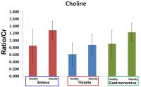 |
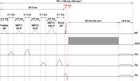
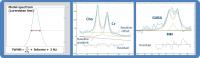
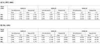
Assessment of Lipid Changes in Obese Calf Using Muti-Echo
Echo-planar Correlated Spectroscopic Imaging
Rajakumar Nagarajan1, Raissa Souza1,
Edward Xu1, Manoj K Sarma1, S. Sendhil
Velan2, Cathy C Lee3, Theodore Hahn3,
Catherine Carpenter4, Vay-Liang Go5,
and M.Albert Thomas1
1Radiological Sciences, University of California
Los Angeles, Los Angeles, CA, United States, 2Laboratory
of Molecular Imaging, Singapore Bioimaging Consortium,
Singapore, Singapore, 3Geriatrics,
VA Greater Los Angeles Healthcare System, Los Angeles, CA,
United States, 4UCLA
Schools of Nursing, Medicine, and Public Health, Los
Angeles, CA, United States, 5UCLA
Department of Medicine, Los Angeles, CA, United States
Obesity is a serious public health problem associated with
high rates of morbidity and mortality. One-dimensional MR
spectroscopy suffers from overlapping spectral resonances
which can complicate metabolite identification and
quantitation. Two-dimensional spectroscopic techniques have
been demonstrated in calf muscle to reduce the problem of
spectral overlap. In this study, we used the four
dimensional (4D) multi-echo echo planar correlated
spectroscopic imaging (ME-EPCOSI) technique to quantify the
lipids and metabolites in soleus, tibialis anterior and
gastrocnemius calf muscles of obese and normal healthy
subjects. The 4D ME-EPCOSI acquired data enabled less
ambiguous quantitation of metabolites, unsaturated and
saturated fatty acids in different calf muscle regions using
IMCL ratios and unsaturation indices.
|
|
2369.
 |
Simultaneous modeling of spectra and apparent diffusion
coefficients.
Victor Adalid Lopez1, André Doering1,
Sreenath Pruthviraj Kyathanahally 1,
Christine S. Bolliger1, and Roland Kreis1
1Depts. Radiology and Clinical Research,
University Bern, Bern, Switzerland
Diffusion weighted spectroscopy can provide information on
the diffusion of metabolites and the microstructure of brain
tissue. A method for simultaneous fitting of spectra related
by mono-exponential diffusion weighting is introduced, which
is similar to simultaneous fitting of a 2DJ or inversion
recovery data set. As shown for simulated white matter data,
the method improves both, accuracy and precision of ADC
estimation for all metabolites. It is also illustrated with
diffusion data obtained from human gray matter at 3T.
|
|
2370.
 |
Novel Triple-refocusing 1H
MRS at 3T for detection of GABA in human brain in
vivo
Zhongxu An1, Sandeep Ganji1, Vivek
Tiwari1, and Changho Choi1
1Advanced Imaging Research Center, University of
Texas Southwestern Medical Center, Dallas, TX, United States
Reliable detection of GABA is important for research studies
in neuro-psychiatric diseases. In
vivo 1H
GABA resonances extensively overlap with the neighboring
resonances of glutamate and glutamine. We present an
optimized single-shot triple-focusing 1H MRS method which
fully resolved GABA 2.29-ppm signal at 3T.
|
|
2371.
 |
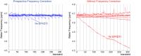
Prospective frequency correction for TE-averaged semi-LASER
Chu-Yu Lee1, In-Young Choi1,2,3, Peter
Adany1, and Phil Lee1,3
1Hoglund Brain Imaging Center, University of
Kansas Medical Center, Kansas city, KS, United States, 2Department
of Neurology, University of Kansas Medical Center, Kansas
City, KS, United States, 3Department
of Molecular & Integrative Physiology, University of Kansas
Medical Center, Kansas City, KS, United States
Frequency drifts during MRS acquisition results in broad and
distorted spectral lineshapes, a reduced SNR and
quantification errors. The consequence of frequency drifts
is particularly significant in spectral-editing sequences,
because spectral editing critically relies on narrow-band
frequency selective pulses or accurate spectral alignments
among scans for subtraction/addition of spectra. Frequency
drift can occur due to subject’s movement and/or MR system
instability. Even in advanced MR systems with self-shielded
gradients, significant frequency drifts occur due to eddy
current-induced heating and cooling of passive shim
materials, particularly after MR scans with heavy gradient
duty cycles. The effects of frequency drifts can be
mitigated through prospective and retrospective frequency
corrections. Currently, most spectral-editing methods use
post-processing approaches to correct the effects of
frequency drifts retrospectively. In this study, we have
developed a prospective frequency correction method and
implemented it in a semi-LASER based TE-averaged sequence
for glutamate detection.
|
|
2372.
 |
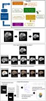
Automatic Multi-layer Classification System of Brain Tumor Based
on Multi-modality MRI and Clinical Information
Yafei Wang1, Yue Zhang1, Lingyi Xu1,
Yu Sun1, Lei Xiang2, Meiping Ye2,
Suiren Wan1, Bing Zhang2, and Bin Zhu2
1The Laboratory for Medical Electronics, School
of Biological Sciences and Medical Engineering, Southeast
University, Nanjing, China, People's Republic of, 2Department
of Radiology, The Affiliated Drum Tower Hospital of Nanjing
University Medical School, Nanjing, China, People's Republic
of
Classification or grading of brain tumor alone would not be
enough for clinical use, therefore we designed a
comprehensive multi-layer system combining the two functions
together. Firstly, we designed it as a three-layer system
according to clinic workflow. Then, we extracted new
features from multi-modality MRI and patients’ clinical
information, which were easily ignored or difficult found by
eyes. And then we implemented SVM and Tumor Model to
classify tumor type and tumor grade. This study proposed a
novel multi-layer system for clinic use by reducing the
diagnosis uncertainty.
|
|
2373.
 |
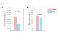
Reproducibility and gender-related effects on macromolecule
suppressed GABA and Glx metabolites
Muhammad Gulamabbas Saleh1, A Alhamud1,
Jamie Near2, Frances Robertson1, André
J.W. van der Kouwe3, and Ernesta M Meintjes1
1Human Biology, MRC/UCT Medical Imaging Research
Unit, University of Cape Town, Cape Town, South Africa, 2Douglas
Mental Health University Institute and Department of
Psychiatry, McGill University, Montreal, QC, Canada, 3Athinoula
A. Martinos Center for Biomedical Imaging, Massachusetts
General Hospital, Charlestown, MA, United States
Several studies have characterized short and long term
reproducibility of Glx and GABA+, but not macromolecule (MM)
suppressed GABA. Further, gender-related differences have
been observed in GABA+, but these may, in part, be due to
inter-individual variations of MM. Motion and magnetic field
inhomogeneity can hamper the consistent application
of frequency-selective pulses at 1.7ppm necessary for
effective GABA editing. We demonstrate that the shim and
motion-navigated MEGA-SPECIAL sequence produces well-edited
GABA and Glx spectra. LCModel quantification yields the best
reproducibility. Observed gender-related differences in GABA
highlight the need for gender-matching in studies
investigating differences in GABA concentrations.
|
|
2374.
 |
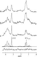
1H-MRS of Human Liver at 3 T: Relaxation Times and Metabolite
Concentrations
Jan Weis1, Fredrik Rosqvist2, Joel
Kullberg1, Ulf Risérius2, and Håkan
Ahlström1
1Department of Radiology, Uppsala University,
Uppsala, Sweden, 2Department
of Public Health and Caring Sciences, Uppsala University,
Uppsala, Sweden
Proton MR spectroscopy of healthy human liver was performed
at 3 T MR scanner. The purpose of this study was to estimate
glycogen (Glycg), choline-containing compounds (CCC), water,
and lipid (-CH2-)nrelaxation times T1,
T2, and absolute concentration of Glycg, CCC, and
fat. Experiments were performed using multiple breath-hold
technique. Spectra were processed by LCModel. T1 and
T2 values
were obtained by mono-exponential fitting spectral
intensities versus repetition or echo times. Quantification
of liver Glycg, CCC and lipids is important for
understanding changes in lipid and glucose metabolism due to
metabolic disorders.
|
|
2375.
 |
Gradient-heavy sequences degrade the quality of subsequent
spectroscopy acquisitions
Benjamin C Rowland1, Fatah Adan1,
Huijun Liao1, and Alexander P Lin1
1Centre for Clinical Spectroscopy, Brigham and
Women's Hospital, Boston, MA, United States
B0 frequency drift is a well-known phenomenon which can have
a significant impact on MR spectroscopy, affecting both peak
resolution and metabolite quantification. B0 drift is
particularly associated with gradient-heavy EPI sequences
like DTI. In a study of 53 subjects receiving DTI and MRS,
the mean FWHM more than doubled as a result of frequency
drift and metabolite concentrations were often misestimated
by LC Model.
|
|
2376.
 |
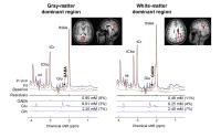
Elucidation of the downfield spectrum of human brain at 7T using
multiple inversion recovery delays and echo times
Nicole D Fichtner1,2, Anke Henning2,3,
Niklaus Zoelch2, Chris Boesch1, and
Roland Kreis1
1Depts. Radiology and Clinical Research,
University of Bern, Bern, Switzerland, 2Institute
for Biomedical Engineering, UZH and ETH Zurich, Zurich,
Switzerland, 3Max
Planck Institute for Biological Cybernetics, Tuebingen,
Germany
Characterization of the full 1H
spectrum may allow for better monitoring of pathologies and
metabolism in humans. The downfield part (5-10ppm) is
currently less well characterized than upfield; this work
aims to benefit from higher field strength in order to
quantify T1 and
T2 in
the downfield spectrum in human grey matter at 7T. We fitted
downfield spectra to a heuristic model and obtained
relaxation times for twelve peaks of interest. The T1’s
are higher than those at 3T downfield; peaks with lower T1’s
may include macromolecules. The T2’s are mostly
shorter than those reported for upfield peaks at 7T.
|
|
2377.
 |
Tissue correction strategy impacts GABA quantification: a study
in healthy aging
Ashley D Harris1,2,3,4, Eric Porges5,
Adam J Woods5,6, Damon G Lamb5,7,
Ronald A Cohen5, John B Williamson5,8,
Nicolaas AJ Puts3,4, and Richard AE Edden3,4
1Radiology, University of Calgary, Calgary, AB,
Canada, 2CAIR
Program, Hotchkiss Brain Institute and Alberta Children's
Hospital Research Institute, Calgary, AB, Canada, 3Russell
H Morgan Department of Radiology and Radiological Science,
The Johns Hopkins University, Baltimore, MD, United States, 4FM
Kirby Center for Functional Brain Imaging, Kennedy Krieger
Institute, Baltimore, MD, United States, 5Center
for Cognitive Aging and Memory (CAM), McKnight Brain
Institute, Department of Aging and Geriatric Research,
University of Florida, Gainesville, FL, United States, 6Department
of Neuroscience, University of Florida, Gainesville, FL,
United States, 7Brain
Rehabilitation and Research Center, Malcom Randall Veterans
Affairs Medical Center, Gainesville, FL, United States, 8Brain
Rehabilitation and Research Center, Brain Rehabilitation and
Research Center, Gainesville, FL, United States
There are various strategies for tissue correction for MRS.
Here, using data from a healthy aging cohort, we show that
the selection of tissue correction method can change the
conclusions that are drawn from data.
|
|
2378.
 |
Towards low power EPR Imaging using Frank poly-phase pulse
sequence
Nallathamby Devasahayam1, Randall H. Pursley2,
Thomas J. Pohida2, Shingo Matsumoto3,
Keita Saito4, Sankaran Subramanian5,
and Murali C. Krishna4
1Radiation Biology Branch, National Cancer
Institute, Bethesda, MD, United States, 2Center
for Information Technology, Bethesda, MD, United States, 3Graduate
School of Information Science and Technology, Sapporo,
Japan, 4National
Cancer Institute, Bethesda, MD, United States, 5Indian
Institute of Technology Madras, Chennai, India
Electron Paramagnetic Resonance (EPR) imaging is suited
well for small animal physiological imaging with its unique
capability of generating in vivo quantitative oxygen maps.
The main bottleneck in scaling up pulsed EPR imaging to
human anatomy is that the required RF power of US federal
food and drug administration (FDA), specific absorption rate
(SAR) limits. In Frank Sequence we are using power levels on
the order of 250 microwatts in a crossed coil resonator with
~35 dB isolation. Using a 256 pulse polyphase Frank
Sequence, it was possible to obtain images with good SNR.
|
|
2379.
|
Hitchhikers guide to voxel segmentation for partial volume
correction of in-vivo magnetic resonance spectroscopy
Scott Quadrelli1,2, Carolyn Mountford3,
and Saadallah Ramadan2
1Queensland University of Technology, Brisbane,
Australia, 2The
University of Newcastle, Newcastle, Australia, 3The
Translational Research Institute, Brisbane, Australia
Whilst many studies have detailed the impact of partial
volume effects on proton magnetic resonance spectroscopy
quantification, there is a paucity of literature explaining
how voxel segmentation can be achieved using freely
available neuroimaging packages. Here we aim to demonstrate
a practical guide to magnetic resonance spectroscopy (MRS)
voxel segmentation, partial volume correction and detail how
to extract other MR metrics (such as DTI, fMRI) from a MRS
voxel.
|
|
2380.
 |
Enhancement of signal intensity using a wireless coil for FT-EPR
oximetry study
Ayano Enomoto1, Gadisetti V. R. Chandramouli2,
Alan P Koretsky3, Chunqi Qian4, Murali
K Cherukuri1, and Nallathamby Devasahayam1
1Radiation Biology Branch, National Cancer
Institute, Bethesda, MD, United States, 2GenEpria
Consulting Inc., Columbia, MD, United States, 3National
Institute of Neurological Disorders and Stroke, National
Institutes of Health, Bethesda, MD, United States, 4Department
of Radiology, Michigan State university, East Lansing, MI,
United States
Sensitivity enhancement is required to detect the weak
signals with Fourier transform Electron Paramagnetic
Resonance (FT-EPR). In the proposed method, a small amount
of sample was placed at a distance less than half the
diameter of the receiving surface coil. The signal was
enhanced by a wirelessly pumped coil. Presently, we used the
TCNQ for our studies to study signal enhancement. Here, we
achieved 7-fold of improvement in signal intensity in
compared with conventional FT-EPR acquisition. We will show
the results of in vivo oximetry using oxygen sensing solids
LiPc and LiNc in in
vivo applications
to measure tissue oxygenation.
|
|
2381.
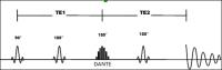 |
Assessment of serine quantification reproducibility using
advanced 1H-MRS in the human brain at 3T
Homa Javadzadeh1,2 and
Jean Théberge1,2,3
1Department of Medical Biophysics, University of
Western Ontario, London, ON, Canada, 2Imaging
Division, Lawson Health Research Institute, London, ON,
Canada, 3Diagnostic
Imaging Department, St. Joseph's Health Care, London, ON,
Canada
D-serine supplements alleviate some of the most debilitating
features of schizophrenia believed to be associated with glutamatergic abnormalities.
Assessment of endogenous serine is impossible using standard
proton Magnetic Resonance Spectroscopy (1H-MRS).
This work employs a novel?1H-MRS sequence called
DANTE-PRESS (D-PRESS) and presents test-retest reliability
study for serine levels acquired at 3T in phantoms and
initial data in two human subjects. We conclude that
reproducibility and precision of serine measurements on a
3.0T scanner is sufficient to assess endogenous levels in
vivo and is
a valuable tool to examine abnormalities
in schizophrenia and monitor supplementation.
|
|
2382.
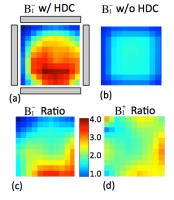 |
Large Improvements of RF field Transmission Efficiency and
Detection Sensitivity for Ultrahigh-field In vivo 31P MRS using
Emerging Technology of Ultrahigh Dielectric Constant Material
Byeong-Yeul Lee1, Xiao-Hong Zhu1,
Sebastian Rupprecht2, Michael T. Lanagan3,
Qing X. Yang2,4, and Wei Chen1
1Center for Magnetic Resonance Research,
Radiology, University of Minnesota, Minneapolis, MN, United
States, 2Center
for NMR Research, Radiology, The Pennsylvania State College
of Medicine, Hershey, PA, United States, 3Engineering
Science and Mechanics, The Pennsylvania State College of
Engineering, University Park, PA, United States, 4Neurosurgery,
The Pennsylvania State College of Medicine, Hershey, PA,
United States
Compared to 1H
MRS, X-nuclei MRS for human application faces two
challenges: higher requirement of RF power (thus, higher
SAR) for achieving the same RF pulse flip angle due to a
relatively lower gyromagnetic ratio, and still limited SNR
even at high/ultrahigh field. In this report, we demonstrate
that up to 200% SNR gain was achieved with ultra high
dielectric constant (uHDC) materials incorporated into the
RF volume coil for 31P
MRS at 7T. Concomitantly, the RF power optimized for
acquiring the spectra was significantly reduced by 200%. Our
data demonstrated that incorporating uHDC with RF coil can
significantly boost SNR and reduce RF transmission power
X-nuclei MRS applications on top of using high field
strength magnet that has approached to its technologic
limits.
|
|
2383.
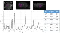 |
9.4 Tesla 1H-MRS of Glutamate and GABA in a 3.6 cubic-mm volume
using an optimized UTE-STEAM sequence
Nicola Bertolino1, Paul Polak1,
Marilena Preda1,2, Robert Zivadinov1,2,
and Ferdinand Schweser1,2
1Buffalo Neuroimaging Analysis Center, Department
of Neurology,Jacobs School of Medicine and Biomedical
Sciences, The State University of New York at Buffalo,
Buffalo, NY, United States, 2MRI
Molecular and Translational Research Center, Jacobs School
of Medicine and Biomedical Sciences, The State University of
New York at Buffalo, Buffalo, NY, United States
In-vivo 1H-MR spectroscopy is a non-invasive technique able
to detect metabolites providing important information from
investigated tissue. GABA and Glutamate are two metabolites
altered in many neurological diseases, although challenging
to quantify in vivo because of a number of technical issues:
voxel localization, low concentration, short T2, overlapping
peaks and spin-spin coupling. In this work we developed an
optimized parameter set for an ultra-short TE STEAM.
|
|
2384.
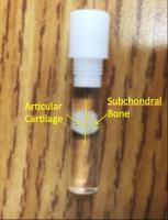 |
Effects of Storage Conditions on Transverse Relaxation in Bovine
Articular Cartilage
Kyle W. Sexton1, Hasan Celik1, Kenneth
W. Fishbein1, and Richard G. Spencer1
1National Institute on Aging, National Institutes
of Health, Baltimore, MD, United States
Quantification of cartilage matrix components with nuclear
magnetic resonance has potential applications to the early
diagnosis of osteoarthritis. Ex-vivo cartilage samples are
often used to observe the MR parameters of healthy and
degraded cartilage. To ensure the accuracy of MR parameters,
the storage of the explants is extremely important. DPBS is
often used to immerse cartilage tissue specimens during
imaging, with the assumption that it prevents dehydration.
In this study it was found that storing BAC tissue explants
in DPBS can rapidly and significantly increase the observed
T2 values. An alternative storage medium to maintain T2
stability is Fluorinert.
|
|
2385.
 |
Frequency correction based on interleaved water acquisition
improves spectral quality in MM-suppressed GABA measurements in
vivo
Nicolaas AJ Puts1,2, Kimberly L Chan1,
Ashley D Harris1,2,3,4, Peter B Barker1,2,
and Richard AE Edden1,2
1Radiology and Radiological Science, Johns
Hopkins University, Baltimore, MD, United States, 2F.M.
Kirby Center for Functional Brain Imaging, Kennedy Krieger
Institute, Baltimore, MD, United States, 3Alberta
Children's Hospital Research Institute, University of
Calgary, Calgary, AB, Canada, 4Radiology,
University of Calgary, Calgary, AB, Canada
MM-suppressed GABA measurements use symmetric editing of
both MM and GABA signals. Frequency drift, either by
gradient induced heating/cooling, or motion, significantly
affects the editing efficiency of GABA and MM. To stabilize
the center frequency, we interleaved the unsuppressed water
acquisition throughout the scan and used it to correct the
frequency, in eight healthy participants, and compared this
to a condition without frequency correction. Frequency
correction improves spectral quality of MM-suppressed GABA
editing in vivo.
|
|
2386.
 |
Single volume localization without RF refocusing for dynamic
hyperpolarized 13C MR spectroscopy
Albert P Chen1, Ralph E Hurd2, Angus Z
Lau3, and Charles H Cunningham4,5
1GE Healthcare, Toronto, ON, Canada, 2GE
Healthcare, Menlo Park, CA, United States, 3Physiology,
Anatomy and Genetics, University of Oxford, Oxford, United
Kingdom, 4Physical
Sciences, Sunnybrook Health Sciences Centre, Toronto, ON,
Canada, 5Medical
Biophysics, University of Toronto, Toronto, ON, Canada
A method for single volume dynamic hyperpolarized 13C MRS
acquisition is proposed. Using a slice selective
pulse-acquire pulse sequence with 2D spiral readout this
technique enables 3D localization of the MRS data. By
confining the readout trajectory to each dwell time, the raw
data sampled during the trajectory are averaged by the
digital filter, thus the output data represent only the
center voxel and no k-space data sorting and reconstruction
are required. This sequence can be used practically the
same way as a standard pulse-acquire acquisition for HP13C
experiments, but the spectrum will be localized to a 3D
volume.
|
|
2387.
 |
Metabolic ratios can increase or decrease sample size
requirements and statistical significance in magnetic resonance
spectroscopy
Sarah E. Hoch1, Ivan I. Kirov2, and
Assaf Tal3
1Radiology, Sheba Medical Center, Ramat-Gan,
Israel, 2Radiology,
New York University Langone Medical Center, New York, NY,
United States, 3Chemical
Physics, Weizmann Institute of Science, Rehovot, Israel
Metabolite ratios are often used to simplify metabolic
quantification. It is often implicitly assumed that they are
also statistically favorable when both numerator and
denominator metabolites change in opposing manners. Herein,
we show that even for such cases, both sample size
requirements and statistical significance depend
non-trivially on taking the ratio. We conclude that care
must be taken when deciding between ratios and absolute
quantification during study design.
|
|
2388.
 |
Improved semi-LASER sequence with short echo time for ultra-high
field using selective GOIA refocusing pulses
Michal Považan1,2, Lukas Hingerl1,
Bernhard Strasser1, Gilbert Hangel1,
Eva Heckova1, Stephan Gruber1,
Siegfried Trattnig1,2, and Wolfgang Bogner1
1High Field MR Center, Department of Biomedical
Imaging and Image-guided Therapy, Medical University Vienna,
Vienna, Austria, 2Christian
Doppler Laboratory for Clinical Molecular MR Imaging,
Vienna, Austria
MR spectroscopy (MRS) profits from ultra-high field (UHF)
with higher SNR and enhanced spectral resolution. However,
the higher demand on bandwidth of RF pulses together with
power limitations complicate the utilization of localization
sequences such as PRESS or STEAM. A semi-LASER sequence
appears to be a suitable candidate for UHF MRS if properly
optimized. We aimed to implement selective GOIA refocusing
pulses and optimize the gradient scheme to yield shortest
echo time possible on a volume coil. Our semi-LASER sequence
outperformed the conventional sequences in terms of SNR and
chemical shift displacement artifact and proved to be
applicable at UHF.
|
|
2389.
 |
Assessment of intracellular lipids of non-adipose pancreatic
cells
Jan Weis1, Lina Carlblom1, Lars
Johansson1, Olle Korsgren2, and Håkan
Ahlström1
1Department of Radiology, Uppsala University,
Uppsala, Sweden, 2Department
of Immunology, Genetics and Pathology, Uppslala University,
Uppsala, Sweden
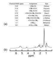
A 1.5 T clinical scanner was used for proton MR spectroscopy
(1H-MRS) of human pancreas allografts. The main
purpose was to estimate intracellular lipid content in
non-adipose pancreatic cells. The secondary aim was to
quantify total fat and choline-containing compounds.
Spectrum processing was performed in the time domain using
MRUI software package. It was demonstrated that 1H-MRS
is an effective method for non-invasive estimation of
intracellular lipid content in non-adipose pancreatic cells.
This knowledge could be helpful in studies of various
aspects of β-cell function (insulin production).
|
|
2390.
 |
Assessment and retrospective correction of rotation-induced
signal attenuation in diffusion-weighted spectroscopy
Michael Dacko1, Benjamin Knowles1,
Patrick Hucker1, Maxim Zaitsev1, and
Thomas Lange1
1Medical Physics, University Medical Center
Freiburg, Freiburg, Germany
Diffusion-weighted spectroscopy of the brain is a highly
motion-sensitive MR method as a consequence of the large
voxel size and low metabolite diffusion coefficients. In
this work, we correct for voxel displacement during DWS
experiments with prospective motion correction and
investigate the signal attenuation due to rotation-induced
intra-voxel dephasing. Phantom experiments with 'synthetic'
rotations confirmed the theoretically predicted signal
attenuation. High correlation between rotational motion and
attenuation of the residual water peak was observed in vivo.
Retrospective rejection criteria based on the recorded
motion tracking data and on the residual water peak
amplitude are compared.
|
|
|
|
|
2391.
 |
Fast automatic voxel positioning with non-rigid registrations
for improved between-subject consistency in MRS
Young Woo Park1, Dinesh K. Deelchand2,
James M. Joers2, Brian J. Soher3,
Peter B. Barker4, HyunWook Park1,
Gülin Öz2, and Christophe Lenglet2
1School of Electrical Engineering, Korea Advanced
Institute of Science and Technology, Daejeon, Korea,
Republic of, 2Department
of Radiology, Center for Magnetic Resonance Research,
University of Minnesota Medical School, Minneapolis, MN,
United States, 3Department
of Radiology, Duke University Medical Center, Durham, NC,
United States, 4Department
of Radiology, Johns Hopkins University School of Medicine,
Baltimore, MD, United States
During the typical acquisition of single-voxel Magnetic
Resonance Spectroscopy (MRS) the corresponding voxel-of-interest
(VOI) must be selected manually, which induces some degree
of variability. To address this, several automated VOI
positioning methods, using rigid registration and aimed at
follow-up scans of the same subject, have been proposed.
This approach can be generalized to cross-subject scans, but
with additional considerations for the anatomical
variability. We hypothesized that non-rigid registration
methods will minimize inter-subject variability in the
tissue content of the VOI. Here, we present an analysis of
registration strategies aimed at a reliable cross-subject
automatic VOI positioning for MRS data acquisition.
|
|
2392.
 |
MM-suppressed GABA measurements are highly susceptible to B0
field instability
Richard Anthony Edward Edden1,2, Ashley D. Harris1,2,3,4,
Nicolaas Puts1,2, Kimberly L. Chan1,2,5,
Michael Schar1, and Peter B. Barker1,2
1Russell H. Morgan Department of Radiology and
Radiological Science, The Johns Hopkins University,
Baltimore, MD, United States, 2F.M.
Kirby Center for Functional Brain Imaging, Kennedy Krieger
Institute, Baltimore, MD, United States, 3Department
of Radiology, University of Calgary, Calgary, AB, Canada, 4Hotchkiss
Brain Institute and Alberta Children's Hospital Research
Institute, Calgary, AB, Canada, 5Department
of Biomedical Engineering, The Johns Hopkins University,
Baltimore, MD, United States
J-difference-edited measurements of GABA are usually
contaminated up to 50% by macromolecular (MM) signal. It is
possible to suppress this signal using a symmetrical editing
motif, which relies upon partially inverting the MM signals
to an equal degree in the two halves of the edited
experiment. In the event of B0 field offset,
the symmetry breaks down and either positive or negative MM
signal rapidly contaminates the measured GABA signal. Here,
we investigate this issue using simulations and in vivo
experiments.
|
|
2393.
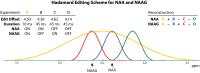 |
HERMES: Hadamard Encoding and Reconstruction of MEGA-Edited
Spectroscopy
Kimberly L Chan1,2,3, Nicolaas AJ Puts2,3,
Peter B Barker2,3, and Richard AE Edden2,3
1Biomedical Engineering, Johns Hopkins School of
Medicine, Baltimore, MD, United States, 2Radiology
and Radiological Science, Johns Hopkins School of Medicine,
Baltimore, MD, United States, 3F.M.
Kirby Center for Functional Brain Imaging, Kennedy Krieger
Institute, Baltimore, MD, United States
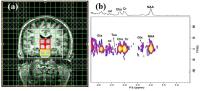
Hadamard Encoding and Reconstruction of MEGA-Edited
Spectroscopy, HERMES, is a novel method of the simultaneous,
separable detection of overlapping metabolite signals.
Classic J-difference editing involves the acquisition of two
subspectra, with editing pulses applied to the target
molecule (ON) or not (OFF). HERMES edits multiple
metabolites simultaneously by acquiring all combinations of
OFF/ON for each (i.e. four experiments to edit two
metabolites) and uses a Hadamard-like addition-subtraction
reconstruction to generate separate edited spectra for each
target metabolite. In this abstract, we describe the method
and demonstrate its application to NAA/NAAG editing, using
simulations, and phantom and in vivo experiments.
|
|
2394.
 |
Resolving Choline from Taurine in In-Vivo Magnetic Resonance
Spectra at 9.4 T
Marissa E. Fisher1, Brennen J. Dobberthien1,
Anthony G. Tessier1,2, and Atiyah Yahya1,2
1Department of Oncology, University of Alberta,
Edmonton, AB, Canada, 2Department
of Medical Physics, Cross Cancer Institute, Edmonton, AB,
Canada
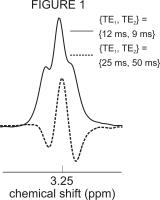
The Cho peak at 3.2 ppm contains significant signal
contamination from the taurine (Tau) resonance in rat and
mouse brain spectra, even at the high field strength of 9.4
T. The purpose of this work it to optimise TE1and
TE2 (echo
times) of a Point RESolved Spectroscopy (PRESS) sequence to
minimize Tau signal in the Cho spectral region at 9.4 T by
exploiting the J-coupling evolution of the Tau protons. The
determined optimal {TE1, TE2}
combination was found to be {25 ms, 50 ms}. The efficacy of
the timings was verified on rat brain in
vivo.
|
|
2395.
 |
Diffusion weighted MR spectroscopy without water suppression
allows to use water as inherent reference signal to correct for
motion-related signal drop
André Döring1, Victor Adalid Lopez1,
Vaclav Brandejsky1, Roland Kreis1, and
Chris Boesch1
1Depts. Radiology and Clinical Research,
University Bern, Bern, Switzerland
A non-water suppressed diffusion-weighting MR spectroscopy
sequence based on metabolite-cycling and STEAM is presented
and tested in-vitro and in-vivo. The water peak as an
inherent reference facilitates a post processing correction
of the signal drop induced in individual acquisitions by
cardiac and other motion. The correction leads to improved
spectral resolution on one hand, but more importantly also
to more accurate fitting of ADC values that are found to be
smaller than without correction and most likely closer to
the true values - and hence better suited for physiological
interpretation.
|
 |
2396.
 |
Sustained GABA reduction induced by anodal Transcranial Direct
Current Stimulation (tDCS) in motor cortex: A Proton Magnetic
Resonance Spectroscopy Study
Harshal Jayeshkumar Patel1, Sandro Romanzetti2,3,
Antonello Pellicano1, Kathrin Reetz2,3,
and Ferdinand Binkofski1,4
1Division of Clinical Cognitive Sciences,
Department of Neurology, RWTH Aachen University Hospital,
Aachen, Germany, 2Department
of Neurology, RWTH Aachen University Hospital, Aachen,
Germany, 3Jülich
Aachen Research Alliance (JARA) — Translational Brain
Medicine, Aachen and Jülich, Germany, 4Research
Center Jülich GmbH, Institute of Neuroscience and Medicine,
Jülich, Germany
Transcranial direct current stimulation (tDCS) modulates
cortical excitability. In this study we investigated long
term effects of anodal stimulation on inhibitory
neurotransmitter concentration using proton magnetic
resonance spectroscopy (MRS). Our results indicates that
excitatory tDCS cause locally reduction in GABA and it
remains in decreased state over a period of 60 minutes
presumably due to the decrease of activity of glutamic acid
decarboxylase(GAD)67.
|
|
2397.
 |
In Vivo Detection of Omega-3 Fatty Acids at 7 T with MEGA-sLASER
Lukas Hingerl1, Martin Gajdošík1,
Michal Považan1, Bernhard Strasser1,
Gilbert Hangel1, Martin Krššák1,
Siegfried Trattnig1,2, and Wolfgang Bogner1
1High Field MR Centre, Department of Biomedical
Imaging and Image-guided Therapy, Medical University of
Vienna, Vienna, Austria, 2Christian
Doppler Laboratory for Clinical Molecular MR Imaging,
Medical University of Vienna, Vienna, Austria
We present a method for detecting omega-3 fatty acids (FA)
at 7 T by 1H-MR spectroscopy (MRS) using a MEGA-sLASER
editing sequence with 12 kHz AFP GOIA-WURST(16,4) pulses for
localization. sLASER localization offers reduced sensitivity
to B1 inhomogeneities, lowers pulse power
requirements compared to PRESS or STEAM and the localization
pulses substantially reduce the 4-compartment effect. The
spectra of in vivo measurements at the echo times TE=332 ms
,465.4 ms and 1130 ms show the omega-3 signal very well.
|
|
2398.
 |
Cerebral Acetate Transport and Utilization in the Rat Brain in
vivo using 1H MRS: Consequences of a revised acetate volume of
distribution value
Masoumeh Dehghani M.1, Bernard Lanz1,
Nicolas Kunz2, Pascal mieville3, and
Rolf Gruetter1,2,4,5
1Laboratoire d'imagerie fonctionnelle et
métabolique(LIFMET), École Polytechnique Fédérale de
Lausanne (EPFL), Lausanne, Switzerland, 2Centre
d’Imagerie Biomedicale(CIBM), École Polytechnique Fédérale
de Lausanne (EPFL), Lausanne, Switzerland, 3Institute
of Chemical Sciences and Engineering, École Polytechnique
Fédérale de Lausanne (EPFL), Lausanne, Switzerland, 4Department
of Radiology, University of Lausanne, Lausanne, Switzerland, 5Department
of Radiology, University of Geneva, Geneva, Switzerland
Metabolic modeling of metabolite 13C
turnover curves in brain with 13C-labeled
acetate infused as tracer substrate requires prior knowledge
of the transport and uptake kinetics of Ace. The aim of this
study was to determine the kinetics of transport and
utilization for acetate uptake in the rat brain using
specific distribution volume of Ace(Vd) in the
rat brain. The dependency of estimated CMRace to
distribution volume of Ace in the rat brain highlights the
importance about a refined determination of Vd for
Ace in brain metabolic studies.
|
|
2399.
 |
Interleaved measurements of BOLD response and energy metabolism
in exercising human calf muscle
Adrianus J. Bakermans1, Chang Ho Wessel2,
Paul F.C. Groot1, Erik S.G. Stroes2,
and Aart J. Nederveen1
1Department of Radiology, Academic Medical
Center, Amsterdam, Netherlands, 2Department
of Vascular Medicine, Academic Medical Center, Amsterdam,
Netherlands
Typically, dynamic MR studies of exercising skeletal muscle
are limited to measurements of only one parameter. Obtaining
multiple parameters simultaneously during a single
experiment would provide more insight into
(patho-)physiology. Here, we report on interleaved
acquisitions of quantitative T2* maps
for assessments of the BOLD response, and 31P-MR
spectra for measuring phosphocreatine recovery kinetics
during an exercise-recovery protocol in healthy subjects and
peripheral artery disease (PAD) patients. We demonstrate
that with such interleaved acquisitions, it is feasible to
dynamically assess both tissue oxygenation as well as muscle
energy metabolism in the human calf muscle during a single
exercise session.
|
|
2400.
 |
Artificial intelligence for high-resolution nuclear MRS under
inhomogeneous magnetic fields
Qiu Wenqi1, Wei Zhiliang1, Ye Qimiao1,
Chen Youhe2, Lin Yulan1, and Chen
Zhong1
1Department of Electronic Engineering, Xiamen
University, Xiamen, China, People's Republic of, 2Department
of Mechanical and Electrical Engineering, Xiamen University,
Xiamen, China, People's Republic of
High-resolution multi-dimensional nuclear magnetic resonance
(NMR) spectroscopy serves as an irreplaceable and versatile
tool in various chemical investigations. In this study, a
method based on the concept of partial homogeneity is
developed to offer two-dimensional (2D) high-resolution NMR
spectra under inhomogeneous fields. Oscillating gradients
are exerted to encode the high-resolution information, and a
field-inhomogeneity correction algorithm based on pattern
recognition is designed to recover high-resolution spectra.
The proposed method improves performances of 2D NMR
spectroscopy under inhomogeneous fields without increasing
the experimental duration or significant loss in
sensitivity, and thus may open important perspectives for
studies of inhomogeneous chemical systems.
|
|
2401.
 |
2D Relaxometry and Diffusivity of Human Knee Synovial Fluid
after ACL-injuries Studied Using HR-MAS NMR
Kaipin Xu1, Subramaniam Sukumar1, John
Kurhanewicz1, and Xiaojuan Li1
1Radiology, University of California, San
Francisco, San Francisco, CA, United States
To better understand the pathological progression of
osteoarthritis (OA), techniques based on high resolution
magic angle spinning (HR-MAS) NMR spectroscopy are developed
for the study of relaxation times (T1, T2, and T1ρ) and
diffusion coefficient (D) of human knee synovial fluids (SF)
harvested from 1 OA and 8 anterior cruciate ligament (ACL)
injured patients.
|
|
2402.
 |
Towards fast and highly localized spectroscopy using
miniaturized coils in a 14.1T animal scanner
Marlon Arturo Pérez Rodas1,2, Jörn Engelmann1,
Hellmut Merkle1, Rolf Pohmann1, and
Klaus Scheffler1,3
1Ultra High-field Magnetic Resonance Center, Max
Planck Institute for Biological Cybernetics, Tübingen,
Germany, 2Graduate
Training Centre of Neuroscience, IMPRS for Cognitive and
Systems Neuroscience, University of Tübingen, Tübingen,
Germany, 3Department
for Biomedical Magnetic Resonance, University of Tübingen,
Tübingen, Germany
The distinction of functional activity between cortical
layers in the brain by MRI or MRS requires high spatial and
temporal resolution. High spatial resolution can be achieved
by increasing the gradient strength or by using the
intrinsic volume selectivity of miniature coils, even in
conventional animal scanner. In the present work, initial
results for highly-localized spectroscopy within seconds are
presented, for a phantom metabolite solution and cell
cultures in a 14.1T animal scanner using a 2mm-diameter
circular coil. The larger signals from the major metabolites
in ~1.5µL were detected in 24sec on the phantom solution
with an acceptable SNR.
|
|
2403.
 |
DRESS localized FAST technique at 7T uncovers the relation
between mitochondrial capacity and ATP synthase flux in
exercising gastrocnemius medialis muscle
Marjeta Tušek Jelenc1,2, Marek Chmelík1,2,
Barbara Ukropcová3,4, Wolfgang Bogner1,2,
Siegfried Trattnig1,2, Jozef Ukropec4,
Martin Krššák1,2,5, and Ladislav Valkovic1,2,6,7
1High Field MR Centre, Department of Biomedical
Imaging and Image-guided Therapy, Medical University of
Vienna, Vienna, Austria, 2Christian
Doppler Laboratory for Clinical Molecular MR Imaging,
Vienna, Austria,3Institute of pathophysiology,
Faculty of Medicine, Comenius University, Bratislava,
Slovakia, 4Obesity
section, Diabetes and Metabolic Disease Laboratory,
Institute of Experimental Endocrinology, Slovak Academy of
Sciences, Bratislava, Slovakia, 5Division
of Endocrinology and Metabolism, Department of Internal
Medicine III, Medical University of Vienna, Vienna, Austria, 6Department
of Imaging Methods, Institute of Measurement Science, Slovak
Academy of Sciences, Bratislava, Slovakia, 7University
of Oxford Centre for Clinical Magnetic Resonance Research,
University of Oxford, John Radcliffe Hospital, Oxford,
United Kingdom
The aim of the study was to investigate the relation between
the maximum oxidative flux (Qmax), a valid
measure of muscular mitochondrial capacity and ATP synthase
flux (FATP) measured in exercising gastrocnemius
medialis muscle in healthy young and elderly subjects.
Furthermore, we explored the possibility of direct
measurement of both, Qmax and
FATP_ex, in a single experiment. The dynamic
experiment consisted of the acquisition of baseline data
during two minutes of rest, six minutes of aerobic plantar
flexion exercise (during which a 3.5 minutes long FAST
measurement was performed), and six minutes of recovery. Our
data showed significant correlation between ATP synthase
flux in exercising muscle and maximal oxidative flux.
|
|