|
Combined Educational & Scientific Session: Rapid Three-Dimensional (3D) MSK Imaging
Skill Level: Intermediate
to Advanced
Organizers: Jenny T. Bencardino, M.D., Eric Y. Chang, M.D., Christine Chung, M.D., Ravinder R. Regatte, Ph.D., Philip Robinson, M.D. & Siegfried Trattnig, M.D.
Thursday 12 May 2016 |
16:00
|
|
Rapid 3D-MSK Imaging: Techniques & Challenges 
Martijn Cloos1
1Bernard and Irene Schwartz Center for
Biomedical Imaging, and Center for Advanced Imaging
Innovation and Research (CAI2R), Department of
Radiology, New York University School of Medicine, New
York, NY, United States
In this talk we will discuss the pros and cons of 3D MSK
imaging from a technical prospective. Using select
examples, we will explore how the transition from 2D
(slice-selective) to 3D (volumetric) imaging influences
the contrast, resolution and acquisition time. The
presentation will start with the fundamental principles
of 3D imaging from which we will buildup to the latest
developments, such as compressed sensing and magnetic
resonance fingerprinting.
|
16:30
|
|
Clinical Applications of 3D-MSK Imaging
Richard Kijowski
This lecture will review the clinical applications of
three-dimensional sequences FSE sequences in
musculoskeletal MR imaging.
|
17:00
 |
1060.
 |
Fast single sequence comprehensive 4D pediatric knee MRI
with T2 Shuffling
Shanshan Bao1, Jonathan I. Tamir2,
Umar Tariq3, Martin Uecker4, Peng
Lai5, Weitian Chen5, Michael
Lustig2, and Shreyas S. Vasanawala1
1Radiology, Stanford University, Stanford,
CA, United States, 2University
of California, Berkeley, Berkeley, CA, United States, 3Geisinger
Medical Center, Danville, PA, United States, 4University
Medical Center Göttingen, Göttingen, Germany, 5GE
Healthcare, Menlo Park, CA, United States
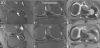
Clinical application of volumetric joint MR imaging has
been hampered by blurring due to T2 decay. A
redesigned volumetric fast spin-echo
acquisition technique termed T2 shuffling corrects for
T2 decay and yields effectively a four-dimensional
reconstruction with varying degrees of T2 weighting. Our
work assesses the clinical application of T2 shuffling
for pediatric knee MRI. Our results show that T2
shuffling has the potential to suffice as a single
sequence MR examination. This is especially relevant for
pediatric imaging where streamlined protocols greatly
improve clinical operations and patient experience.
|
17:15
 |
1061.
 |
Rapid Three-Dimensional Fast Spin-Echo Knee Imaging Using
Compressed Sensing
Fang Liu1, Humberto Rosas1, James
Holmes2, Kevin King2, Rob Peters2,
and Richard Kijowski1
1Department of Radiology, University of
Wisconsin-Madison, Madison, WI, United States, 2Applied
Science Laboratory, GE Healthcare, Waukesha, WI, United
States
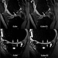
A Cube 3D-FSE sequence was performed with and without
compressed sensing (CS) twice on the knees of 10
asymptomatic volunteers to assess signal-to-noise- ratio
(SNR) and once on the knees of 25 symptomatic patients
to assess diagnostic performance for detecting knee
joint pathology. CS k-space acceleration provided a 30%
reduction in scan time without a corresponding decrease
in SNR. The use of CS resulted in mild increased image
blurring which did not influence diagnostic performance
with near perfect to perfect agreement between Cube and
Cube-CS for detecting knee joint pathology.
|
17:30
|
1062.
 |
A Variable-TE Stack-of-Spirals Sequence for 3D UTE Imaging
Samuel Fielden1, John Mugler2,
Wilson Miller2, Alto Stemmer3,
Josef Pfeuffer3, Berthold Kiefer3,
and Craig Meyer1,2
1Biomedical Engineering, University of
Virginia, Charlottesville, VA, United States, 2Radiology
& Medical Imaging, University of Virginia,
Charlottesville, VA, United States, 3Application
Development, Siemens Healthcare, Erlangen, Germany
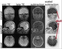
While 3D radial-based methods have become established in
recent years for ultrashort-echo-time (UTE) imaging,
these acquisitions are generally slow due to the
inefficiency of radial k-space trajectories. The purpose
of this work was to implement a fast UTE acquisition
based on an optimized 3D stack-of-spirals acquisition
and to perform a proof-of-concept evaluation of the
method for bone imaging of the skull and cartilage
imaging of the knee.
|
17:45
|
1063.
 |
High spatial and temporal resolution DCE-MRI of
intervertebral disc endplates using GRAPPA accelerated
3D-Linogram acquisition
L. Tugan Muftuler1,2, Ali Ersoz3,
and Volkan Emre Arpinar1
1Department of Neurosurgery, Medical College
of Wisconsin, Milwaukee, WI, United States, 2Center
for Imaging Research, Medical College of Wisconsin,
Milwaukee, WI, United States, 3Department
of Biophysics, Medical College of Wisconsin, Milwaukee,
WI, United States
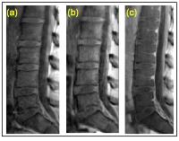
It is suggested that disruption of nutrient delivery
through the intervertebral disc endplates could lead to
physiological and morphological changes in the discs.
Our earlier DCE-MRI studies demonstrated major changes
in endplate regions. However, we had to sacrifice
temporal resolution to obtain high spatial resolution to
image the thin endplates. Higher temporal resolution is
needed for quantitative analysis of tracer kinetics.
Therefore, we developed and tested 3D-Linogram
acquisition technique that allowed higher temporal
resolution and reduced motion artifacts. Tofts’ tracer
kinetic model was implemented and Ktrans values from
vertebral endplates were estimated.
|
18:00
|
|
Adjournment & Meet the
Teachers |
|