16:00
|
|
Compositional Mapping
Techniques 
Xiaojuan Li
|
16:30
|
|
Advanced Cartilage Imaging: Clinical Applications
Michel Crema
Advanced MRI techniques enable evaluation of the
biochemical composition of articular cartilage.
Compositional MRI techniques have the potential to
supplement clinical MRI sequences in identifying
cartilage degeneration at an earlier stage than is
possible today using morphologic sequences only.
Although there is some evidence regarding the
relationship with some compositional MRI techniques
(mainly T2 mapping, T1rho, and dGEMRIC) with symptoms
and progression of disease, additional work is needed to
isolate the role of the different compositional MRI
techniques in predicting structural and clinical
outcomes taking into account feasibility of application,
reliability and responsiveness of the different
techniques available today.
|
17:00
|
0536.
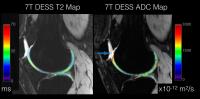 |
Comparison of DESS T2 Relaxation Times and Apparent
Diffusion Coefficient in Articular Cartilage at 3T and 7T
Garry E Gold1, Bragi Sveinsson2,
Kevin Eppersson2, Akshay Chaudhari3,
Marcus Alley2, Daehyun Yoon2,
Brian A Hargreaves3, and Feliks Kogan2
1Radiology, Bioengineering, and Orthopedic
Surgery, Stanford University, Stanford, CA, United
States, 2Radiology,
Stanford University, Stanford, CA, United States, 3Radiology
and Bioengineering, Stanford University, Stanford, CA,
United States
Double-echo Steady-Sate (DESS) is an efficient 3D
approach to measure cartilage thickness, T2, and
apparent diffusion coefficient (ADC). We tested the
DESS sequence at 3T and 7T in healthy volunteers. DESS
can acquire accurate cartilage T2 and ADC values at both
3T and 7T, with more consistent ADC measurements at 7T,
likely due to less image noise in the fit.
|
17:12
|
0537.
 |
DESS T2 mapping in Knee Cartilage at Supine and Standing
Positions in an Upright MR Scanner
Andrew C Yung1, Reza Nickmanesh2,
Piotr Kozlowski1,3, and David R Wilson2,4
1UBC MRI Research Centre, University of
British Columbia, Vancouver, BC, Canada, 2Centre
for Hip Health and Mobility, University of British
Columbia, Vancouver, BC, Canada, 3Radiology,
University of British Columbia, Vancouver, BC, Canada, 4Department
of Orthopaedics, University of British Columbia,
Vancouver, BC, Canada
With the use of an upright open MR scanner, we
demonstrate knee cartilage T2 mapping using DESS in a
true standing position for the first time, and have
shown preliminary evidence that there may be differences
between loading the joint in the standing position
versus the supine loaded and unloaded case. The
volumetric DESS T2 maps were acquired with short
acquisition time which is critical for imaging
weightbearing postures, while maintaining a range of T2
values that were similar to gold-standard T2 maps
generated by a multi-spin-echo sequence.
|
17:24
|
0538.
 |
Texture analysis of T2 relaxation time maps reveals
degenerative changes in articular cartilage: Oulu Knee
Osteoarthritis study
Arttu Peuna1,2,3, Joonas Hekkala1,3,
Marianne Haapea1,2, Jana Podlipska1,3,
Ali Guermazi4, Miika T Nieminen1,2,3,
Simo Saarakkala1,2,3, and Eveliina
Lammentausta1,2
1Medical Research Center, University of Oulu
and Oulu University Hospital, Oulu, Finland, 2Department
of Diagnostic Radiology, Oulu University Hospital, Oulu,
Finland, 3Research
group of Medical Imaging, Physics and Technology,
University of Oulu, Oulu, Finland, 4Department
of Radiology, Boston University School of Medicine,
Boston, MA, United States
Gray level co-occurrence matrix based texture analysis
is a sensitive image processing method that probes the
spatial information from knee MR T2 maps and of the
changes caused by osteoarthritis (OA). Texture analysis
can distinguish symptomatic patients from healthy
control subjects more sensitively than regional mean T2
analysis, and provides additional information also when
compared to clinical evaluations such as MOAKS. Advanced
learning algorithms can be further utilized to classify
asymptomatic and OA subjects.
|
17:36
|
0539.
 |
Use of comprehensive MRI to assess cartilage composition in
patients with acute cartilage injury
Didier Laurent1, Stefan Zbyn2,
Vladimir Mlynarik2, Markus Schreiner2,
Pavol Szomolanyi2, Nicole Getzmann1,
Harry Haber1, Joerg Goldhahn1,
Stefan Marlovits3, and Siegfried Trattnig2
1Novartis Institutes fo Biomedical Research,
Basel, Switzerland, 2Department
of Biomedical Imaging and Image-Guided Therapy, Medical
University of Vienna, Vienna, Austria, 3Department
of Traumatology, Medical University of Vienna, Vienna,
Austria
A comprehensive MRI approach was implemented to assess
cartilage macromolecular composition in patients with
acute cartilage injury. Differences in T2 relaxation and
gagCEST asymmetry values were observed between the
defective and adjacent regions in the tibio-femoral
cartilage. Preliminary results indicate that the
combination of T2 mapping with gagCEST scans at 7T may
be reproducible and sensitive enough to monitor early
cartilage degeneration, and thus may be considered as a
good alternative to cartilage biopsies in future
clinical trials on new therapies aimed at cartilage
regeneration.
|
17:48
|
0540.
 |
A New High-resolution 3D gagCEST Imaging method for In Vivo
Human Knee Cartilage at 7T
Guruprasad Krishnamoorthy1, Ravi Prakash
Reddy Nanga1, Puneet Bagga1, Hari
Hariharan1, and Ravinder Reddy1
1Center for Magnetic Resonance and Optical
Imaging, Department of Radiology, University of
Pennsylvania, Philadelphia, PA, United States
Osteoarthritis (OA), one of the most prevalent
musculoskeletal conditions, affects a large number of
people around the world with an increased risk on an
even larger number of people getting affected by it in
the future [1]. GAG chemical exchange saturation
transfer (gagCEST) is a promising MRI technique to
non-invasively quantify GAG content present in the
cartilages [2]. In this study, a new burst mode
magnetization preparation 3D gagCEST technique was
developed which provided high-resolution gagCEST maps of
knee cartilages in practically achievable scan times at
7T with more than twice the sensitivity of the
previously reported steady-state saturation 3D gagCEST
study [5].
|
18:00
|
|
Adjournment & Meet the
Teachers |
|