ISMRM 24th Annual Meeting & Exhibition • 07-13 May 2016 • Singapore |
|
Weekend Educational Course: Clinical Cancer MRI: Case-Based
Skill Level: Basic to Intermediate
Organizers: Linda Moy, M.D. & Valeria Panebianco, M.D.
Sunday 08 May 2016 |
Overview
MRI has a major role in a number of cancer sites. Reporting standards
and guidelines have been established. Recognition of when to use MRI,
how to report it and where MRI may address unmet needs are the focus of
this course. This will be illustrated through case-based learning.Target Audience
Physicians, Imaging scientists/engineers, technologists and other health
professionals with a developing need for utilizing MRI applications in
cancer and personalized care.
Educational Objectives
Upon completion of this course, participants should be able to:
- Understand guidelines and
standards for MRI of multiple tumour types;
- Recognize potential
applications of new MRI technology to better diagnose and assess
therapy response in cancer patients; and
- Understand the tumour
microenvironment and the potential of MRI to assess
microenvironment.
|
|
PROGRAM |
| |
|
|
Guidelines & Reporting Standards |
|
| |
|
|
Moderators:
Linda Moy, Valeria Panebianco |
|
|
|
| |
|
|
|
|
| |
|
|
Addressing Clinical Needs |
|
| |
|
|
Moderators:
Linda Moy, Valeria Panebianco |
|
10:15
|
|
Overdiagnosis & Over Treatment - Permission Withheld
Christiane Kuhl
Overdiagnosis is an important issue in oncologic
radiology. Avoiding diagnosis of disease altogether in
order to avoid overdiagnosis is, however, probably not
the best solution to the problem. Choosing appropriate
Treatment based on Imaging as well as proteomic and
genomic Information is probably more useful. Moreover,
overdiagnosis is not the most important concern of
current Screening programs - rather, under-diagnosis is.
MRI is probably the best method to avoid both, over- as
well as underdiagnosis
|
10:45
|
|
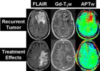
Tumour Recurrance & Pseudo-Progression in Glioma 
Alberto Bizzi
|
11:15
|
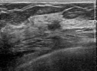 |
Finding Cancer in the Dense Breast: Ultrasound & MRI - Permission Withheld
Nariya Cho1
1Department of Radiology, Seoul National
University College of Medicine, Seoul National
University Hospital, Seoul, Korea, Republic of
Features of undiagnosed breast cancers on prior
screening US and screening MRI of patients with breast
cancers diagnosed on subsequent screening examinations
will be presented.
|
11:45
|
|
Roundtable |
12:00
|
|
Lunch & Meet the Teachers |
|
| |
|
|
|
|
| |
|
|
Tumour Microenvironment |
|
| |
|
|
Moderators:
Utaroh Motosugi, Harriet Thoeny |
|
13:30
|
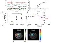 |
Tumour Microenvironment
Ralph Peter Mason1
1Radiology, University of Texas Southwestern
Medical Center, Dallas, TX, United States
Historically, radiology/imaging has served to identify
tumors in terms of location, size, and metastatic
spread. It is increasingly recognized that tumors may
exhibit very different micro environmental
characteristics, which can influence therapeutic
success. A new goal is precision oncology, whereby
individual tumors are further characterized based on
potential prognostic imagine biomarkers. Tumor hypoxia
is associated with aggressive phenotypes and resistance
to therapy and may be the most significant factor
influencing therapy outcomes for solid tumors. Many NMR
approaches are being developed and evaluated to measure
tumor oxygenation. This review will consider human
applications of oxygen sensitive MRI in the context of
pre-clinical developments. Strengths and weaknesses in
terms of temporal and spatial resolution, precision and
accuracy, ease of implementation and robustness of
observations will be considered. Methods may provide
qualitative or quantitative insights including dynamic
response to interventions.
|
14:00
|
|
Collagen & Stroma - Permission Withheld
Kristine Glunde, Samata Kakkad, and Zaver M. Bhujwalla
The tumor stroma, and in particular the Col1 fiber
meshwork, plays an important role in cancer migration
and metastasis. Novel MRI approaches such as
macromolecular contrast agent based DCE MRI and DTI can
be applied to noninvasively detect critical features of
the Col1 fiber network in tumors.
|
14:30
|
 |
Tumor Associated Inflammation: Biology & Imaging
Heike Elisabeth Daldrup-Link1
1Department of Radiology, Stanford
University, Stanford, CA, United States
|
15:00
|
|
Roundtable |
15:15
|
|
Break & Meet the Teachers |
|
| |
|
|
|
|
| |
|
|
New Horizons |
|
| |
|
|
Moderators:
Utaroh Motosugi, Harriet Thoeny |
|
15:30
|
 |
A semi-quantitative overview of tumor CEST MRI
Phillip Zhe Sun1
1Martinos Center, MGH and Harvard Medical
School
Tumor CEST MRI has emerged as a molecular imaging
approach to characterize complex microenvironment,
including protein/peptide, glutamate, exogenous glucose
and artificial reporter gene MRI. Despite their diverse
names, variant CEST imaging methods provide
complementary information about the underlying tumor
pathophysiology and it is helpful to provide a
semi-quantitative overview to understand their potential
clinical applications.
|
16:00
|
 |
Radiomics the New Buzzword
Radka Stoyanova1
1University of Miami, FL, United States
“Radiomics” refers to the extraction and analysis of
large amounts of advanced quantitative imaging features
from medical images using high throughput methods. In
this syllabus MRI radiomics features and extraction are
described; second, examples of applications of radiomics
in glioblastoma multiforme (GBM) and prostate cancer are
reviewed and lastly the importance of incorporating
radiomics features in clinical databases is discussed.
|
16:30
|
|
Interventional MRI of
Cancer
Carlo Catalano
|
17:00
|
|
Roundtable |
17:15
|
|
Adjournment & Meet the
Teachers |
|
| |
|
|
|
|
| |
The International Society for Magnetic Resonance in Medicine is accredited by the Accreditation Council for
Continuing Medical Education to provide continuing medical education for physicians. |